
Aortic stenosis its pre anaesthesia evaluation and anaesthetic management for non cardiac surgery 20 mark answer
aortic stenosis pathophysiology diagram LV hypertrophy
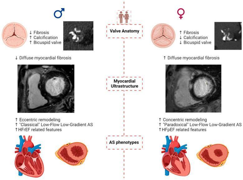
A comparative medical infographic detailing the sex-based differences in the pathophysiology and presentation of aortic stenosis (AS). The diagram is divided into three main rows: Valve Anatomy, Myocardial Ultrastructure, and AS Phenotypes, with males on the left and females on the right. In 'Valve Anatomy', males exhibit a higher prevalence of bicuspid valves with greater calcification and less fibrosis, as shown in anatomical diagrams and axial CT scans. Females show less calcification but higher levels of valvular fibrosis. Under 'Myocardial Ultrastructure', cardiac MRI frames illustrate that males typically present with diffuse myocardial fibrosis and eccentric left ventricular (LV) remodeling. Females demonstrate increased diffuse myocardial fibrosis and concentric LV remodeling. The 'AS Phenotypes' section contrasts 'Classical' Low-Flow Low-Gradient AS in males, often associated with Heart Failure with reduced Ejection Fraction (HFrEF), against 'Paradoxical' Low-Flow Low-Gradient AS in females, associated with Heart Failure with preserved Ejection Fraction (HFpEF). Cross-sectional heart diagrams reinforce these remodeling patterns, showing a dilated LV in the male phenotype and a thickened, small-cavity LV in the female phenotype.
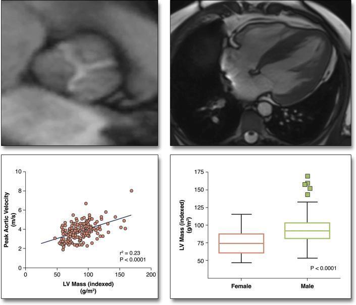
A multi-panel figure illustrating the hypertrophic response in patients with aortic stenosis. Top left: A Cardiac Magnetic Resonance (CMR) image in the short-axis view shows a thickened, calcified aortic valve. Top right: A four-chamber CMR view demonstrates significant left ventricular (LV) wall thickening consistent with concentric hypertrophy. Bottom left: A scatter plot correlates peak aortic velocity (m/s) with indexed LV mass (g/m²). It shows a modest positive correlation (r² = 0.23, p < 0.0001), indicating that while higher stenosis severity generally relates to higher LV mass, it only partially explains the variance in the hypertrophic response. Bottom right: A box-and-whisker plot compares indexed LV mass between sexes. The data indicates that males have significantly higher indexed LV mass compared to females (p < 0.0001), with a higher median and more frequent high-value outliers. Together, these panels illustrate that sex and stenosis severity are key determinants of LV remodeling in aortic stenosis.
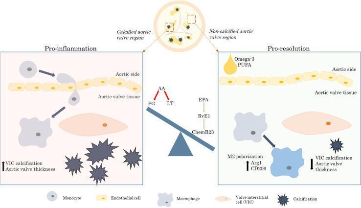
This pathophysiology diagram illustrates the cellular and molecular mechanisms of aortic valve stenosis, specifically contrasting 'Pro-inflammation' and 'Pro-resolution' pathways. The central overview shows a cross-section of an aortic valve with highlighted calcified and non-calcified regions. In the 'Pro-inflammation' panel, the diagram depicts monocyte transendothelial migration from the aortic side into the valve tissue, where they differentiate into macrophages that interact with valvular interstitial cells (VICs). This state is characterized by increased VIC calcification (shown as dark starburst shapes) and increased aortic valve thickness, driven by arachidonic acid (AA) metabolites like prostaglandins (PG) and leukotrienes (LT). Conversely, the 'Pro-resolution' panel demonstrates the therapeutic signaling axis involving Omega-3 polyunsaturated fatty acids (PUFA), eicosapentaenoic acid (EPA), resolvin E1 (RvE1), and the ChemR23 receptor. This axis promotes M2 macrophage polarization (marked by Arg1 and CD206), leading to a reduction in VIC calcification and valve thickness. The visual comparison highlights the role of specialized pro-resolving mediators in mitigating valvular disease progression by shifting the balance away from inflammatory lipid mediators.
Aortic Stenosis: Pre-Anaesthesia Evaluation and Anaesthetic Management for Non-Cardiac Surgery
1. Introduction and Pathophysiology
| Parameter | Mild | Moderate | Severe |
|---|---|---|---|
| Peak AV velocity | 2.6-3.0 m/s | 3-4 m/s | >4 m/s |
| Mean pressure gradient | <20 mmHg | 20-40 mmHg | >40 mmHg |
| Aortic valve area (AVA) | >1.5 cm² | 1.0-1.5 cm² | <1.0 cm² |
- Diastolic dysfunction - reduced LV compliance, elevated LVEDP
- Myocardial ischaemia risk - increased LV mass raises O2 demand; elevated LVEDP reduces coronary perfusion pressure (CPP = Aortic diastolic pressure - LVEDP); subendocardial ischaemia can occur even without coronary artery disease
- Preload dependence - stroke volume depends heavily on adequate preload; reduced LV compliance means the "atrial kick" of sinus rhythm contributes up to 40% of total cardiac output
- Fixed afterload - the obstructing valve determines afterload; compensatory increase in stroke volume is impossible
- Angina - prognosis ~5 years after onset
- Syncope - ~3 years
- Dyspnoea / Heart failure - ~2 years (worst prognosis)
2. Pre-Anaesthesia Evaluation
A. History
- Presence and duration of cardinal symptoms (angina, syncope, dyspnoea, reduced exercise tolerance)
- Functional status: METS (metabolic equivalents) - if patient cannot climb one flight of stairs (>4 METS), risk is high
- Existing cardiac comorbidities: coronary artery disease, hypertension, arrhythmias, prior valve intervention (SAVR or TAVR)
- Medications: anticoagulants, antiplatelet agents, beta-blockers, diuretics
- History of infective endocarditis or rheumatic fever
B. Physical Examination
- Murmur: Harsh crescendo-decrescendo systolic ejection murmur, best heard at right upper sternal border, radiating to the neck and carotid arteries. Absence of radiation to right clavicle has negative likelihood ratio of 0.1 for AS
- Pulse: Delayed carotid upstroke (pulsus parvus et tardus), low-volume pulse, narrow pulse pressure (may be 50 mmHg or less even with normal EF)
- Paradoxically split S2 (in severe AS)
- Signs of LV failure: elevated JVP, pulmonary crepitations, S3 gallop, peripheral oedema
C. Investigations
- Left ventricular hypertrophy (LVH), often with strain pattern (ST depression, T-wave inversion in lateral leads)
- Left axis deviation
- Left bundle branch block (LBBB)
- Cardiomegaly in decompensated disease
- Aortic valve calcification (lateral view)
- Post-stenotic dilatation of ascending aorta
- Pulmonary venous congestion in heart failure
- The cornerstone of pre-operative evaluation
- Confirms diagnosis, quantifies severity (AVA, mean gradient, peak velocity), assesses LVEF
- Evaluates LV wall thickness, diastolic function, any coexistent valvular disease
- If LV dysfunction is present, dobutamine stress echo distinguishes true severe AS from "low-flow, low-gradient" pseudo-AS
- Full blood count, coagulation screen - acquired von Willebrand syndrome occurs in 67-92% of patients with severe AS (due to mechanical disruption of VWF multimers by turbulent flow); may cause significant perioperative bleeding
- Renal function, electrolytes, BNP/NT-proBNP
- Coronary angiography if symptoms suggest CAD or before AVR
D. Risk Stratification and Decision to Proceed
| Clinical Scenario | Recommendation |
|---|---|
| Asymptomatic severe AS, preserved EF | Proceed with elective non-cardiac surgery with appropriate monitoring |
| Symptomatic severe AS | Consider AVR (surgical or TAVR) before elective non-cardiac surgery |
| Asymptomatic severe AS + EF <50% scheduled for high-risk surgery | Prior AVR recommended (European guidelines) |
| Emergency non-cardiac surgery | Proceed with surgery regardless of AS severity; optimise haemodynamics |
| High-risk / ineligible for AVR | Percutaneous balloon aortic valvuloplasty (PBAV) as bridge; TAVR if feasible |
3. Anaesthetic Management
A. Haemodynamic Goals
| Parameter | Goal | Rationale |
|---|---|---|
| Preload | Maintain or increase | AS is preload-dependent; hypovolaemia reduces stroke volume drastically |
| Heart rate | Normal (60-80 bpm) | Tachycardia: increases O2 demand, reduces diastolic filling time; Bradycardia: CO falls (fixed stroke volume) |
| Rhythm | Sinus | Atrial kick contributes up to 40% of CO; AF is very poorly tolerated |
| Afterload | Maintain or increase | Hypotension reduces CPP; the hypertrophied LV is critically dependent on diastolic blood pressure |
| Contractility | Normal to mildly decreased | Preserved early; may be reduced late. Avoid negative inotropes |
| Myocardial O2 consumption | Minimise | Avoid tachycardia; treat hypotension promptly |
B. Pre-operative Optimisation
- Continue beta-blockers (do NOT stop abruptly)
- Continue cardiac medications up to the morning of surgery
- Correct anaemia, electrolyte disturbances
- Optimise heart failure if present (diuresis cautiously - maintain adequate preload)
- Anxiolysis with careful premedication (avoid respiratory depression causing hypoxia which increases myocardial demand; benzodiazepines in small doses are acceptable)
C. Monitoring
- Intra-arterial blood pressure line - place before induction (preinduction arterial monitoring is strongly recommended given the haemodynamic sensitivity)
- ECG with ST-segment monitoring (leads II and V5)
- SpO2, ETCO2, temperature
- Invasive haemodynamic monitoring:
- Intra-arterial line is standard
- Pulmonary artery catheter (PAC): considered for major high-risk surgery or LV dysfunction; note - arrhythmias during PAC insertion can be catastrophic in AS (coronary perfusion can be severely compromised)
- Transoesophageal echocardiography (TEE): preferred for real-time monitoring of preload, wall motion abnormalities, valve function, and CO. The midesophageal short-axis and long-axis views assess AV, while transgastric views allow Doppler gradient measurement
D. Induction of Anaesthesia
- The most dangerous phase - sudden vasodilatation causes hypotension, reducing CPP
- Slow, careful titrated induction is preferable over rapid bolus techniques
- Drug choices for induction:
- Etomidate - most haemodynamically stable; preferred for compromised patients (minimal effect on SVR and CO)
- Ketamine - sympathomimetic; maintains SVR; useful if LV is decompensated (beware tachycardia)
- Propofol - use cautiously in reduced doses; causes significant vasodilatation and hypotension; avoid large boluses
- Thiopentone - avoid (profound vasodilatation)
- Opioids - high-dose opioid-based induction (fentanyl/sufentanil) blunts the sympathetic response to laryngoscopy with minimal haemodynamic effects; use as an adjunct
- Avoid succinylcholine if tachycardia is a concern; rocuronium preferred
E. Maintenance of Anaesthesia
-
Neuraxial blocks (spinal/epidural): Used with caution
- Spinal anaesthesia causes abrupt sympathectomy and vasodilatation - sudden SVR drop is poorly tolerated; may precipitate cardiovascular collapse in severe AS
- Epidural anaesthesia: if used, slow titration of epidural with dilute local anaesthetics is safer; allows gradual onset of block; preferred over single-shot spinal for patients with moderate-severe AS
- Some guidelines consider neuraxial blockade a relative contraindication in severe AS
-
General anaesthesia:
- Volatile agents (isoflurane, sevoflurane, desflurane): cause dose-dependent vasodilatation; sevoflurane is preferred due to smoother induction; maintain at lowest effective concentration
- Isoflurane has the advantage of coronary vasodilation (ischaemic preconditioning) but causes SVR reduction
- Total IV anaesthesia (TIVA) with propofol/remifentanil: propofol must be used cautiously; remifentanil provides excellent analgesia and haemodynamic control
- Muscle relaxants: vecuronium and rocuronium are preferred (minimal cardiovascular effects); avoid pancuronium (tachycardia); avoid atracurium (histamine release, vasodilatation)
-
Key intraoperative principles:
- Maintain adequate preload - liberal fluid administration; avoid hypovolaemia
- Treat hypotension promptly with phenylephrine (vasopressor of choice - pure alpha-agonist, raises diastolic BP and CPP without causing tachycardia; avoids reflex tachycardia seen with ephedrine)
- Vasopressin is an alternative vasopressor
- Avoid epinephrine-containing local anaesthetics (tachycardia risk)
- Treat tachycardia (HR >90 bpm) aggressively - IV esmolol, metoprolol
- Maintain sinus rhythm - DC cardioversion if AF with haemodynamic compromise; amiodarone for pharmacological rhythm control
- Avoid sudden large increases in heart rate (airway manipulation, surgical stimulation); deepen anaesthesia or use opioids before stimulating manoeuvres
F. Post-operative Management
- Extended monitoring in HDU/ICU for moderate-severe AS undergoing major surgery
- Continue invasive arterial monitoring for early detection of haemodynamic changes
- Maintain analgesia (pain causes sympathetic activation and tachycardia)
- Adequate IV fluids to maintain preload; avoid dehydration from prolonged fasting or diuretics
- Vigilance for postoperative arrhythmias (AF is common post-operatively)
- Early resumption of cardiac medications
- Anticoagulation if prosthetic valve or AF
- Watch for heart failure exacerbation, myocardial infarction, and renal dysfunction in the postoperative period
4. Special Considerations
| Scenario | Management |
|---|---|
| Emergency surgery | Proceed regardless of AS severity; aggressive invasive monitoring, haemodynamic optimisation, ICU post-op |
| TAVR/SAVR prior to elective surgery | If symptomatic severe AS needs major surgery, TAVR/SAVR first; otherwise proceed with caution |
| Acquired vWF syndrome | Screen coagulation; have FFP/cryoprecipitate available; AVR corrects the vWF deficiency |
| Concomitant CAD | 50% of severe AS patients have significant CAD; may require coronary revascularisation simultaneously or prior to surgery |
| Low-flow, low-gradient AS | Use dobutamine stress echo to differentiate true vs pseudo-AS; affects surgical risk stratification |
Summary: Key Principles
- Classify AS severity pre-operatively using echocardiography
- Symptomatic severe AS - consider valve replacement before elective surgery
- Haemodynamic goals: normal heart rate (sinus rhythm), maintain/increase preload and afterload, avoid vasodilation and tachycardia
- Preinduction arterial line is standard; TEE strongly recommended
- Etomidate is the induction agent of choice; avoid thiopentone
- Phenylephrine is the vasopressor of choice for hypotension
- Spinal anaesthesia is relatively contraindicated in severe AS; epidural with slow titration is safer if regional technique needed
- ICU/HDU care post-operatively for major surgery