
I have a doubt in large intestine the longitudinal muscle is modified so it is completely modified I mean the longitudinal muscle wholelyis modified into teaniea vc li or only a few parts of it is modified and a few parts still remain as longitudinal muscle?
taeniae coli longitudinal muscle large intestine colon anatomy
Taeniae Coli and the Longitudinal Muscle Layer
What exactly happens:
-
A thin, continuous sheet of longitudinal muscle that runs all the way around the circumference of the colon (this is the true background layer that persists everywhere)
-
Three thickened/condensed bands called the taeniae coli - these are areas where the longitudinal muscle is packed together and reinforced with extra collagen and elastic tissue
"The outer layer is thin, except where it is condensed into three narrow bands called the taeniae coli that contain more collagen and elastic tissue than muscle."
- Grainger & Allison's Diagnostic Radiology, Large Intestine Anatomy section
To directly answer your doubt:
| Component | What happens |
|---|---|
| Most of the longitudinal muscle | Remains as a thin, continuous sheet all around |
| Three specific zones | Condense/thicken into the taeniae coli |
| The taeniae coli themselves | Contain less muscle and more collagen/elastic tissue |
Why this matters clinically:
-
Because the thin background longitudinal muscle still exists between the taeniae, the colon wall is not completely bare between the bands
-
The taeniae run from the base of the appendix to the rectosigmoid junction, after which (in the rectum) the longitudinal layer spreads out again to form a complete, uniform layer around the rectum - this is why the rectum has no taeniae and no haustrations
-
The taeniae pull the colon wall inward, creating the characteristic haustrations (sacculations) seen on X-ray
-
Bailey and Love's Surgery 28th Ed., Anatomy of the Large Intestine
-
Grainger & Allison's Diagnostic Radiology, Anatomy section
Can u give me.an image of longitudinal muscles along with teaniea voli
taeniae coli longitudinal muscle colon cross section anatomy diagram
A detailed anatomical diagram showing a cross-section and external view of the large intestine (colon), clearly labeling: the three taeniae coli (taenia libera, taenia mesocolica, taenia omentalis) as three thick longitudinal bands running along the outer surface, the thin continuous longitudinal muscle layer underneath and between the taeniae, the inner circular muscle layer, the haustra (sacculations) formed between the taeniae, and the appendices epiploicae. Use a cut-away view to show the wall layers. Medical textbook style illustration with clear labels and arrows.
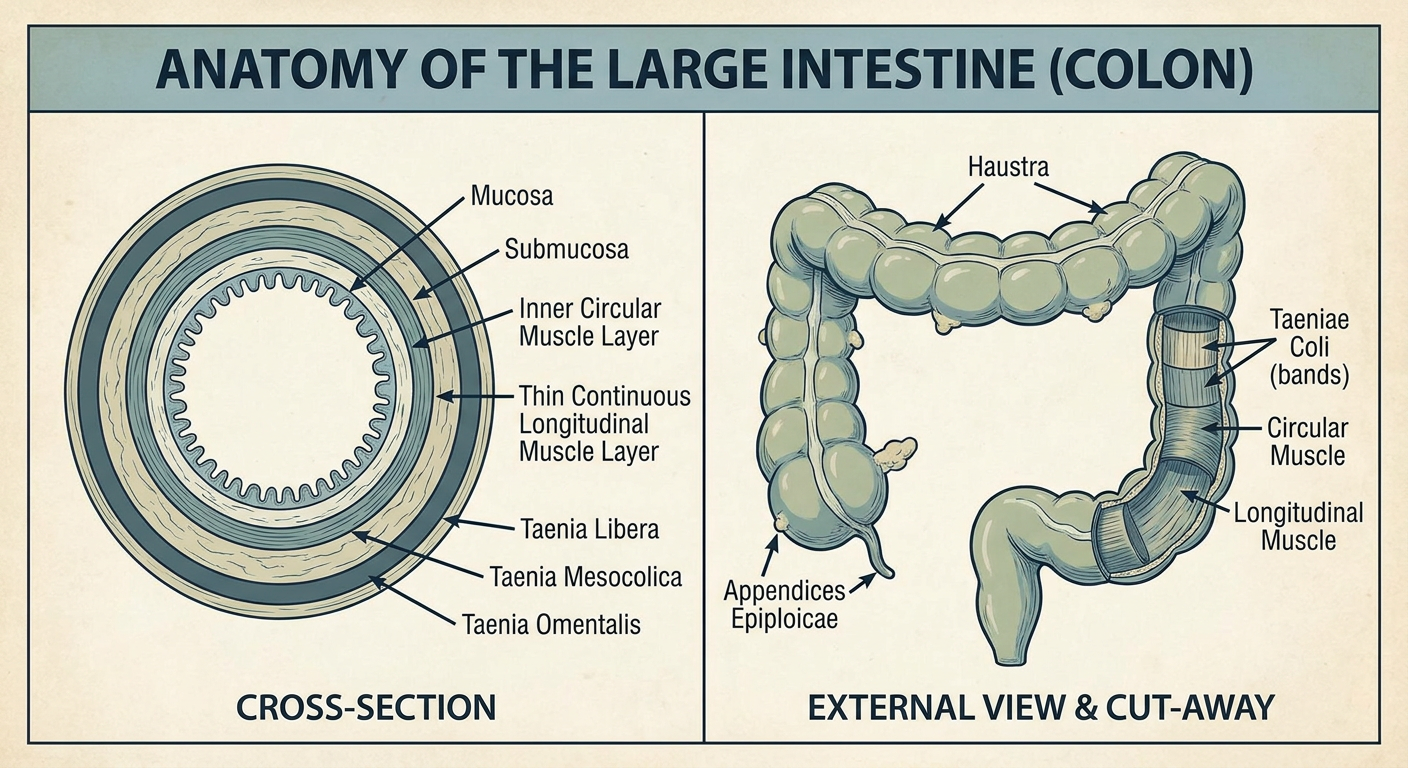
- Taenia libera - free/anterior surface
- Taenia mesocolica - where the mesentery attaches
- Taenia omentalis - where the omentum attaches
- The taeniae are thick, condensed bands
- Between them, the thin continuous longitudinal muscle still exists (just much thinner)
- The inner circular muscle runs all the way around uniformly
- The haustra (pouches/sacculations) form between the taeniae because the taeniae are shorter than the total colon length, bunching the wall up
1 more doubt so the teaniea coli is divided in 3 names yea msocolic, omentalis and libre is that only with regards to the transverse colon or throughout the colony but that part is takes as reference?