
Audiometric findings in otosclerosis in detail for 20 marker long answer according to new scott brown edition 9 in simplified manner
I'll start by reading the medical library skill, then search for otosclerosis audiometric findings in the textbooks.
Searching sources...
Reading File
Now let me search the library for otosclerosis audiometric findings in Scott Brown's:
Finding Sources
Finding Sources
Scott-Brown's Vol 2 has the dedicated otosclerosis chapter. Let me now read the key blocks in parallel - the main otosclerosis chapter and the audiometric-specific sections:
Reading File
Finding Sources
Now let me read the main otosclerosis chapter blocks - the Vol 2 block12 (dedicated chapter) and the audiometric section in block10:
Reading File
Finding Sources
Let me read the main Scott-Brown Vol 2 block 10 which has the otosclerosis chapter - searching for the audiometric/diagnosis sections:
Finding Sources
Finding Sources
Excellent! I found the key sections. The main audiometric content is in block10 around lines 8811-8949 (Pure-tone audiometry, Air-bone gap, Carhart notch). Let me read all these critical sections in parallel:
Reading File
Reading File
Excellent content! Let me now read more of the Carhart notch section and the impedance/tympanometry/immittance sections:
Reading File
Finding Sources
Now let me read the tympanometry section in block10 and the acoustic reflex/far-advanced sections:
Reading File
Reading File
Now let me read the important section between lines 9025-9320 (which likely covers acoustic reflex, speech audiometry, cochlear otosclerosis/SNHL) and also fetch the key audiogram image:
Reading File
Loading Image
I now have a comprehensive picture of the audiometric findings from Scott-Brown's Vol 2. Let me also fetch the second audiogram (post-operative showing Carhart notch resolution) and check the acoustic reflex section:
Loading Image
Reading File
I now have all the necessary content from Scott-Brown's to write a comprehensive 20-mark answer. Let me also fetch the Carhart notch audiogram for illustration:
Loading Image
I now have all three parts of Figure 89.5 from Scott-Brown's: (a) pre-op audiogram showing conductive loss with ABG greatest at low frequencies, (b) post-op audiogram showing closed ABG with Carhart notch resolved, and (c) the normal post-op result. I have comprehensive content for a 20-mark answer. Let me now compose the full answer.
Audiometric Findings in Otosclerosis
(Scott-Brown's Otorhinolaryngology Head & Neck Surgery, 9th Edition - Chapter 89)
INTRODUCTION
Otosclerosis is characterized by abnormal bone remodeling of the otic capsule, most commonly causing stapes fixation. The audiological hallmark is a progressive conductive hearing loss with a normal tympanic membrane. Audiometry is the cornerstone of diagnosis and surgical eligibility assessment.
1. PURE TONE AUDIOMETRY (PTA)
a) Air Conduction (AC)
- Shows elevated thresholds - the patient hears poorly via air conduction because the fixed stapes cannot transmit sound vibrations effectively to the inner ear.
- The hearing loss is typically bilateral (though often asymmetric), gradually progressive, and commences in early adulthood (2nd-4th decade).
- The loss is greatest at low frequencies initially (the classic low-frequency conductive pattern), gradually becoming flat as disease progresses.
b) Bone Conduction (BC)
- In early/uncomplicated otosclerosis, bone-conduction thresholds are normal or near-normal, reflecting intact cochlear function.
- The air-bone gap (ABG) is therefore present: AC thresholds are elevated while BC thresholds remain near 0 dB HL.
Figure 89.5(a) - Pre-operative audiogram showing classical otosclerosis:
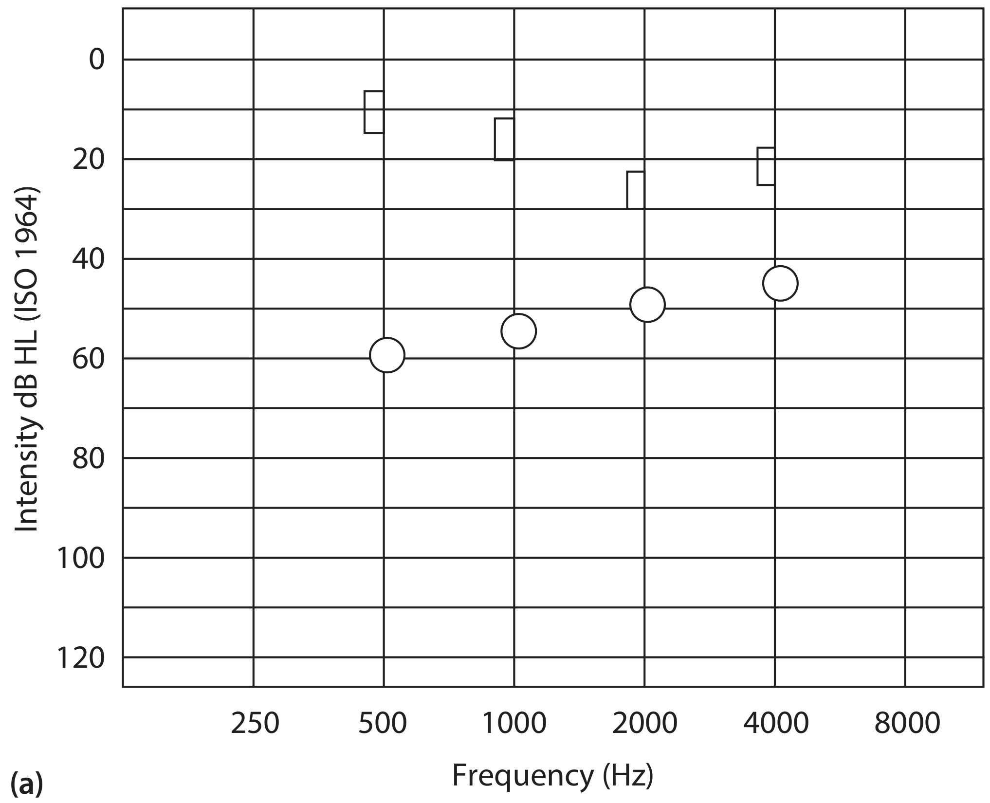
Figure 89.5(a) - Pre-operative audiogram: open circles = air conduction, squares = bone conduction. The large gap between them (ABG) is greatest at low frequencies (0.5-2 kHz), typical of otosclerosis.
2. THE AIR-BONE GAP (ABG)
The ABG is the defining audiometric feature of otosclerosis. It directly quantifies the degree of conductive impairment from stapes fixation.
Calculating the ABG
- Measured as the difference between average AC and average BC thresholds.
- Three-frequency average over 0.5, 1 and 2 kHz is traditionally recommended, as the ABG is greatest at these frequencies.
- Some use a four-frequency average over 0.5, 1, 2 and 4 kHz, as the AC average more accurately reflects monaural hearing disability. The American Academy of Otolaryngology-Head and Neck Surgery recommends averaging over 0.5, 1, 2 and 3 kHz.
Pattern of the ABG
- The gap is characteristically greater at low frequencies (0.5-2 kHz), producing a "rising" pattern on audiogram (worse at low frequency, relatively better at high frequency).
- As stapes fixation becomes complete and severe, the ABG may extend across all frequencies.
- The maximum conductive loss from total stapes fixation is approximately 60-65 dB - hearing cannot be worse than this by a purely conductive mechanism alone.
Clinical Significance of ABG Size
- ABG ≥ 20 dB at 0.5, 1 and 2 kHz: traditional threshold to recommend stapes surgery.
- ABG 10-20 dB: experienced stapes surgeons may still operate, as patients appreciate even small gains especially in bilateral disease.
- ABG < 10 dB: some surgeons now operate even with a small gap, as the Carhart effect (see below) artificially depresses bone conduction thresholds, and the true ABG may be larger than measured.
- Scott-Brown's Otorhinolaryngology Head & Neck Surgery, Vol 2, Chapter 89
3. THE CARHART NOTCH AND CARHART EFFECT
This is the most characteristic and examined audiometric finding in otosclerosis.
What is the Carhart Notch?
- A dip (notch) in the bone-conduction thresholds, classically at 2000 Hz (by approximately 15 dB), with smaller dips at 500 Hz (~5 dB), 1000 Hz (~10 dB), and 4000 Hz (~5 dB).
- It is a mechanical artifact - not a true loss of cochlear sensitivity, but a reduction in the efficiency of bone-conduction sound transmission caused by the fixed ossicular chain.
Mechanism - The Carhart Effect
Normally, when the skull vibrates (bone-conduction testing), sound reaches the cochlea via three routes:
- Route A: Directly through skull bone to the cochlea (osseous route)
- Route B: Via the tympanic membrane and ossicular chain to the cochlea (ossicular inertia route)
- Route C: Via air in the external auditory canal
In otosclerosis, the fixed stapes blocks Route B (ossicular contribution). The ossicular system has a natural resonance frequency near 2000 Hz, so the loss of ossicular contribution is maximal at this frequency, producing the characteristic 2 kHz notch. The other frequencies are affected to a lesser degree.
Carhart Effect Values (from various studies - Table 89.2, Scott-Brown's):
| Frequency | Carhart (1950) | Gatehouse & Browning (1982) |
|---|---|---|
| 500 Hz | 5 dB | 5 dB |
| 1000 Hz | 10 dB | 8 dB |
| 2000 Hz | 15 dB | 12 dB |
| 4000 Hz | 5 dB | 5 dB |
Key Points about the Carhart Notch
- It is a mechanical/conductive phenomenon, NOT a sensorineural loss.
- After successful stapes surgery, the Carhart notch disappears - bone-conduction thresholds improve especially at 2 kHz, explaining the phenomenon of "overclosure" (post-op AC thresholds better than pre-op BC thresholds).
- Average improvement in BC over 0.5, 1 and 2 kHz after stapes surgery = at least 12 dB.
- The Carhart notch can be seen in any ossicular fixation, not just otosclerosis - so it is not pathognomonic.
- A recent literature review showed limited predictive value of the Carhart notch specifically for diagnosing otosclerosis.
Figure 89.5(b) - Post-operative audiogram after successful stapedectomy:
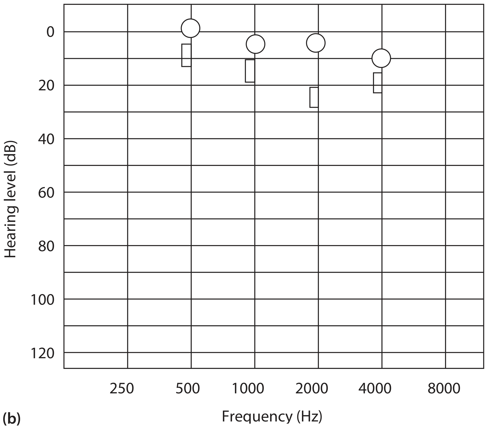
Figure 89.5(b) - Post-operative audiogram: note closure of ABG and improvement of bone conduction (especially at 2 kHz), confirming the Carhart effect was mechanical.
4. TYMPANOMETRY
- Tympanometry shows a peaked, Type A tympanogram (normal middle ear pressure) because the tympanic membrane is normal and mobile, and the Eustachian tube functions normally.
- The compliance (admittance) in surgically confirmed otosclerosis is less than normal due to the stiffened ossicular chain, but the range overlaps so widely with normal that it does not aid diagnosis in an individual ear.
- The same limitation applies to multifrequency/multicomponent tympanometry.
- Tympanometry is useful to rule out otitis media with effusion (Type B/flat tympanogram) as an alternative cause of conductive hearing loss.
Key point: Tympanometry does NOT generally aid the diagnosis of clinical otosclerosis. - Scott-Brown's Vol 2, Chapter 89
5. ACOUSTIC REFLEX
- In otosclerosis with stapes fixation, the acoustic (stapedial) reflex is absent because the stapes footplate cannot move to generate the reflex response.
- This is important: an absent reflex in the presence of a conductive hearing loss with normal tympanogram strongly supports stapes fixation.
- In very early disease with minimal fixation, the reflex may still be present but with increased threshold.
6. SPEECH AUDIOMETRY
- Speech discrimination scores are typically well-preserved in uncomplicated (purely conductive) otosclerosis, because cochlear function is intact.
- The patient hears speech poorly due to the conductive block, but when sound is amplified to overcome the conductive component, word recognition is excellent (close to 100%).
- In the British National Study of Hearing, clinical otosclerosis was defined as: normal tympanic membrane + peaked tympanogram in normal pressure + ABG ≥ 15 dB at 0.5, 1 and 2 kHz.
- Poor speech discrimination (<30% pre-operatively) suggests significant cochlear involvement (sensorineural component) and is a predictor of poorer outcome from stapedotomy in far-advanced otosclerosis.
7. MIXED HEARING LOSS - COCHLEAR OTOSCLEROSIS
In some patients, otosclerosis extends beyond the oval window to involve the cochlea (retrofenestral/cochlear otosclerosis), producing a mixed hearing loss (conductive + sensorineural components):
- BC thresholds are also elevated (above the ABG and beyond what the Carhart effect alone can explain).
- Two scenarios exist:
- Mixed impairment: conductive loss from stapes fixation + SNHL from cochlear involvement.
- Pure SNHL: rare - otosclerotic focus surrounds the cochlea without oval window involvement (very few documented cases in temporal bone studies).
- For accurate assessment of cochlear reserve, BC thresholds must be corrected for the Carhart effect before judging the degree of sensorineural impairment.
- Browning and Gatehouse found that on average otosclerotic patients have no worse BC thresholds than the general population; however, subsequent studies (Topsakal et al., Redfors and Moller) showed significantly greater SNHL in non-operated otosclerotic ears than age-matched controls.
8. FAR-ADVANCED OTOSCLEROSIS (FAO)
First described by House and Sheehy (1961):
- Defined as average AC threshold > 85 dB HL with bone-conduction thresholds unmeasurable at the limits of audiometric equipment.
- Despite absent measurable BC, a significant conductive component may still be present (up to ~60 dB of latent ABG beneath the severe SNHL).
- Example: a patient may have 65 dB SNHL + 60 dB conductive loss = total AC ~125 dB (unmeasurable), yet after stapedectomy, AC improves to 65 dB HL (the residual SNHL).
- Audiological clues suggesting FAO rather than pure SNHL: prior audiograms showing ABG, family history of otosclerosis, previous stapes surgery, preserved (though reduced) speech discrimination, and CT evidence of cochlear otosclerosis.
- Pre-operative speech discrimination <30% and small ABG on CT suggest the patient will do poorly with stapedotomy alone and should be considered for cochlear implantation.
9. AUDIOLOGICAL ELIGIBILITY FOR SURGERY - SUMMARY
| ABG | Recommendation |
|---|---|
| ≥ 20 dB (traditional) | Standard indication for stapes surgery |
| 10-20 dB | Experienced surgeons may operate (bilateral disease) |
| < 10 dB | Carhart effect may mask true gap; some surgeons still operate |
| FAO (AC > 85 dB, BC unmeasurable) | Stapedectomy + hearing aid, or cochlear implantation |
10. KEY AUDIOMETRIC POINTS - SUMMARY TABLE
| Test | Finding in Otosclerosis |
|---|---|
| Air Conduction | Elevated, worse at low frequencies |
| Bone Conduction | Normal in early disease; may be depressed by Carhart effect |
| Air-Bone Gap | Present, greatest at 0.5-2 kHz |
| Carhart Notch | BC notch at 2 kHz (~15 dB dip), mechanical artifact |
| Tympanogram | Type A (normal compliance) - peaked, normal pressure |
| Acoustic Reflex | Absent (stapes fixed) |
| Speech Discrimination | Excellent (pure conductive), reduced if cochlear involved |
| Post-op BC | Improves - especially 2 kHz (Carhart effect reversal) |
CONCLUSION
The audiometric fingerprint of otosclerosis is:
- Progressive conductive hearing loss with low-frequency predominance
- Normal BC thresholds (corrected for Carhart effect)
- Air-bone gap maximal at 0.5-2 kHz
- Carhart notch at 2 kHz on BC audiogram (mechanical, not cochlear)
- Type A tympanogram with absent acoustic reflex
- Excellent speech discrimination (in pure conductive form)
- Post-op resolution of Carhart notch confirming mechanical basis
All content from: Scott-Brown's Otorhinolaryngology Head & Neck Surgery, 9th Edition, Vol 2, Chapter 89 (Browning et al.) - the authoritative source for this topic.
This is a shared conversation. Sign in to Orris to start your own chat.