
Flexor retinaculum short note with diagram
Flexor Retinaculum - Short Note
1. Flexor Retinaculum of the Wrist (Transverse Carpal Ligament)
Definition
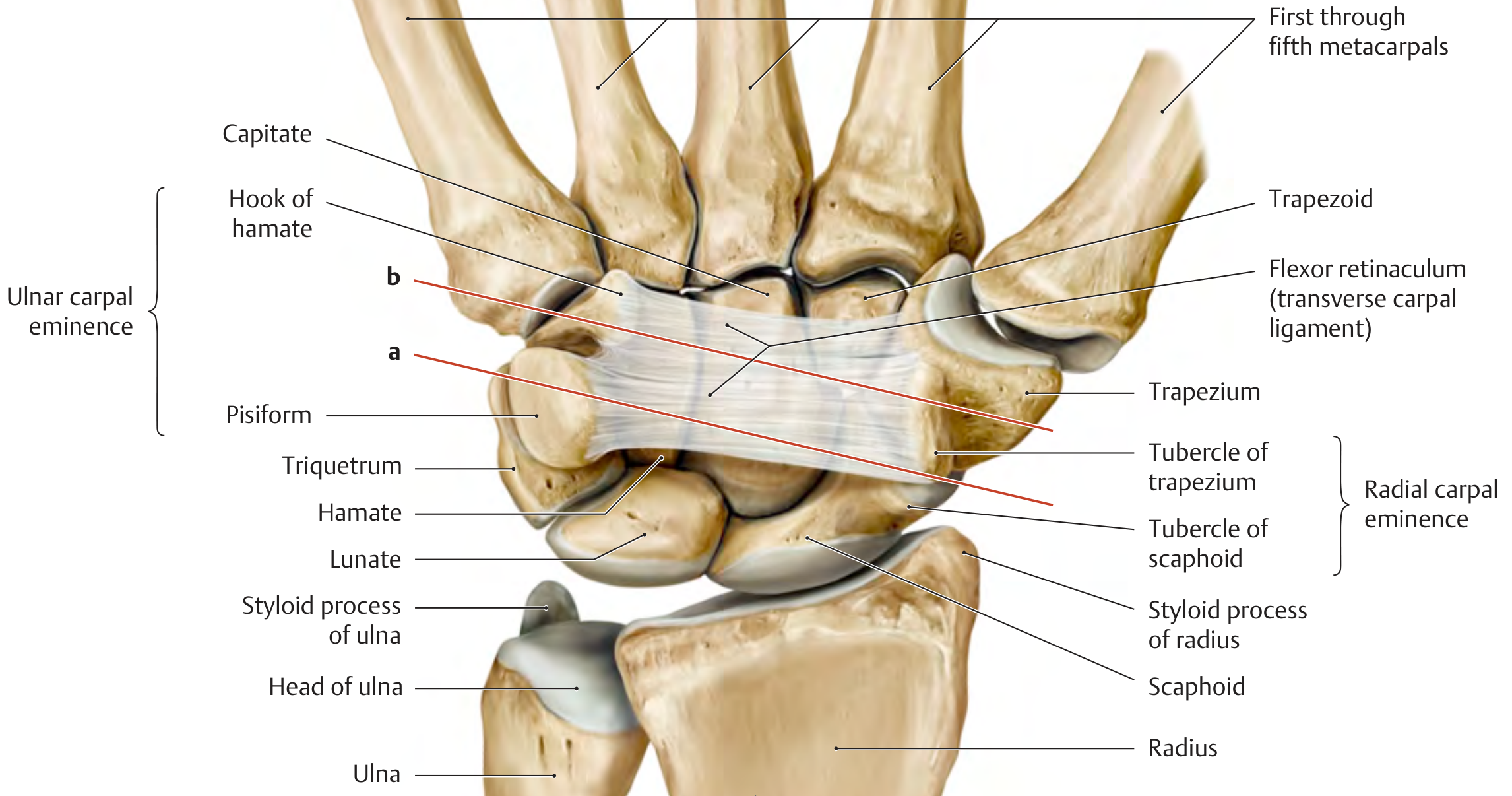
Attachments
| Side | Bony Attachments |
|---|---|
| Medial (ulnar) | Pisiform and hook of hamate |
| Lateral (radial) | Tubercle of scaphoid and tubercle (ridge) of trapezium |
Surface Landmarks
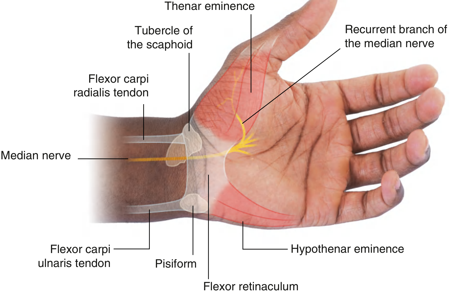
- The proximal margin lies along an imaginary line connecting the pisiform (palpable at the distal end of the flexor carpi ulnaris tendon) to the tubercle of the scaphoid (palpable at the distal end of the flexor carpi radialis tendon).
- The distal margin lies approximately at the junction of the thenar and hypothenar eminences near the base of the palm.
Carpal Tunnel - Contents (pass through the retinaculum)
- Four tendons of flexor digitorum superficialis
- Four tendons of flexor digitorum profundus
- Tendon of flexor pollicis longus (in its own synovial sheath)
- Median nerve (anterior to the tendons)
- Total = 9 tendons + 1 nerve
Structures passing anterior to the retinaculum (not through the tunnel)
- Ulnar artery and ulnar nerve (in Guyon's canal)
- Tendon of palmaris longus
- Superficial branch of radial artery
Special note - Flexor carpi radialis
Function
- Holds flexor tendons against the bony plane at the wrist, preventing "bowstringing"
- Provides origin for muscles of the thenar eminence (abductor pollicis brevis, flexor pollicis brevis, opponens pollicis) and hypothenar eminence (abductor digiti minimi, flexor digiti minimi, opponens digiti minimi)
Recurrent (Thenar) Branch of the Median Nerve
- In 46% of cases: arises distal to the retinaculum (safe zone)
- In 31%: subligamentous course
- In 23%: pierces the retinaculum (most vulnerable to injury during surgery)
Clinical Significance - Carpal Tunnel Syndrome
2. Flexor Retinaculum of the Ankle (Laciniate Ligament / Medial Retinaculum)
Definition
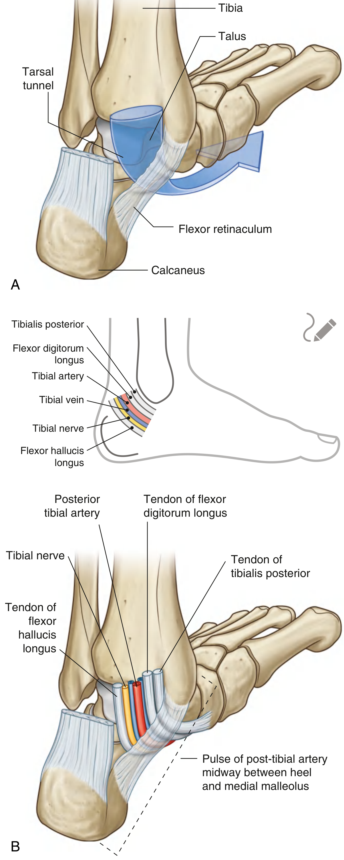
Attachments
- Above: medial malleolus
- Below and behind: inferomedial margin of the calcaneus
- Continuous above with the deep fascia of the leg and below with the plantar aponeurosis
Fibro-osseous Tunnels (Tom, Dick ANd Very Nervous Harry - mnemonic)
| Order (medial to lateral) | Structure |
|---|---|
| 1st (medial malleolus groove) | Tibialis posterior tendon |
| 2nd | Flexor digitorum longus tendon |
| 3rd (middle) | Posterior tibial artery + veins + Tibial nerve |
| 4th (under sustentaculum tali/talus) | Flexor hallucis longus tendon |
Clinical Significance - Tarsal Tunnel Syndrome
Quick Comparison Table
| Feature | Wrist (Carpal) | Ankle (Tarsal) |
|---|---|---|
| Attachments | Scaphoid, trapezium (lateral); pisiform, hamate (medial) | Medial malleolus (above); calcaneus (below) |
| Tunnel formed | Carpal tunnel | Tarsal tunnel |
| Nerve enclosed | Median nerve | Tibial nerve |
| Entrapment syndrome | Carpal tunnel syndrome | Tarsal tunnel syndrome |
| Key artery pulse | - | Posterior tibial artery |
- Gray's Anatomy for Students, pp. 919, 948, 753
- THIEME Atlas of Anatomy: General Anatomy and Musculoskeletal System, p. 309
- Firestein & Kelley's Textbook of Rheumatology
Diagram for hip joint
Hip Joint - Diagrams
Diagram 1 - Articular Surfaces & Rotation
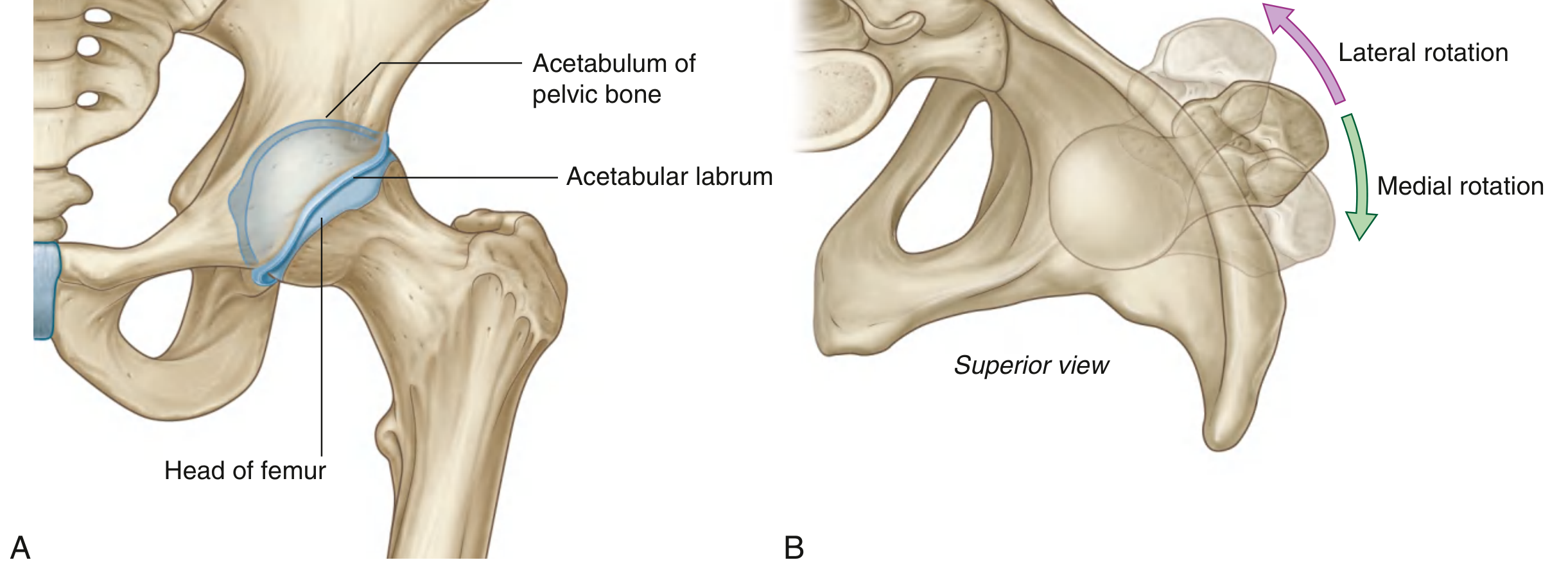
Diagram 2 - Acetabular Labrum, Transverse Ligament & Ligament of the Head of Femur
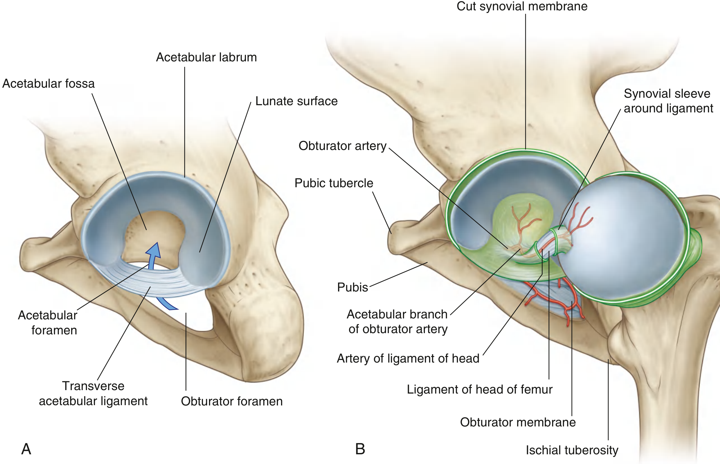
Diagram 3 - Synovial Membrane
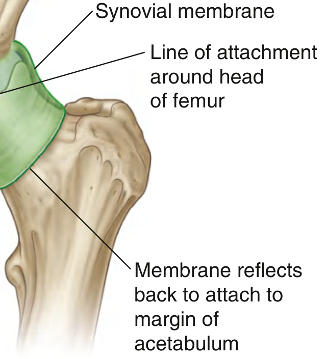
Diagram 4 - Ligaments of the Hip Joint (Capsule, Iliofemoral, Pubofemoral, Ischiofemoral)
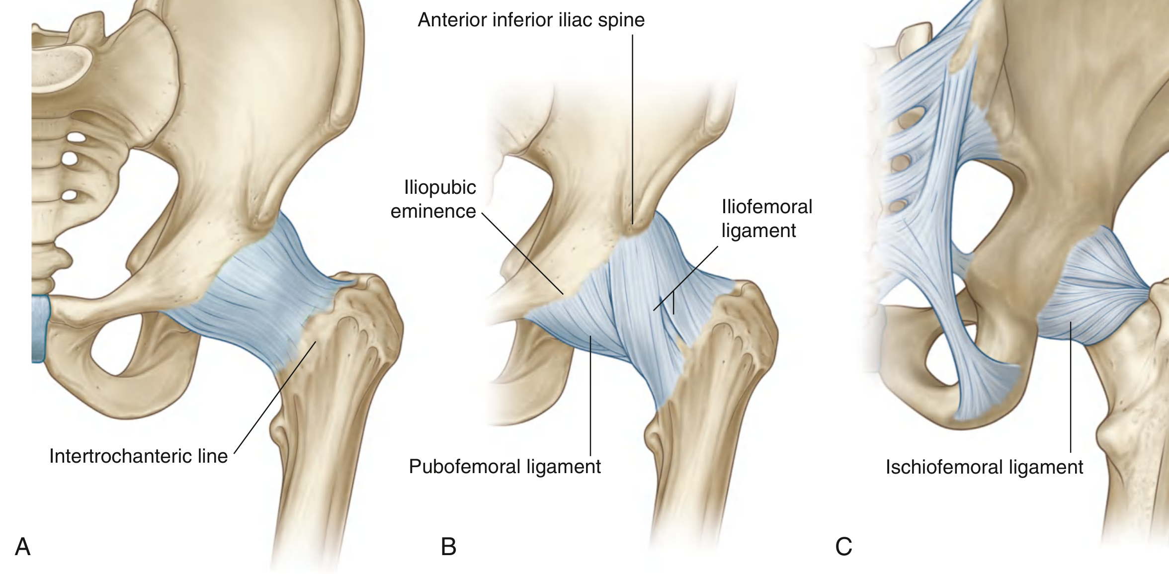
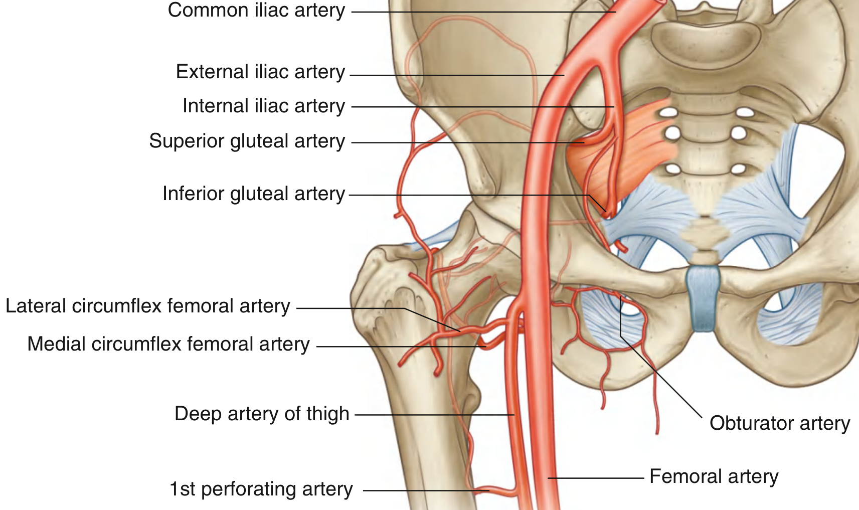
Diagram 5 - Blood Supply of the Hip Joint
Key Summary Points (for each diagram)
| Feature | Detail |
|---|---|
| Type | Multiaxial ball-and-socket synovial joint |
| Articulation | Head of femur + lunate surface of acetabulum |
| Deepened by | Acetabular labrum (fibrocartilage) + transverse acetabular ligament |
| Capsule | Attaches to acetabular rim proximally; to intertrochanteric line (anterior) & neck of femur (posterior) |
| Ligaments | Iliofemoral (strongest, Y-shaped), Pubofemoral, Ischiofemoral - all spiral, tighten in extension |
| Intracapsular ligament | Ligament of head of femur (carries artery to head - branch of obturator artery) |
| Blood supply | Obturator a., medial & lateral circumflex femoral aa., superior & inferior gluteal aa., 1st perforating a. |
| Nerve supply | Femoral, obturator, superior gluteal nerves + nerve to quadratus femoris |
| Movements | Flexion (0-120°), Extension, Abduction, Adduction, Medial/lateral rotation, Circumduction |
Hip joint diagram for exam
Detailed labeled anatomical diagram of the hip joint for medical exam, showing: anterior and posterior views. Include all key structures clearly labeled: - Head of femur (ball) - Acetabulum of pelvic bone (socket) - Acetabular labrum (fibrocartilage rim) - Acetabular fossa and lunate surface - Transverse acetabular ligament (bridging acetabular notch) - Ligament of head of femur (ligamentum teres) with artery - Joint capsule / fibrous membrane - Iliofemoral ligament (Y-ligament of Bigelow) - anterior, from AIIS to intertrochanteric line - Pubofemoral ligament - anteroinferior - Ischiofemoral ligament - posterior - Synovial membrane - Greater trochanter, lesser trochanter, intertrochanteric line, femoral neck - Zona orbicularis (circular fiber zone) Clean white background, medical textbook illustration style, black label lines with text, color-coded structures (blue for ligaments, yellow for nerves, red for arteries), professional anatomical art suitable for MBBS/MD exam preparation