
Decompensated features of cold
Decompensated features of cld
Decompensated Features of Chronic Liver Disease (CLD)
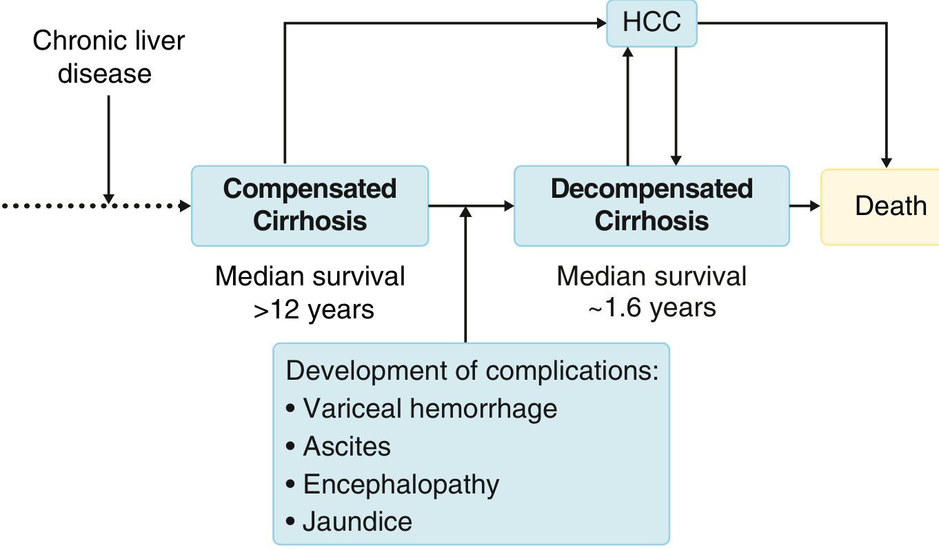
Definition of Decompensation
Clinical Staging (Baveno Classification)
| Stage | Description |
|---|---|
| Stage 1 (Compensated) | No ascites, no varices |
| Stage 2 (Compensated) | Varices present, no bleeding, no ascites |
| Stage 3 (Decompensated) | Ascites ± varices |
| Stage 4 (Decompensated) | Variceal bleeding ± ascites |
Cardinal Features of Decompensation
1. Ascites (Most Common - 80%)
- Most frequent sign of decompensation; occurs at 7-10% per year in compensated cirrhosis
- Caused by portal hypertension + splanchnic vasodilation + sodium/water retention
- Clinical signs: increased abdominal girth, bulging flanks, flank dullness, shifting dullness
- Hyponatremia (serum Na <130 mEq/L) is present in ~25% of patients with cirrhosis and ascites - a marker of disease severity
- Complications: Spontaneous Bacterial Peritonitis (SBP) - infection without an obvious surgical source; Refractory ascites - unresponsive to diuretics, requires repeated paracentesis or TIPS
2. Variceal Hemorrhage
- Gastroesophageal varices present in ~50% of newly diagnosed cirrhosis
- Prevalence ranges from 40% (Child A) to 85% (Child C)
- Small varices bleed at ~5%/year; large varices bleed at ~15%/year
- Predictors: large variceal size, severe liver disease, red wale markings
- Presents as hematemesis, melena, or both
- Gastric varices (especially fundal) carry higher bleeding risk than esophageal
3. Hepatic Encephalopathy (HE)
- Brain dysfunction caused by liver insufficiency and/or portosystemic shunting
- Pathogenesis: Ammonia accumulates (due to portosystemic collaterals + decreased hepatic metabolism) → damages astrocytes (Alzheimer type II astrocytosis) → GABA-mediated cortical depression
- Also: manganese deposition in globus pallidus → impaired motor function
- Grades I-IV: from subtle personality changes/sleep disturbance → confusion → stupor → coma
- Precipitants: GI bleed, infection, constipation, sedatives, electrolyte imbalance, dehydration
4. Jaundice
- Results from inability to excrete bilirubin (liver insufficiency)
- In cholestatic disease (PBC, PSC), may reflect biliary damage rather than pure liver failure
- Accompanied by coagulopathy (prolonged INR) and hypoalbuminaemia
Secondary/Progressive Decompensation Features
5. Hepatorenal Syndrome (HRS)
- Functional renal failure in setting of advanced cirrhosis; no intrinsic kidney pathology
- Results from extreme splanchnic vasodilation → renal vasoconstriction → decreased GFR
- HRS Type 1 (AKI type): Rapid deterioration (creatinine doubles to >2.5 mg/dL within 2 weeks); very poor prognosis
- HRS Type 2: Slower, associated with refractory ascites
6. Spontaneous Bacterial Peritonitis (SBP)
- Infection of ascitic fluid without a surgical cause
- Diagnosis: ascitic fluid PMN count >250 cells/mm³
- Common organisms: E. coli, Klebsiella, Streptococcus
- Associated with 30-50% in-hospital mortality if untreated
7. Cardiopulmonary Complications
- Cirrhotic cardiomyopathy: High-output heart failure with decreased peripheral O₂ utilization
- Hepatopulmonary syndrome (HPS): Pulmonary capillary dilation → right-to-left shunt → hypoxemia (PaO₂ <80 mmHg, or increased A-a gradient)
- Portopulmonary hypertension (PoPH): Vasoconstrictive substances bypass hepatic metabolism → irreversible pulmonary HTN
8. Coagulopathy
- Decreased synthesis of clotting factors (II, V, VII, IX, X)
- Thrombocytopenia (from hypersplenism + decreased thrombopoietin)
- Results in prolonged PT/INR; bleeding tendency
9. Other Advanced Features
- Hepatocellular carcinoma (HCC): Can develop at any stage and precipitate/accelerate decompensation
- Acute-on-Chronic Liver Failure (ACLF): A distinct syndrome - acute deterioration on a background of cirrhosis, with organ failures; high short-term mortality
- Infection/Sepsis: Now recognized as a distinct stage in the natural history; poor survival even after infection clearance
- Muscle wasting/Sarcopenia and malnutrition: Progressive loss of muscle mass
- Hepatic hydrothorax: Transudative pleural effusion (usually right-sided) from ascitic fluid passing through diaphragmatic defects
Physical Signs of Decompensation
| Sign | Mechanism |
|---|---|
| Jaundice / scleral icterus | Bilirubin excretion failure |
| Ascites (bulging flanks, shifting dullness) | Portal HTN + aldosterone activation |
| Spider naevi, palmar erythema | Hyperestrogenism |
| Caput medusae | Portal-systemic collaterals |
| Splenomegaly | Portal hypertension |
| Hepatic flap (asterixis) | Hepatic encephalopathy |
| Gynecomastia, testicular atrophy | Hormonal imbalance |
| Peripheral oedema | Hypoalbuminaemia |
| Fetor hepaticus | Portosystemic shunting (mercaptans) |
| Dupuytren's contracture | Alcohol-related (especially) |
Prognostic Scoring
- Child-Turcotte-Pugh (CTP): Scores ascites, encephalopathy, bilirubin, albumin, INR (range 5-15). Child A (5-6) = good; Child C (10-15) = poor
- MELD/MELD-Na score: Incorporates creatinine, bilirubin, INR, and sodium - used for liver transplant prioritization
Median survival in compensated cirrhosis: 9-12 years. Median survival in decompensated cirrhosis: ~1.6-2 years. - Goldman-Cecil Medicine, Sleisenger & Fordtran's GI and Liver Disease
Liver span normal range and abnormal
liver span normal range percussion midclavicular line midsternal hepatomegaly
https://www.ncbi.nlm.nih.gov/books/NBK421
liver span percussion hepatomegaly normal range diagram
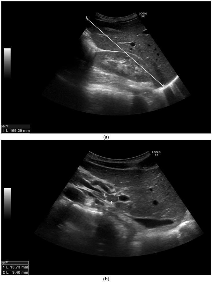
Diagnostic abdominal ultrasound images demonstrating hepatomegaly and regional lymphadenopathy. (a) Longitudinal scan of the right upper quadrant showing the liver parenchyma with a speckled, homogeneous echotexture. A measurement calipers line indicates a liver span of 169.29 mm in the midclavicular line, exceeding the normal threshold of 150 mm and confirming hepatomegaly. (b) Targeted ultrasound of the porta hepatis region revealing multiple hypoechoic, well-defined rounded structures consistent with enlarged lymph nodes. Two nodes are measured at 13.73 mm and 9.40 mm, indicating portal hepatic lymphadenopathy. These findings are presented in the clinical context of a patient with elevated liver enzymes and systemic infection, such as coccidioidomycosis, highlighting the use of ultrasound for evaluating visceral involvement and identifying lymph node enlargement in infectious or inflammatory disease processes.
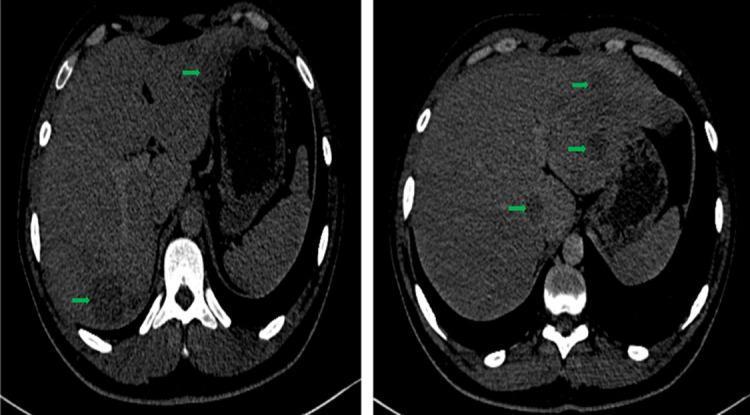
This diagnostic image consists of two axial abdominal CT scan slices demonstrating hepatomegaly and diffuse liver involvement. The liver is significantly enlarged, with a measured hepatic span of approximately 17 cm. The parenchyma is characterized by multiple, scattered, hypodense tissue-like lesions (highlighted by green arrows). These lesions are rounded, well-circumscribed, and appear widespread across various hepatic segments, notably involving segment I (21 x 16mm) and segment V (36 x 27mm). Post-contrast administration, these nodules show no significant enhancement, maintaining a hypodense appearance relative to the surrounding normal hepatic tissue. The findings are indicative of an infiltrative or multifocal process, which clinically correlated with hepatic tuberculosis in this case. No deep lymphadenopathy is visible in these sections. The image serves as a clinical example of infectious granulomatous liver disease presenting as multiple hypodense nodules on computed tomography.
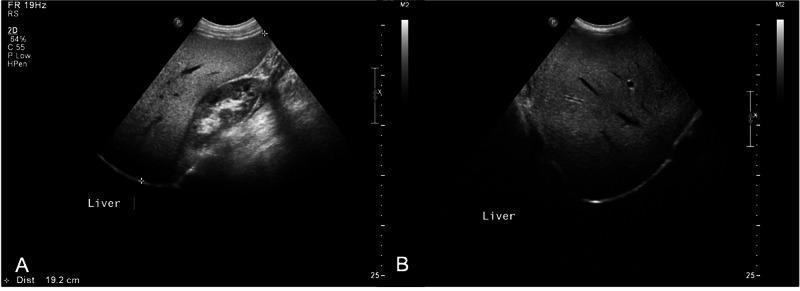
Abdominal diagnostic ultrasound images demonstrating hepatomegaly and hepatic steatosis. Image A is a longitudinal gray-scale view of the right upper quadrant showing an enlarged liver with a span measured at 19.2 cm in the midclavicular line. The liver parenchyma exhibits diffuse hyperechogenicity (bright liver) compared to the adjacent renal cortex, characteristic of fatty liver disease. The right kidney is partially visualized and appears morphologically normal. Image B is a transverse view showing the same hyperechoic pattern throughout the liver parenchyma with some visualization of hepatic vasculature. The increased echogenicity causes significant attenuation of the ultrasound beam in the deeper tissues. These findings are clinically significant for diagnosing hepatic steatosis and monitoring liver size in the context of metabolic or endocrine disorders.
Liver Span - Normal Range and Abnormal Values
How to Measure Liver Span
- Upper border: Use heavy percussion from the mid-thorax downward in the MCL - the point where lung resonance changes to hepatic dullness marks the upper liver border (normally at the level of the right nipple, ~5th intercostal space)
- Lower border: Use light percussion (examiner's ear close to the abdomen) from the right lower quadrant upward - the point where tympany changes to dullness marks the lower border (normally at the right costal margin)
- Can also be percussed at the midsternal line and anterior axillary line
- Palpation of the lower border below the costal margin alone is unreliable for determining span - percussion is superior
Normal Liver Span by Percussion
Adult Values (Mid-Clavicular Line)
| Parameter | Value |
|---|---|
| General adult range | 6-12 cm |
| Mean in men | 10.5 cm |
| Mean in women | 7 cm |
| A span 2-3 cm above/below these means is considered abnormal |
- Goldman-Cecil Medicine states: normal liver span in the MCL is 6 to 12 cm; a span <12 cm reduces the likelihood of hepatomegaly
- Clinical Methods (NCBI): mean percussion span is 7 cm (women) and 10.5 cm (men)
Average Liver Span by Percussion Line (Adults)
| Line | Normal Span |
|---|---|
| Mid-clavicular line (MCL) | 6-12 cm (mean ~10.5 cm men; ~7 cm women) |
| Midsternal line | 4-8 cm |
| Anterior axillary line | Variable; not routinely measured |
By Sex (Clinical vs Ultrasound - Research Data)
| Method | Males | Females |
|---|---|---|
| Clinical percussion (MCL) | ~12.0 cm ± 1.6 cm | ~11.0 cm ± 1.5 cm |
| Ultrasound (MCL) | ~14.2 cm ± 1.3 cm | ~12.8 cm ± 1.4 cm |
Note: Ultrasound consistently gives larger values than clinical percussion (statistically significant, p<0.001), because percussion underestimates the true hepatic extent.
Paediatric Values (by percussion, MCL - Johns Hopkins study)
| Age | Approximate Liver Span |
|---|---|
| 1 week (neonate) | ~1.9 cm |
| 5 years | ~5 cm |
| 12 years (male) | ~6 cm |
| 15 years | Adult size reached |
| 20 years (male) | up to 7.7 cm |
| 20 years (female) | up to 6.3 cm |
Abnormal Values
Hepatomegaly (Enlarged Liver)
| Grade | Definition |
|---|---|
| Span >12 cm (MCL) | Hepatomegaly |
| Liver edge >2 cm below right costal margin | Suggests hepatomegaly (but may be displaced) |
| Span >2-3 cm above sex-specific mean | Abnormal |
- Vascular/Congestive: Right heart failure, Budd-Chiari syndrome, cardiac cirrhosis
- Metabolic/Infiltrative: Non-alcoholic fatty liver disease (NAFLD), haemochromatosis, Wilson's disease, amyloidosis, glycogen storage disorders
- Infective/Inflammatory: Viral hepatitis (acute), hepatic abscess, schistosomiasis, malaria, infectious mononucleosis
- Neoplastic: Primary HCC, metastatic carcinoma (most common cause of massive hepatomegaly), lymphoma, haemangioma
- Cholestatic: Primary biliary cholangitis, primary sclerosing cholangitis
- Haematological: Sickle cell disease, thalassaemia, polycythaemia vera
- Smooth + tender = acute viral/alcoholic hepatitis, hepatic congestion (CCF)
- Hard + irregular = cirrhosis, malignancy
- Smooth + non-tender = early cirrhosis, fatty liver, infiltrative disease
Shrunken/Small Liver
| Span | Interpretation |
|---|---|
| <6 cm (MCL) | Small liver |
| <9 cm (some sources) | Suggests cirrhosis (end-stage, shrunken) |
| Diminished span | Advanced cirrhosis with hepatic atrophy |
Important Caveats
| Factor | Effect on Measurement |
|---|---|
| Pleural effusion (right) | Can give false upper border (overestimates span) |
| Right lower lobe pneumonia | Falsely elevates upper border |
| Emphysema | Pushes liver downward (falsely appears enlarged by palpation) |
| Ascites | Liver edge harder to palpate; use palpation with quick pressure |
| Obesity | Percussion and palpation less reliable |
| Riedel's lobe | Tongue-like projection of right lobe - mimics hepatomegaly on palpation |
| Examiner variability | Estimates of the same liver span can vary by up to 8 cm between examiners |
Clinical Pearl
"A liver edge felt up to 2 cm below the right costal margin is considered normal, but a normal-sized liver can be displaced downward by other abnormalities such as emphysema."
- Goldman-Cecil Medicine
Give me only the points in gi examination for mbbs final year practical exam
GI Examination — MBBS Final Year Practical
1. GENERAL EXAMINATION (Before touching the abdomen)
- Built, nourishment, BMI
- Pallor (anaemia — GI bleed, malabsorption)
- Icterus / Jaundice
- Cyanosis
- Clubbing (IBD, cirrhosis, malabsorption)
- Koilonychia (iron deficiency), Leuconychia (hypoalbuminaemia)
- Lymphadenopathy — especially left supraclavicular (Virchow's node) = gastric ca
- Oedema (pedal — hypoalbuminaemia, portal HTN)
- Parotid enlargement (alcoholic liver disease)
- Fetor hepaticus
- Flapping tremor / Asterixis (hepatic encephalopathy)
- Gynaecomastia, testicular atrophy (CLD)
- Loss of axillary/pubic hair (CLD)
2. HANDS & UPPER LIMBS
- Palmar erythema (CLD, pregnancy)
- Dupuytren's contracture (alcoholic liver disease)
- Leuconychia (hypoalbuminaemia)
- Koilonychia (iron deficiency)
- Clubbing
- Asterixis (flapping tremor) — ask patient to dorsiflex wrists
- Spider naevi on arms/chest (CLD) — >5 above the nipple line is significant
- Muscle wasting
3. FACE & NECK
- Scleral icterus (jaundice)
- Conjunctival pallor
- Kayser-Fleischer rings (Wilson's disease — slit lamp needed)
- Angular stomatitis, glossitis (nutritional deficiency)
- Parotid enlargement
- Fetor hepaticus
- Virchow's node — left supraclavicular fossa
- JVP — raised in right heart failure causing congestive hepatomegaly
4. CHEST WALL (relevant)
- Spider naevi — >5 significant (CLD)
- Gynaecomastia
- Dilated veins on chest wall
5. ABDOMINAL EXAMINATION
A. INSPECTION (Patient supine, arms by side, knees slightly flexed)
- Scaphoid (sunken) — malnutrition, advanced malignancy
- Distended — ascites, obesity, organomegaly, gas, pregnancy (5 F's + 1 F: Fat, Fluid, Flatus, Faeces, Fetus + Fictitious)
- Localised bulge — organomegaly, hernia, mass
- Dilated veins:
- Caput medusae (radiates from umbilicus) — portal HTN
- Inferior vena cava obstruction (flow upward in flanks)
- Superior vena cava obstruction (flow downward)
- Striae (silvery/white = old; purple = Cushing's)
- Visible peristalsis — gastric outlet obstruction, intestinal obstruction
- Scars (note operative, laparoscopic port scars)
- Umbilicus — inverted (normal) vs everted (ascites, obesity)
- Sister Mary Joseph nodule (periumbilical nodule = metastatic malignancy)
- Hernia — umbilical, inguinal, incisional
- Jaundice of skin
- Ecchymosis — Grey Turner's sign (flanks = retroperitoneal bleed/pancreatitis), Cullen's sign (periumbilical = same)
B. PALPATION
Superficial Palpation
- Tenderness (localise to quadrant)
- Guarding (voluntary vs involuntary)
- Rigidity (board-like = peritonitis)
Deep Palpation
- Deeper tenderness
- Masses (note: position, size, shape, surface, margins, consistency, mobility, tenderness, pulsatility, movement with respiration)
Liver Palpation
- Start RIF, move toward right costal margin
- Ask patient to breathe deeply
- Normal: liver edge may be felt up to 2 cm below right costal margin
- Note: size below costal margin, surface (smooth/nodular), edge (sharp/rounded/irregular), consistency (soft/firm/hard), tenderness
- Hepatomegaly = >2 cm below costal margin
Spleen Palpation
- Start RIF, move toward left costal margin (diagonally)
- Ask patient to breathe deeply
- Normal spleen: not palpable
- If not felt supine → palpate in right lateral decubitus position
- Splenic notch felt on medial border = confirms spleen
- Differentiate from left kidney: cannot get above it (spleen), moves with respiration, notch present, dull to percussion, bimanual ballotability absent
Kidney Palpation (Bimanual)
- Ballotable, can get above it, resonant (colonic gas over it), does not move with respiration much
Other Masses
- Murphy's sign (cholecystitis): arrest of deep inspiration on palpating RHC
- McBurney's point tenderness (appendicitis)
- Rebound tenderness (peritonism)
- Rovsing's sign (appendicitis)
C. PERCUSSION
Liver
- Liver span in MCL:
- Normal: 6-12 cm
-
12 cm = hepatomegaly
- <6 cm = shrunken liver (cirrhosis/acute liver failure)
- Upper border: 5th ICS (MCL) / level of right nipple
- Lower border: right costal margin
Spleen
- Traube's space (left lower chest, bounded by 6th rib above, MAL laterally, left costal margin below) — normally resonant
- Dull Traube's space = splenomegaly or pleural effusion
Ascites
- Shifting dullness (most reliable clinical sign):
- Percuss from umbilicus to flank - note where dullness begins
- Patient turns to opposite side - wait 30 seconds
- Re-percuss - if dullness shifts to dependent side = positive
- Detects >500 mL of fluid
- Fluid thrill (large ascites >2L):
- One hand on flank, flick opposite flank
- Assistant places edge of hand in midline to dampen fat vibrations
- Feel transmitted wave
General
- Tympanic over bowel (gas)
- Dull over solid organs/masses
- Obliteration of hepatic dullness = gas under diaphragm (perforation)
D. AUSCULTATION
- Bowel sounds:
- Normal: 5-12/minute, gurgling
- Absent: paralytic ileus, peritonitis
- High-pitched/tinkling: mechanical obstruction
- Hyperactive: gastroenteritis, early obstruction
- Bruits:
- Over liver = hepatocellular carcinoma, hepatic arteriovenous fistula
- Over aorta = aortic aneurysm
- Renal bruits = renovascular HTN
- Venous hum in epigastrium/umbilicus = portal HTN (Cruveilhier-Baumgarten syndrome)
- Rub over liver/spleen = hepatic/splenic infarction, perihepatitis (Fitz-Hugh-Curtis syndrome)
- Succussion splash (heard 3+ hours after meals) = gastric outlet obstruction
6. GROIN & GENITALIA
- Inguinal lymph nodes (enlargement)
- Inguinal/femoral hernia
- Testicular atrophy (CLD)
- Scrotal oedema (ascites, hypoalbuminaemia)
7. RECTAL EXAMINATION (PR) — mention even if not performed
- Inspect perianal area: haemorrhoids, fistula, fissure, skin tags
- Tone of sphincter
- Palpate rectal wall — mass, tenderness
- Prostate in males
- Blood, mucus on glove
8. TO COMPLETE EXAMINATION
"To complete my examination, I would like to:
- Examine the hernial orifices
- Perform a per rectal examination
- Check external genitalia
- Examine the back and renal angles (costovertebral angle tenderness)
- Do relevant investigations: LFT, USG abdomen, endoscopy, ascitic tap, etc."
QUICK SUMMARY TABLE — What Examiner Will Ask
| Sign | What it indicates |
|---|---|
| Caput medusae | Portal HTN |
| Spider naevi >5 | CLD |
| Palmar erythema | CLD |
| Leuconychia | Hypoalbuminaemia |
| Shifting dullness | Ascites (>500 mL) |
| Fluid thrill | Massive ascites (>2L) |
| Dull Traube's space | Splenomegaly |
| Murphy's sign +ve | Acute cholecystitis |
| Virchow's node | Gastric/abdominal malignancy |
| Asterixis | Hepatic encephalopathy |
| Grey Turner's sign | Retroperitoneal bleed / pancreatitis |
| Cullen's sign | Periumbilical haemorrhage (pancreatitis) |
| Liver span <6 cm | Shrunken liver (end-stage cirrhosis) |
| Liver span >12 cm | Hepatomegaly |
| Tinkling BS | Mechanical obstruction |
| Succussion splash | Gastric outlet obstruction |
Also give history taking for liver pathology in similar way
History Taking — Liver Pathology
MBBS Final Year Practical Exam
OPENING
"Good morning sir/madam. I am a final year MBBS student. I would like to ask you some questions about your illness. May I proceed?"
- Name, Age, Sex, Occupation, Address, Religion, Marital status, Date of admission
1. CHIEF COMPLAINTS
- Yellowish discolouration of eyes/skin (jaundice)
- Swelling of abdomen (ascites)
- Swelling of feet (pedal oedema)
- Pain/discomfort in right upper abdomen
- Nausea, vomiting
- Loss of appetite (anorexia)
- Weakness, fatigue
- Fever
- Altered behaviour / confusion (encephalopathy)
- Itching (pruritus — cholestatic)
- Dark urine / clay-coloured stools
- Haematemesis / melaena (variceal bleed)
- Weight loss
2. HISTORY OF PRESENTING ILLNESS (HOPI)
For each complaint ask: SOCRATES
A. JAUNDICE (if present)
- Sudden vs gradual
- Duration
- Colour of urine: dark (bilirubinuria = obstructive/hepatocellular) vs normal
- Colour of stools: clay/pale (obstructive) vs normal/dark
- Itching (pruritus) — prominent in obstructive/cholestatic jaundice
- Fever + rigors before jaundice → Charcot's triad = cholangitis (fever + jaundice + RUQ pain)
- Painless progressive jaundice → carcinoma head of pancreas
- Jaundice + anorexia + low-grade fever → viral hepatitis
- Jaundice + abdominal pain + vomiting + alcohol → alcoholic hepatitis
- Jaundice + weight loss + anorexia → malignancy
- Jaundice that waxes and wanes → haemolytic or stone disease
- Preceded by prodrome (malaise, nausea, low-grade fever 1-2 wks before) → viral hepatitis
B. ASCITES / ABDOMINAL SWELLING (if present)
- Onset: sudden or gradual
- Duration
- Rate of progression: rapid (malignancy, SBP) vs slow (cirrhosis)
- Symmetrical or asymmetrical
- Associated with:
- Pedal oedema
- Breathlessness (diaphragm splinting, hepatic hydrothorax)
- Decreased urine output (hepatorenal syndrome)
- Fever + abdominal pain → spontaneous bacterial peritonitis (SBP)
- Weight gain
- Previous tapping (paracentesis) — how many times, how much drained
- Response to diuretics
C. PAIN ABDOMEN (if present)
- Site: Right hypochondrium / epigastrium / generalised
- Onset: sudden (perforation, bleed) vs gradual
- Character:
- Dull aching (hepatomegaly, capsule distension)
- Colicky (biliary colic — comes in waves, radiates to right shoulder/back)
- Constant severe (cholecystitis, abscess, malignancy)
- Radiation: right shoulder/scapula tip (biliary/diaphragm irritation)
- Aggravating: fatty food (biliary disease)
- Relieving: posture, antacids
- Associated: fever, nausea, vomiting, jaundice
D. HAEMATEMESIS / MELAENA (if present)
- Amount of blood vomited (teaspoon/cup/bowl)
- Fresh red blood vs coffee-ground
- Melaena (black tarry stools) — confirms upper GI bleed
- Haematochezia (fresh blood PR) — lower GI or massive upper GI bleed
- Associated with known portal HTN, varices, previous episodes
- Precipitated by: alcohol binge, vomiting (Mallory-Weiss if after prolonged retching)
- Haemodynamic status: giddiness, palpitations, syncope
E. ALTERED SENSORIUM / CONFUSION (if present)
- Onset: sudden or gradual
- Fluctuating or constant (encephalopathy fluctuates)
- Precipitants:
- GI bleed
- Infection
- Constipation
- Excess protein intake
- Sedatives / tranquillizers
- Electrolyte disturbance
- Dehydration
- Renal failure
- Previous episodes
- Sleep disturbance (reversal of sleep-wake cycle — early HE)
- Change in personality, behaviour (family members' observation)
- Fetor hepaticus noticed
F. FEVER (if present)
- Duration, pattern
- Rigors/chills
- Associated with jaundice → cholangitis
- Associated with ascites → SBP
- Associated with tender hepatomegaly → amoebic/pyogenic liver abscess
- Evening rise of temperature → TB (also causes hepatomegaly)
G. PRURITUS (if present)
- Duration, generalised or localised
- Worse at night
- Relieved by scratching or not
- Suggests cholestatic jaundice (PBC, PSC, obstructive)
- Scratch marks on skin
H. WEIGHT LOSS & ANOREXIA
- Amount of weight lost (in kg) over what period
- Anorexia: specific aversion to fat (cholecystitis), meat (hepatitis), alcohol
- Associated fatigue
- Malignancy: rapid weight loss + anorexia + jaundice = red flags
3. PAST HISTORY
- Previous episodes of jaundice
- Previous liver disease / hepatitis (type A, B, C, E)
- Previous abdominal surgeries (cholecystectomy, splenectomy, hepatectomy, Whipple's)
- Blood transfusions (risk for HBV, HCV)
- Diabetes mellitus (NAFLD)
- Hypertension
- Tuberculosis (hepatic TB, anti-TB drug hepatotoxicity — RIPE drugs)
- Malaria (hepatomegaly, Blackwater fever)
- Sickle cell disease / haemolytic anaemia
- Known cardiac disease (right heart failure → congestive hepatomegaly)
- Wilson's disease / haemochromatosis (hereditary)
- Autoimmune disorders (AIH, PBC)
- Previous hospital admissions and treatment
4. DRUG HISTORY
| Drug Category | Examples |
|---|---|
| Anti-TB drugs | Isoniazid, Rifampicin, Pyrazinamide (most hepatotoxic) |
| NSAIDs | Paracetamol (overdose = acute liver failure), Diclofenac |
| Antibiotics | Amoxicillin-clavulanate, Flucloxacillin, Tetracycline |
| Statins | Atorvastatin (transaminitis) |
| Antifungals | Ketoconazole, Fluconazole |
| Cardiac drugs | Amiodarone, Methyldopa |
| Hormonal | OCPs (cholestasis, hepatic adenoma, Budd-Chiari) |
| Herbal/Ayurvedic | Very common cause of drug-induced liver injury (DILI) in India |
| Methotrexate | Hepatic fibrosis |
| Valproate | Microvesicular steatosis |
- Always ask about herbal/traditional medicines (common in India, often not volunteered)
- Duration of drug use
5. PERSONAL HISTORY
Alcohol History (CAGE Questionnaire + Quantification)
- Have you ever felt you should Cut down on drinking?
- Have people Annoyed you by criticising your drinking?
- Have you ever felt Guilty about your drinking?
- Have you ever had a drink first thing in the morning (Eye-opener)?
2 or more YES = significant alcohol use disorder
- Type of alcohol consumed (country liquor, beer, wine, spirits)
- Amount per day (standard units): 1 unit = 10 mL pure alcohol
- Duration (in years)
- Daily vs binge drinking
- Last drink consumed (important for withdrawal risk)
- Hepatotoxic threshold: >80 g/day in men, >40 g/day in women for >5 years
Smoking / Tobacco
- Smoking (number of pack-years) — risk for malignancy
- Tobacco chewing
Diet History
- Vegetarian or non-vegetarian
- Fat intake (biliary disease)
- Protein intake (relevant in encephalopathy)
- Contaminated water / street food (Hepatitis A, E — faeco-oral)
- Raw shellfish consumption (Hepatitis A, E)
- Malnutrition
Sexual History (sensitively)
- Multiple sexual partners (risk for HBV, HCV, HIV)
- Commercial sex worker contact
- Homosexual behaviour (HBV risk)
IV Drug Use
- Intravenous drug abuse (major risk factor for HBV, HCV, HIV)
- Needle sharing
Occupation
- Healthcare worker (needle-stick injury, HBV/HCV risk)
- Exposure to chemicals/solvents (carbon tetrachloride, vinyl chloride = hepatotoxic)
- Farmer (Weil's disease/leptospirosis)
- Travel history (endemic areas for Hepatitis A/E, malaria, schistosomiasis, echinococcus)
Tattooing / Body Piercing / Ear Piercing
- HBV and HCV transmission risk
Menstrual History (in females)
- Last menstrual period (rule out pregnancy — AFLP, hyperemesis)
- OCP use (cholestasis, hepatic adenoma, Budd-Chiari syndrome)
6. FAMILY HISTORY
- Similar illness in family members:
- Jaundice in siblings/parents (viral hepatitis clusters — Hep A/E outbreaks)
- Haemolytic anaemia, sickle cell, thalassaemia
- Wilson's disease (autosomal recessive)
- Haemochromatosis (autosomal recessive)
- Alpha-1 antitrypsin deficiency (autosomal codominant)
- Hepatocellular carcinoma in family (HBV-related vertical transmission)
- Gilbert's syndrome (benign familial jaundice)
7. SOCIOECONOMIC HISTORY
- Socioeconomic status (lower = risk for Hep A/E, malnutrition, alcoholism)
- Housing — overcrowding (Hep A/E)
- Sanitation and clean water access
- Financial impact of illness
- Social support system (important for counselling in chronic disease)
8. IMMUNISATION HISTORY
- Hepatitis B vaccination status (3 doses)
- Hepatitis A vaccination
- Important for healthcare workers and contacts
CLOSING THE HISTORY
"Thank you for answering my questions. Is there anything else you would like to tell me that you think is important?"
- General examination
- Systemic examination (GI — as above)
QUICK POINTER TABLE — History Clues to Diagnosis
| History Finding | Think of |
|---|---|
| Young patient + jaundice + prodrome + contaminated water/travel | Hepatitis A or E |
| IV drug user / blood transfusion + chronic jaundice | Hepatitis B or C |
| Heavy alcohol + jaundice + tender liver | Alcoholic hepatitis |
| Painless progressive jaundice + weight loss + elderly | Ca head of pancreas |
| Jaundice + fever + RUQ pain (Charcot's triad) | Cholangitis |
| Fever + tender hepatomegaly + travel to endemic area | Amoebic liver abscess |
| Young patient + neuropsychiatric symptoms + jaundice | Wilson's disease |
| Middle-aged woman + pruritus + jaundice + AMA positive | Primary biliary cholangitis |
| Anti-TB drugs + jaundice | Drug-induced liver injury (DILI) |
| Herbal/Ayurvedic medicine + jaundice | DILI |
| OCP use + acute hepatic vein occlusion + ascites | Budd-Chiari syndrome |
| Ascites + jaundice + variceal bleed + alcohol | Decompensated cirrhosis |
| Rapid ascites + weight loss + anorexia | Malignant ascites |
| Confusion + known cirrhosis + GI bleed/infection | Hepatic encephalopathy |
Make a little crisp which i can produce by writing in 20 minutes
History Taking — Liver Pathology
(20-minute exam version)
BIODATA
CHIEF COMPLAINTS (with duration)
- Jaundice / Yellow eyes
- Abdominal swelling
- Pedal oedema
- RUQ pain
- Nausea, vomiting, anorexia
- Fever
- Dark urine / pale stools
- Haematemesis / melaena
- Confusion / altered behaviour
- Pruritus, weight loss, fatigue
HOPI
Jaundice
- Onset, duration, progressive or fluctuating
- Urine: dark | Stool: clay-coloured → obstructive
- Pruritus → cholestatic
- Fever + RUQ pain + jaundice → Charcot's triad (cholangitis)
- Painless + progressive + elderly → Ca pancreas
- Prodrome + contaminated water → viral hepatitis
- Alcohol + vomiting → alcoholic hepatitis
Ascites
- Onset, rate of progression
- Breathlessness, decreased urine output
- Fever + abdominal pain → SBP
- Previous tapping — how many times
Pain Abdomen
- Site, character (colicky = biliary; dull = capsule distension)
- Radiation to right shoulder (biliary)
- Aggravated by fatty food
Haematemesis
- Amount, fresh red vs coffee-ground
- Melaena present or not
- Giddiness / syncope (haemodynamic instability)
Confusion
- Fluctuating → hepatic encephalopathy
- Precipitants: GI bleed / infection / constipation / sedatives / electrolyte imbalance
- Sleep reversal (early sign)
PAST HISTORY
- Previous jaundice / hepatitis / liver disease
- Blood transfusions
- Abdominal surgeries
- DM (NAFLD), TB (drug hepatotoxicity), cardiac disease (congestive hepatomegaly)
- Malaria, sickle cell, haemolytic anaemia
DRUG HISTORY (hepatotoxic drugs)
- Anti-TB — INH, Rifampicin, PZA ← most important
- Herbal/Ayurvedic ← ask specifically
- NSAIDs (paracetamol overdose)
- Statins, Amiodarone, OCP
- Methotrexate, Valproate
PERSONAL HISTORY
- Cut down? | Annoyed? | Guilty? | Eye-opener? (≥2 = significant)
- Amount/day, duration, last drink
- Threshold: >80 g/day men, >40 g/day women for >5 yrs
- IV drug use / needle sharing
- Multiple sexual partners
- Blood transfusion / tattooing / piercing
- Healthcare worker (needle-stick)
FAMILY HISTORY
- Similar jaundice (Hep A/E outbreaks, Gilbert's)
- Wilson's disease / Haemochromatosis / α1-AT deficiency (AR inheritance)
- HBV (vertical transmission → HCC in family)
SOCIOECONOMIC HISTORY
- Overcrowding, poor sanitation → Hep A/E
- Financial status, social support
IMMUNISATION
- Hepatitis B — 3 doses given or not
- Hepatitis A
QUICK CLUE TABLE
| Clue | Diagnosis |
|---|---|
| Young + water-borne + prodrome | Hep A / E |
| IV drugs / transfusion + chronic | Hep B / C |
| Alcohol + tender liver + jaundice | Alcoholic hepatitis |
| Painless jaundice + weight loss | Ca head pancreas |
| Fever + RUQ + jaundice | Cholangitis |
| Tender hepatomegaly + travel | Amoebic abscess |
| Young + neuro + jaundice | Wilson's disease |
| OCP + acute ascites | Budd-Chiari |
| Anti-TB / herbal drugs + jaundice | DILI |
| Cirrhosis + bleed/infection + confusion | Hepatic encephalopathy |
| Cirrhosis + ascites + variceal bleed | Decompensated CLD |
End with: "To complete history I would ask about review of systems and then proceed to general and systemic examination."
History taking of respiratory case for medicine final year practical exam
History Taking — Respiratory Case
MBBS Final Year Practical Exam
BIODATA
CHIEF COMPLAINTS (in patient's words, with duration)
- Cough
- Breathlessness / Dyspnoea
- Chest pain
- Haemoptysis
- Fever
- Wheezing
- Hoarseness of voice
- Weight loss / Anorexia
- Swelling of face / neck
- Noisy breathing (stridor)
HOPI
1. COUGH
- Onset: Sudden vs gradual
- Duration: Acute (<3 wks) | Subacute (3-8 wks) | Chronic (>8 wks)
- Character:
- Dry / non-productive → ILD, early LVF, ACE inhibitor, pleural effusion
- Productive → bronchiectasis, COPD, lung abscess, TB
- Brassy/loud → tracheal compression, laryngeal
- Barking → croup (children), epiglottitis
- Bovine (no explosive phase) → RLN palsy
- Whooping → pertussis
- Timing:
- Morning → bronchiectasis, COPD (daily morning mucopurulent)
- Night → asthma, LVF, GORD
- On lying down (orthopnoeic cough) → LVF, bronchiectasis
- Positional (one side) → lung abscess, bronchiectasis
- Sputum (if productive):
- Amount per day (bronchiectasis: copious, >200 mL/day)
- Colour: white/clear (viral, asthma) | yellow/green (bacterial) | rust-coloured (pneumococcal pneumonia) | currant jelly (Klebsiella) | pink frothy (pulmonary oedema) | black (coal miners) | anchovy sauce (amoebic abscess)
- Smell: foul/offensive → lung abscess, bronchiectasis
- Three-layer sputum on standing → bronchiectasis
- Blood in sputum → see haemoptysis
2. BREATHLESSNESS / DYSPNOEA
- Onset: Sudden vs gradual
- Grading (MRC scale):
| Grade | Description |
|---|---|
| 1 | Breathless only on strenuous exercise |
| 2 | Breathless hurrying on level or slight hill |
| 3 | Slower than peers on level / stops after 100m |
| 4 | Stops after few minutes on level |
| 5 | Too breathless to leave house |
- Character:
- Episodic + wheezing → asthma
- Progressive + chronic smoker → COPD
- Sudden onset → pneumothorax, PE, acute asthma
- Orthopnoea (flat position) → LVF, severe asthma
- PND (wakes from sleep) → LVF, asthma (3 AM dip)
- Platypnoea (worse on sitting up, better lying) → hepatopulmonary syndrome, ASD
- Trepopnoea (worse on one side) → pleural effusion, unilateral lung pathology
- Diurnal variation: Worse in morning → COPD | Worse at night → asthma
- Precipitants: Exercise | Allergens | Dust | Cold air | Fumes | NSAIDs (aspirin-sensitive asthma) | Beta-blockers
3. CHEST PAIN
- Site: Localised vs diffuse
- Onset: Sudden (pneumothorax, PE) vs gradual
- Character:
- Sharp, stabbing, worse on breathing/coughing → pleuritic (pleuritis, pneumonia, PE, pneumothorax)
- Dull aching, constant → malignancy, mediastinal
- Central crushing → cardiac (distinguish from respiratory)
- Burning retrosternal → GORD
- Radiation: To shoulder (diaphragmatic pleuritis) | To arm/jaw (cardiac)
- Aggravating: Deep breathing, coughing, movement → pleuritic
- Relieving: Leaning forward → pericarditis / pericardial effusion
4. HAEMOPTYSIS
- Onset, duration, episodes
- Amount: Streaky | Cupful | Frank (>200 mL/24h = massive haemoptysis)
- Colour: Bright red (fresh) | Dark (old) | Pink frothy (pulmonary oedema — not true haemoptysis)
- Mixed with sputum or pure blood
- Associated with:
- Cough + weight loss + evening fever → TB ← most common cause in India
- Recurrent + copious sputum → bronchiectasis
- Elderly + smoker + weight loss → lung carcinoma
- Young + mitral stenosis → MS with pulmonary HTN
- Sudden onset + pleuritic pain + risk factors → pulmonary embolism
- Rusty sputum + fever + pleurisy → pneumococcal pneumonia
- Rule out: Haematemesis (mixed with food, pH acidic, melaena) | Epistaxis (blood from nasopharynx)
5. FEVER
- Duration, pattern
- Evening rise + night sweats + weight loss → TB (classic)
- High fever + rigors + rust-coloured sputum → pneumonia
- Low-grade + chronic cough → TB, malignancy
- Fever + pleuritic pain → pleuritis, empyema
- Recurrent fever + purulent sputum → bronchiectasis, lung abscess
6. WHEEZE
- Onset, episodic or continuous
- Precipitants: allergens, exercise, cold, fumes, drugs
- Relieved by bronchodilators → asthma
- Persistent unilateral wheeze (monophonic) → foreign body, tumour (fixed obstruction)
- Polyphonic diffuse wheeze → asthma, COPD
- Inspiratory stridor → upper airway obstruction (larynx, trachea)
PAST HISTORY
- Previous similar episodes (asthma — recurrent from childhood)
- Previous TB and treatment taken (duration, compliance, DOT)
- Previous hospitalisation / ICU admission for breathing difficulty
- Childhood respiratory illness (recurrent LRTI → bronchiectasis)
- Tuberculin test / Mantoux done previously
- Allergic disorders — eczema, allergic rhinitis, urticaria (atopic triad with asthma)
- Cardiac disease (LVF → pulmonary oedema, cardiac asthma)
- Malignancy elsewhere (lung mets)
- Surgery / immobilisation / long travel (DVT → PE)
- HIV status (TB, PCP, fungal infections)
- Diabetes (TB, mucormycosis, susceptibility to infection)
DRUG HISTORY
| Drug | Effect |
|---|---|
| ACE inhibitors (Enalapril, Ramipril) | Dry persistent cough |
| Beta-blockers | Bronchospasm (asthma) |
| Aspirin / NSAIDs | Aspirin-exacerbated asthma |
| Amiodarone | Pulmonary fibrosis / toxicity |
| Methotrexate | Pneumonitis, fibrosis |
| Bleomycin, Busulfan | Pulmonary fibrosis |
| Nitrofurantoin | Pulmonary eosinophilia |
| OCP | Predispose to PE |
| Inhaler use | Type, technique, compliance |
- Current inhalers — SABA, LABA, ICS, LAMA (type and frequency)
- Oral steroids — current / previous use
- Nebuliser use at home
PERSONAL HISTORY
Smoking (most important in respiratory history)
- Quantify in pack-years:
Pack-years = (cigarettes per day ÷ 20) × years smoked
- Type: cigarettes | bidi | hookah | pipe
- Age started, duration
- Current smoker or ex-smoker (if stopped — when and why)
- >10 pack-years → significant risk for COPD, lung Ca
- Passive smoking — household exposure
Occupation (very important — always ask)
| Occupation | Disease |
|---|---|
| Coal miner | Coal worker's pneumoconiosis |
| Silica/stone cutter, sandblaster | Silicosis |
| Asbestos worker, shipbuilder | Asbestosis, mesothelioma |
| Farmer, grain handler | Farmer's lung (hypersensitivity pneumonitis) |
| Pigeon/bird keeper | Bird fancier's lung |
| Baker, cotton worker | Occupational asthma (byssinosis) |
| Healthcare worker | TB exposure |
| Chemical industry | Occupational asthma, toxic inhalation |
- Duration of occupational exposure
- Use of protective equipment (mask)
Residence / Environment
- Urban (pollution → COPD, asthma) vs rural
- Indoor cooking with biomass fuel / wood fire / cowdung cakes → COPD in women (common in India)
- Animal exposure at home (dog, cat, bird → allergic asthma, HP)
- Overcrowding → TB transmission
- Damp housing → mould → asthma
Diet
- Nutritional status (TB — malnourished)
- Alcohol (aspiration pneumonia, TB risk)
Travel history
- Endemic areas for TB, histoplasmosis, coccidioidomycosis
- Long-haul flights (PE risk)
Sexual history
- HIV risk (TB, PCP, CMV pneumonitis)
FAMILY HISTORY
- TB in household contacts (close contact = high risk)
- Asthma (strong genetic predisposition)
- Atopy — eczema, allergic rhinitis, food allergy in family
- Cystic fibrosis (AR — recurrent chest infections from childhood)
- Alpha-1 antitrypsin deficiency (AR — early onset emphysema)
- Malignancy (lung cancer family history)
- ILD (familial pulmonary fibrosis)
SOCIOECONOMIC HISTORY
- Overcrowding, poor ventilation → TB
- Low SES → malnutrition → TB susceptibility
- Biomass fuel use (major COPD risk in Indian women)
- Financial ability for treatment and follow-up
- Compliance with previous treatment (TB — very important)
IMMUNISATION HISTORY
- BCG vaccination (scar present or not) — TB
- Influenza vaccine (annually for COPD, elderly, immunocompromised)
- Pneumococcal vaccine (COPD, elderly, asplenic)
- COVID-19 vaccination
REVIEW OF SYSTEMS (quickly)
- CVS: ankle swelling, palpitations (cor pulmonale, PE)
- GIT: heartburn, regurgitation (GORD → chronic cough, aspiration)
- Neurology: any weakness (neuromuscular → respiratory failure)
- ENT: nasal discharge, post-nasal drip, sinusitis (upper airway → chronic cough)
CLOSING
"Thank you. Is there anything else you would like to tell me? I will now proceed to general and systemic examination."
QUICK CLUE TABLE
| History Finding | Think of |
|---|---|
| Smoker + progressive dyspnoea + productive cough | COPD |
| Episodic wheeze + atopy + night symptoms | Asthma |
| Evening fever + haemoptysis + weight loss + contact | TB |
| Sudden dyspnoea + pleuritic pain + risk for DVT | Pulmonary embolism |
| Copious foul sputum + recurrent LRTI since childhood | Bronchiectasis |
| Elderly smoker + haemoptysis + weight loss | Lung carcinoma |
| Dry cough + ACE inhibitor use | ACE inhibitor cough |
| Aspirin + bronchospasm + nasal polyps | Samter's triad / aspirin asthma |
| Farmer + breathless 4-8 hrs after exposure | Farmer's lung (HP) |
| Asbestos worker + pleural disease | Asbestosis / mesothelioma |
| Biomass fuel exposure + non-smoker woman | COPD |
| Bilateral basal crepitations + orthopnoea + PND | LVF / cardiac asthma |
| HIV + dry cough + bilateral infiltrates | PCP pneumonia |
| Child + recurrent infections + failure to thrive | Cystic fibrosis |
| Unilateral wheeze + sudden onset + child | Foreign body aspiration |
Make it little short except hopi
History Taking — Respiratory Case
MBBS Final Year Practical (Short Version)
BIODATA
CHIEF COMPLAINTS (with duration)
- Cough | Breathlessness | Chest pain | Haemoptysis
- Fever | Wheeze | Weight loss | Hoarseness
HOPI
1. COUGH
- Onset: Sudden vs gradual | Duration: Acute <3wks | Subacute 3-8wks | Chronic >8wks
- Character:
- Dry → ILD, LVF, ACE inhibitor
- Productive → COPD, bronchiectasis, TB, lung abscess
- Bovine (no explosive phase) → RLN palsy
- Barking → croup | Brassy → tracheal compression
- Timing:
- Morning → COPD, bronchiectasis
- Night → asthma, LVF
- Positional → lung abscess, bronchiectasis
- Sputum:
- Amount (>200 mL/day → bronchiectasis)
- Colour: white/clear (viral) | yellow-green (bacterial) | rust (pneumococcal) | currant jelly (Klebsiella) | pink frothy (pulmonary oedema) | anchovy sauce (amoebic abscess) | foul smelling (abscess, bronchiectasis)
- Three-layer on standing → bronchiectasis
2. BREATHLESSNESS
- Onset: Sudden (pneumothorax, PE) vs gradual (COPD, ILD)
- MRC Grading:
| Grade | Description |
|---|---|
| 1 | Only strenuous exercise |
| 2 | Hurrying on level / slight hill |
| 3 | Slower than peers / stops after 100m |
| 4 | Stops after few minutes on level |
| 5 | Too breathless to leave house |
- Type:
- Episodic + wheeze → asthma
- Progressive + smoker → COPD
- Sudden → pneumothorax, PE
- Orthopnoea / PND → LVF
- Platypnoea (worse sitting up) → hepatopulmonary syndrome
- Trepopnoea (worse one side) → pleural effusion
- Precipitants: Allergens | Cold air | Exercise | Dust | NSAIDs | Beta-blockers
- Diurnal variation: Morning worse → COPD | Night worse → asthma
3. CHEST PAIN
- Pleuritic (sharp, worse on breathing/coughing) → pneumonia, PE, pleuritis, pneumothorax
- Dull/constant → malignancy, mediastinal
- Central crushing → cardiac (rule out)
- Radiation to shoulder → diaphragmatic pleuritis
- Relieved leaning forward → pericarditis
4. HAEMOPTYSIS
- Amount: streaky | cupful | massive (>200 mL/24h)
- Cough + weight loss + evening fever → TB ← most common in India
- Recurrent + copious sputum → bronchiectasis
- Elderly + smoker + weight loss → lung carcinoma
- Young + mitral stenosis → MS
- Sudden + pleuritic pain → PE
- Rusty sputum + fever → pneumococcal pneumonia
- Rule out: haematemesis, epistaxis
5. FEVER
- Evening rise + night sweats + weight loss → TB
- High fever + rigors + rust sputum → pneumonia
- Recurrent + purulent sputum → bronchiectasis / lung abscess
- Fever + pleuritic pain → empyema / pleuritis
6. WHEEZE
- Episodic + relieved by bronchodilator → asthma
- Monophonic unilateral → foreign body / tumour
- Polyphonic diffuse → asthma / COPD
- Inspiratory stridor → upper airway obstruction
PAST HISTORY
- Previous similar episodes / hospitalisation / ICU admission
- Previous TB — treatment taken, duration, compliance, DOT
- Atopic triad: asthma + eczema + allergic rhinitis
- Cardiac disease (LVF) | HIV/DM (TB susceptibility)
- Surgery / immobilisation / long travel → PE risk
- Childhood recurrent LRTI → bronchiectasis
DRUG HISTORY
| Drug | Effect |
|---|---|
| ACE inhibitors | Dry cough |
| Beta-blockers | Bronchospasm |
| Aspirin / NSAIDs | Aspirin asthma |
| Amiodarone, Methotrexate, Bleomycin | Pulmonary fibrosis |
| OCP | PE risk |
- Inhalers (type, frequency, compliance, technique)
- Oral steroids — current/previous
PERSONAL HISTORY
- Pack-years = (cigarettes/day ÷ 20) × years smoked
- Type: cigarette / bidi / hookah | Active or ex-smoker
- Passive smoking at home
| Occupation | Disease |
|---|---|
| Coal miner | Pneumoconiosis |
| Stone/silica worker | Silicosis |
| Asbestos worker | Asbestosis / mesothelioma |
| Farmer / grain handler | Farmer's lung (HP) |
| Bird keeper | Bird fancier's lung |
| Cotton / baker | Occupational asthma |
| Healthcare worker | TB exposure |
- Biomass fuel / wood fire / cowdung cooking → COPD in women (very common India)
- Overcrowding + poor ventilation → TB
- Pet/animal exposure → allergic asthma, HP
FAMILY HISTORY
- TB in household contacts
- Asthma / atopy
- Cystic fibrosis (AR — recurrent childhood infections)
- Alpha-1 antitrypsin deficiency (early emphysema)
- Lung malignancy
SOCIOECONOMIC HISTORY
- Overcrowding + poor ventilation → TB
- Biomass fuel use
- Treatment compliance (TB)
IMMUNISATION
- BCG scar present or not
- Influenza / Pneumococcal / COVID vaccines
QUICK CLUE TABLE
| History Finding | Diagnosis |
|---|---|
| Smoker + progressive dyspnoea + morning cough | COPD |
| Episodic wheeze + atopy + night symptoms | Asthma |
| Evening fever + haemoptysis + weight loss + contact | TB |
| Sudden dyspnoea + pleuritic pain + immobilisation | PE |
| Copious foul sputum + recurrent LRTI since childhood | Bronchiectasis |
| Elderly smoker + haemoptysis + weight loss | Lung carcinoma |
| ACE inhibitor + dry cough | ACE inhibitor cough |
| Aspirin + wheeze + nasal polyps | Samter's triad |
| Farmer/bird keeper + breathless after exposure | Hypersensitivity pneumonitis |
| Asbestos worker + pleural disease | Asbestosis / mesothelioma |
| Biomass fuel + non-smoker woman | COPD |
| Orthopnoea + PND + bilateral basal creps | LVF / cardiac asthma |
| HIV + dry cough + bilateral infiltrates | PCP |
| Unilateral wheeze + child + sudden | Foreign body |
"To complete my history I will review other systems and proceed to general and respiratory examination."
Respiratory examination just like you provided before
Respiratory Examination
MBBS Final Year Practical Exam
1. GENERAL EXAMINATION
- Built, nourishment, BMI
- Breathlessness at rest — count RR, use of accessory muscles
- Cyanosis — central (tongue, lips) vs peripheral (fingertips)
- Clubbing — grades I-V
- Causes: bronchiectasis, lung abscess, empyema, ILD, lung Ca, mesothelioma, cystic fibrosis
- Pallor (anaemia → dyspnoea)
- Icterus (pneumonia — haemolysis | liver-lung disease)
- Lymphadenopathy — cervical, supraclavicular (TB, malignancy)
- Pedal oedema (cor pulmonale — right heart failure due to lung disease)
- Hoarseness (RLN palsy — lung Ca, mediastinal mass)
- Fever (infectious cause)
2. HANDS
- Clubbing (grade and describe)
- Cyanosis (peripheral)
- Nicotine staining (smoker — yellow-brown staining of fingers)
- Fine tremor (CO₂ retention / salbutamol excess)
- Flapping tremor (asterixis) — CO₂ narcosis / type 2 respiratory failure
- Wasting of small muscles (Pancoast tumour → T1 root compression)
- Tar staining of fingers
- Pulse: tachycardia (infection, PE, cor pulmonale) | Pulsus paradoxus (severe asthma, cardiac tamponade — fall in SBP >10 mmHg on inspiration)
3. FACE & NECK
- Central cyanosis — lips, tongue, buccal mucosa
- Conjunctival pallor
- Plethoric face — polycythaemia (COPD)
- Pursed lip breathing — COPD (auto-PEEP to prevent airway collapse)
- Nasal flaring — respiratory distress
- Tracheal position — central / deviated (examine from front)
- Deviated AWAY from lesion: pleural effusion, pneumothorax (tension)
- Deviated TOWARD lesion: collapse, fibrosis, pneumonectomy
- JVP — raised in cor pulmonale, SVC obstruction
- SVC obstruction signs: Facial puffiness, engorged neck veins non-pulsatile, chemosis, dilated chest wall veins (blood flows downward) — lung Ca / mediastinal mass
- Cervical / supraclavicular lymph nodes — TB, malignancy
- Horner's syndrome (ptosis + miosis + anhidrosis + enophthalmos) — Pancoast tumour (apex lung Ca)
- Pemberton's sign — raise both arms above head → facial flushing + JVP rise + stridor = retrosternal goitre / superior mediastinal mass
4. CHEST EXAMINATION
Patient position: Sitting upright, chest fully exposed, arms relaxed
A. INSPECTION
| Shape | Cause |
|---|---|
| Barrel chest (AP = lateral diameter) | COPD, emphysema |
| Pigeon chest (pectus carinatum) | Rickets, chronic childhood asthma |
| Funnel chest (pectus excavatum) | Connective tissue disorders |
| Kyphosis | TB spine, osteoporosis |
| Scoliosis | Idiopathic, neuromuscular |
| Flat chest (one side) | Collapse, fibrosis |
| Chest wall asymmetry | Unilateral pathology |
- Symmetrical or asymmetrical
- Reduced on one side → pathology on that side
- Overall reduced → COPD, bilateral fibrosis
- Sternocleidomastoid, scalene → severe COPD, asthma
- Intercostal recession → airway obstruction, stiff lungs
- Normal: 12-18/min
- Tachypnoea (>20) → infection, PE, acidosis
- Bradypnoea (<12) → opioids, raised ICP
- Pursed lip (COPD)
- Kussmaul (deep, rapid — metabolic acidosis)
- Cheyne-Stokes (waxing-waning — LVF, raised ICP)
- Paradoxical breathing (abdomen goes in on inspiration) → diaphragm palsy
- Scars: thoracotomy, VATS ports, chest drain site, mastectomy
- Prominent veins on chest wall (SVC obstruction — downward flow)
- Visible pulsation (aortic aneurysm)
- Subcutaneous emphysema (surgical emphysema — crepitus on palpation)
B. PALPATION
- Central or deviated — use index finger in sternal notch
- Tracheal tug (downward pull on inspiration) → severe airflow obstruction, aortic aneurysm
- Place hands on front of chest / back, thumbs meeting at midline
- Ask to take deep breath
- Thumbs should move equally 3-5 cm apart
- Reduced unilaterally → effusion, pneumothorax, collapse, consolidation, fibrosis on THAT side
- Reduced bilaterally → COPD, bilateral fibrosis, bilateral effusion
- Normally 5th ICS, MCL
- Displaced (mediastinal shift)
- Place ulnar border of hand on chest
- Ask patient to say "99" or "one-one-one"
- Compare symmetrical areas
| TVF | Cause |
|---|---|
| Increased | Consolidation (solid lung transmits better) |
| Decreased | Pleural effusion, pneumothorax, collapse (distal), obesity, pleural thickening |
| Absent | Pneumothorax, large effusion |
- Tenderness (rib fracture, pleuritis, costochondritis)
- Subcutaneous emphysema (crepitus — feels like bubble wrap)
- Lymph nodes (axillary, cervical)
C. PERCUSSION
| Note | Cause |
|---|---|
| Resonant | Normal air-filled lung |
| Dull | Consolidation, collapse, pleural thickening |
| Stony dull | Pleural effusion ← absolute stony dullness |
| Hyper-resonant | Pneumothorax, emphysema, large cavity |
| Tympanic | Large cavity (Wintrich's sign — changes with mouth open/closed) |
- Liver dullness — right side; absent = emphysema / pneumothorax
- Cardiac dullness — left side
- Traube's space (left lower chest) — normally resonant; dull = splenomegaly / left pleural effusion
- Kronig's isthmus — band of resonance over shoulder (reduced in TB apical fibrosis)
- Grocco's triangle (paravertebral dullness on opposite side of effusion)
D. AUSCULTATION
| Sound | Description | Cause |
|---|---|---|
| Vesicular | Soft, rustling, inspiration > expiration, no gap | Normal |
| Bronchial | Loud, tubular, expiration = inspiration, gap between I and E | Consolidation, fibrosis, top of effusion |
| Bronchovesicular | Mixed | Normal in right 2nd ICS / between scapulae |
| Diminished/absent | Reduced air entry | Effusion, pneumothorax, collapse, obesity, COPD |
| Sound | Character | Cause |
|---|---|---|
| Fine crepitations | End-inspiratory, non-cleared by cough | ILD, pulmonary fibrosis, early pulmonary oedema |
| Coarse crepitations | Early-inspiratory, cleared by cough | Bronchiectasis, COPD, pneumonia |
| Wheeze (rhonchi) | High-pitched, musical, expiratory | Asthma, COPD (diffuse); tumour/FB (localised) |
| Pleural rub | Creaking, leather-on-leather, both phases, not cleared by cough, disappears if effusion forms | Pleuritis, PE, pneumonia |
| Stridor | Harsh inspiratory (occasionally biphasic) | Upper airway obstruction — larynx, trachea |
- Ask patient to say "99" — auscultate
- Increased (same as TVF increased) → consolidation
- Bronchophony = loud, clear "99" over consolidation
- Whispering pectoriloquy = whispered "99" heard clearly → consolidation (most sensitive sign)
- Aegophony = "ee" sounds like "aa" (E to A change) → consolidation, top of effusion
- Decreased/absent → effusion, pneumothorax
5. CLINICAL PATTERNS — EXAM FAVOURITE
| Feature | Consolidation | Pleural Effusion | Pneumothorax | Collapse | Fibrosis | COPD/Emphysema |
|---|---|---|---|---|---|---|
| Trachea | Central | Deviated away (large) | Deviated away (tension) | Deviated toward | Deviated toward | Central |
| Expansion | Reduced (same side) | Reduced (same side) | Reduced (same side) | Reduced (same side) | Reduced (same side) | Reduced (bilateral) |
| TVF | Increased | Decreased | Absent | Decreased | Increased | Decreased |
| Percussion | Dull | Stony dull | Hyper-resonant | Dull | Dull | Hyper-resonant |
| Breath sounds | Bronchial | Absent/diminished (bronchial at top) | Absent | Diminished | Bronchial/diminished | Vesicular (diminished) |
| Added sounds | Crepitations | Pleural rub (before fluid) | None | None | Fine creps | Wheeze, coarse creps |
| VR | Increased | Decreased | Absent | Decreased | Increased | Decreased |
6. BACK OF CHEST
- Repeat inspection, expansion, percussion, auscultation
- Costophrenic angles percuss — dull = effusion
- Interscapular region (TB, bronchial breathing upper zones)
- Sacral oedema (cor pulmonale in bedridden)
7. TO COMPLETE EXAMINATION
"To complete my examination I would like to:
- Check SpO₂ and peak flow rate (PEFR)
- Examine the cardiovascular system (cor pulmonale, LVF)
- Look for pedal oedema
- Check lymph nodes — cervical, axillary
- Examine the abdomen (liver — hepatomegaly in cor pulmonale)
- Perform relevant investigations: CXR, spirometry, ABG, sputum AFB, CT thorax, bronchoscopy"
QUICK SIGNS SUMMARY
| Sign | Meaning |
|---|---|
| Trachea deviated away | Effusion / tension pneumothorax |
| Trachea deviated toward | Collapse / fibrosis / pneumonectomy |
| Stony dull + absent BS + reduced TVF | Pleural effusion |
| Hyper-resonant + absent BS | Pneumothorax |
| Dull + bronchial BS + increased TVF | Consolidation |
| Fine end-inspiratory creps (bibasal) | ILD / pulmonary fibrosis / LVF |
| Coarse creps + cleared by cough | Bronchiectasis |
| Barrel chest + hyper-resonant + wheeze | COPD / emphysema |
| Pleural rub | Pleuritis / PE |
| Stridor | Upper airway obstruction |
| Clubbing + coarse creps | Bronchiectasis / ILD |
| Horner's + wasting small muscles hand | Pancoast tumour |
| Facial puffiness + non-pulsatile JVP rise | SVC obstruction |
| Pulsus paradoxus | Severe asthma / cardiac tamponade |
| Whispering pectoriloquy | Consolidation (most sensitive) |
History taking of CVS case
History Taking — CVS Case
MBBS Final Year Practical Exam
BIODATA
CHIEF COMPLAINTS (in patient's words, with duration)
- Chest pain / discomfort
- Breathlessness / Dyspnoea
- Palpitations
- Swelling of feet / legs
- Syncope / fainting
- Fatigue / easy fatigability
- Cough (cardiac)
- Cyanosis
- Fever (rheumatic / infective endocarditis)
- Joint pains (rheumatic fever)
HOPI
1. CHEST PAIN
- Onset: Sudden vs gradual
- Site: Retrosternal | Left chest | Epigastric
- Character:
- Crushing / squeezing / tightening → IHD / ACS
- Sharp stabbing + worse on breathing → pleuritic / pericarditis
- Tearing / ripping, radiates to back → aortic dissection
- Burning retrosternal → GORD (mimic)
- Positional (relieved leaning forward) → pericarditis
- Radiation:
- Left arm / jaw / neck → IHD
- Right shoulder → pericarditis, diaphragm
- Back (interscapular) → aortic dissection
- Duration:
- <20 min, relieved by rest/nitrates → stable angina
-
20 min, not relieved by nitrates → ACS / NSTEMI / STEMI
- Seconds only → musculoskeletal / functional
- Aggravating:
- Exertion → angina
- Cold, emotional stress → angina
- Inspiration → pleuritis, pericarditis
- Lying flat → pericarditis (worse), GORD
- Relieving:
- Rest + nitrates → angina
- Leaning forward → pericarditis
- Antacids → GORD
- Associated:
- Sweating, nausea, vomiting → ACS
- Palpitations + syncope → arrhythmia
- Fever + new murmur → infective endocarditis
- Grading (CCS Angina):
| Class | Description |
|---|---|
| I | Angina only on strenuous exertion |
| II | Slight limitation — walking >2 blocks, climbing >1 flight |
| III | Moderate limitation — walking <2 blocks, climbing <1 flight |
| IV | Angina at rest / any activity |
2. BREATHLESSNESS / DYSPNOEA
- Onset: Sudden vs gradual
- NYHA Grading:
| Class | Description |
|---|---|
| I | No symptoms on ordinary activity |
| II | Slight limitation — comfortable at rest, symptoms on ordinary activity |
| III | Marked limitation — comfortable at rest, symptoms on less than ordinary activity |
| IV | Symptoms at rest, any activity causes discomfort |
- Type:
- Orthopnoea — breathless on lying flat, relieved sitting up; number of pillows used → LVF
- PND (wakes from sleep, 1-2 hrs after sleeping, relieved sitting/standing) → LVF
- Cardiac asthma (wheeze at night) → LVF
- Sudden onset + pleuritic pain → PE (DVT, AF, post-surgery)
- Progressive + exertional → CHF, valvular disease
- Precipitants: Exertion | Lying flat | Emotional stress
- Associated: Ankle swelling, cough with pink frothy sputum (acute pulmonary oedema)
3. PALPITATIONS
- Onset: Sudden or gradual
- Character:
- Fast and regular → SVT, sinus tachycardia
- Fast and irregular → AF ← most common
- Slow and regular → complete heart block
- Missed beats / extra beats → ectopics (PAC, PVC)
- Pounding (awareness of heartbeat) → anaemia, anxiety, hyperthyroidism
- Onset and offset:
- Sudden onset AND sudden offset → SVT (paroxysmal)
- Gradual onset → sinus tachycardia, anaemia
- Duration: Seconds / minutes / hours / days
- Precipitants: Exercise | Caffeine | Alcohol | Stress | Thyroid drugs
- Associated:
- Syncope / pre-syncope → VT, complete heart block, severe AS
- Chest pain → ACS, SVT
- Polyuria after episode → SVT (ANP release)
- Termination: Spontaneous | Valsalva | Required cardioversion
4. SYNCOPE / PRE-SYNCOPE
- Definition: Transient loss of consciousness due to global cerebral hypoperfusion
- Onset: Sudden (cardiac) vs preceded by prodrome (vasovagal)
- Precipitants:
- Prolonged standing, hot environment, emotional stress, pain → vasovagal (most common)
- On exertion → AS, HOCM, pulmonary HTN ← dangerous, always ask
- On neck turning / tight collar → carotid sinus hypersensitivity
- On urination / coughing / defaecation → situational syncope
- Lying to standing → orthostatic hypotension
- Warning (prodrome):
- Nausea, sweating, yawning, blurring before → vasovagal
- No warning, sudden → cardiac (arrhythmia, AS)
- Duration of unconsciousness
- Recovery: Rapid (cardiac, vasovagal) vs prolonged (seizure — mimic)
- During episode: Injury, tongue bite, incontinence → seizure mimic
- Associated palpitations → arrhythmia
5. PEDAL OEDEMA
- Onset: Sudden vs gradual
- Bilateral vs unilateral (bilateral → cardiac/systemic; unilateral → DVT, lymphatic)
- Pitting vs non-pitting (pitting → cardiac, renal, hepatic; non-pitting → lymphoedema, myxoedema)
- Level: Ankle only | Up to knee | Up to thigh | Sacral (bedridden)
- Diurnal variation: Worse in evening, better in morning → cardiac oedema (gravity-dependent)
- Associated:
- Breathlessness + orthopnoea → CCF
- Ascites + jaundice → hepatic
- Facial puffiness in morning → renal
- JVP raised → cardiac / SVC obstruction
- Response to diuretics
6. FEVER / JOINT PAINS
- Age of onset (typically 5-15 years)
- Preceding sore throat / pharyngitis (2-4 weeks before) — Group A Streptococcus
- Joint involvement:
- Migratory polyarthritis — large joints (knees, ankles, elbows, wrists)
- Duration in each joint (days), complete resolution
- Fever: High grade, during acute phase
- Carditis: Breathlessness, palpitations, chest pain during acute episode
- Chorea (Sydenham's): Involuntary movements, emotional lability, in girls
- Skin: Erythema marginatum | Subcutaneous nodules
- Recurrent attacks → each attack damages valves further
- Treatment taken → Penicillin prophylaxis on or not
- Resulting valve lesion: MS most common → MR → AR → AS (in rheumatic heart disease)
7. COUGH (Cardiac)
- Dry, irritating, worse at night / lying flat → LVF
- Pink frothy sputum → acute pulmonary oedema
- Haemoptysis → MS (pulmonary HTN + pulmonary venous hypertension)
PAST HISTORY
- Previous episodes of chest pain / MI / angioplasty / CABG / stenting
- Previous rheumatic fever / joint pains / sore throats in childhood
- Known valvular heart disease / murmur detected before
- Previous infective endocarditis
- Hypertension (duration, treatment, control)
- Diabetes mellitus (major CVS risk factor)
- Dyslipidaemia / hypercholesterolaemia
- Previous stroke / TIA (thromboembolic — AF, valve disease)
- CKD (hypertension, CVD risk, uremic pericarditis)
- Thyroid disease (AF, cardiac failure, pericardial effusion)
- Congenital heart disease (childhood cyanosis, murmur since birth)
- Previous cardiac surgery / pacemaker / ICD / cardiac catheterisation
DRUG HISTORY
| Drug | Cardiac relevance |
|---|---|
| Antihypertensives | Beta-blockers, ACE-I, ARB, CCB, diuretics — compliance |
| Antiplatelets | Aspirin, clopidogrel — ACS, stent |
| Anticoagulants | Warfarin, NOAC — AF, valve replacement, PE |
| Statins | Atorvastatin, rosuvastatin — dyslipidaemia |
| Digoxin | AF, CCF |
| Nitrates | Angina — type, frequency of use |
| Antidiabetics | Metformin, insulin |
| NSAIDs | Fluid retention, precipitate CCF, raise BP |
| OCP | PE, thrombosis risk, hypertension |
| Cocaine / stimulants | Coronary spasm, arrhythmia, cardiomyopathy |
| Chemotherapy (Adriamycin) | Cardiomyopathy |
| Clozapine, antipsychotics | QT prolongation, myocarditis |
- Compliance with current medications
- Any recent changes in medications
PERSONAL HISTORY
Smoking
- Pack-years = (cigarettes/day ÷ 20) × years smoked
- Type: cigarette / bidi / hookah
- Current / ex-smoker (when stopped)
- Major independent risk factor for IHD
Alcohol
- Amount per day, duration
- Heavy alcohol → dilated cardiomyopathy, AF (holiday heart syndrome)
- Moderate → protective (HDL rise)
Diet
- High fat / salt diet (hypertension, dyslipidaemia)
- Dietary compliance in known HTN / DM / dyslipidaemia
Physical Activity
- Sedentary lifestyle → IHD risk
- Exercise-induced symptoms (angina, syncope → AS, HOCM)
Occupation
- Sedentary (IHD risk)
- High stress job
- Shift work (hypertension risk)
- Occupation that requires fitness clearance (pilot, driver — arrhythmia, syncope)
Sexual history
- HIV → cardiomyopathy, pericarditis, pulmonary HTN
- Syphilis → aortitis, AR, coronary ostial stenosis
FAMILY HISTORY
- IHD / MI — father <55 yrs, mother <65 yrs = premature family history (strong risk factor)
- Hypertension (familial tendency)
- Diabetes (familial)
- Sudden cardiac death in young family member → HOCM, long QT, Brugada, arrhythmogenic CMP
- Rheumatic heart disease in siblings
- Congenital heart disease (some familial — Marfan's, HOCM, bicuspid aortic valve)
- Dyslipidaemia — familial hypercholesterolaemia (xanthomas, premature IHD)
- Marfan's syndrome (AR aortic dilatation, AR, MVP, aortic dissection)
SOCIOECONOMIC HISTORY
- Low SES → rheumatic fever (overcrowding, poor sanitation, inadequate treatment)
- Penicillin prophylaxis affordability and compliance
- Dietary habits (high salt, fat)
- Stress and lifestyle
- Treatment compliance and follow-up
IMMUNISATION
- No direct cardiac vaccine but:
- Influenza + pneumococcal vaccines recommended in CCF / valvular disease patients
QUICK CLUE TABLE
| History Finding | Diagnosis |
|---|---|
| Crushing chest pain + radiation to left arm + sweating | ACS / STEMI |
| Chest pain relieved by rest + nitrates | Stable angina |
| Chest pain relieved leaning forward | Pericarditis |
| Tearing chest pain radiating to back | Aortic dissection |
| Young + sore throat 2-4 wks ago + migratory arthritis + carditis | Acute rheumatic fever |
| MS + haemoptysis + AF + young female | Rheumatic MS |
| Syncope on exertion + ejection systolic murmur | Severe AS / HOCM |
| Sudden palpitations + sudden offset + polyuria after | SVT |
| Fast irregular palpitations + stroke + rheumatic valve disease | AF with thromboembolism |
| Bilateral pitting oedema + orthopnoea + PND | CCF |
| Heavy alcohol + progressive dyspnoea + cardiomegaly | Dilated cardiomyopathy |
| Young + sudden cardiac death in family + syncope on exertion | HOCM / Long QT |
| Father MI at 45 yrs + chest pain + smoker | Premature IHD |
| Fever + new murmur + IV drug user / dental procedure | Infective endocarditis |
| HIV + dyspnoea + raised JVP | HIV cardiomyopathy / pericardial effusion |
| Cyanosis since birth + squatting history | Tetralogy of Fallot |
"To complete my history I will review other systems and proceed to general and cardiovascular examination."
CVS examination just like you provided before
CVS Examination
MBBS Final Year Practical Exam
1. GENERAL EXAMINATION
- Built, nourishment, BMI
- Dyspnoea at rest — note RR, use of accessory muscles
- Cyanosis — central (lips, tongue) vs peripheral (fingertips)
- Central from birth → congenital cyanotic HD
- Central acquired → Eisenmenger's, severe LVF
- Clubbing — grades I-V
- Causes: cyanotic CHD (TOF, TGA), infective endocarditis, rarely in CCF
- Pallor (anaemia → high output CCF; worsens existing CCF)
- Icterus (chronic CCF → hepatic congestion → jaundice)
- Oedema — bilateral pitting pedal oedema (CCF, cor pulmonale)
- Lymphadenopathy (infective endocarditis, rheumatic fever rarely)
- Fever (rheumatic fever, infective endocarditis)
- Built: Tall + long limbs + high arched palate → Marfan's (AR, MVP, aortic dissection)
- Short stature + webbed neck + wide carrying angle → Turner's (bicuspid aortic valve, coarctation)
- Down syndrome features → ASD, VSD, AVSD
2. HANDS
- Clubbing (grade and describe)
- Peripheral cyanosis (fingertips, nail beds)
- Splinter haemorrhages (linear, subungual) → infective endocarditis (also trauma)
- Osler's nodes (tender, red-purple nodules on finger pulps) → infective endocarditis (immune complex)
- Janeway lesions (non-tender, erythematous macules on palms/soles) → infective endocarditis (septic emboli)
- Koilonychia (anaemia → high output CCF)
- Xanthomata (tendon xanthomata on extensor tendons) → familial hypercholesterolaemia → IHD
- Nicotine staining (smoker → IHD risk)
- Arachnodactyly (long spider fingers → Marfan's)
- Pulse (detailed — see below)
3. PULSE (Extremely Important in CVS)
Examine radial pulse first, then brachial, then carotid
- Normal: 60-100/min
- Tachycardia >100 → CCF, AF, fever, anaemia, PE, hyperthyroidism
- Bradycardia <60 → complete heart block, beta-blockers, hypothyroidism, sick sinus
- Regular
- Regularly irregular → 2nd degree heart block (Mobitz I/II), bigeminy
- Irregularly irregular → AF ← most common cause
- High volume (bounding) → AR, hyperthyroidism, CO₂ retention, anaemia, PDA, AV fistula, pregnancy
- Low volume → AS, CCF, shock, tamponade, constrictive pericarditis
- Absent → peripheral arterial occlusion
| Character | Description | Cause |
|---|---|---|
| Slow rising / Plateau pulse | Slow upstroke, sustained | Severe AS |
| Collapsing / Water-hammer pulse | Rapid upstroke + rapid fall, best felt with arm raised | AR, PDA, hyperthyroidism |
| Bisferiens pulse | Two systolic peaks | AR + AS combined, HOCM |
| Pulsus alternans | Alternating strong-weak, regular rhythm | Severe LVF |
| Pulsus paradoxus | SBP falls >10 mmHg on inspiration | Cardiac tamponade, severe asthma, constrictive pericarditis |
| Dicrotic pulse | Two peaks — one systolic, one diastolic | Severe CCF, sepsis (low cardiac output) |
| Pulsus bigeminus | Alternating strong-weak, irregular | Bigeminy (every 2nd beat is ectopic) |
- Both radials simultaneously → radio-radio delay (subclavian stenosis, aortic coarctation, Takayasu's)
- Radial vs femoral → radio-femoral delay → coarctation of aorta
- Both arms (difference >15 mmHg → subclavian stenosis, aortic dissection, Takayasu's)
- Postural drop (>20 mmHg systolic on standing) → orthostatic hypotension
- Pulse pressure:
- Wide (>60 mmHg) → AR, PDA, hyperthyroidism
- Narrow (<30 mmHg) → AS, cardiac tamponade, CCF
4. FACE & NECK
Face
- Malar flush (mitral facies) — bilateral dusky red discolouration of cheeks → severe MS (low CO + pulmonary HTN)
- Central cyanosis — lips, tongue, buccal mucosa
- Corneal arcus (white ring around cornea in young <40 yrs) → hypercholesterolaemia
- Xanthelasma (yellow plaques on eyelids) → hypercholesterolaemia → IHD
- High arched palate → Marfan's
- Low set ears, hypertelorism → congenital syndromes
- Pallor of conjunctiva
Neck — JVP (Very Important)
- Seen in right internal jugular vein (between heads of SCM)
- Fills from below (venous), not palpable, obliterated by pressure, varies with respiration (falls on inspiration)
- Normal level: <4 cm above sternal angle (= <9 cm above right atrium)
| Wave | Represents | Cause of Abnormality |
|---|---|---|
| a wave | Atrial contraction | Large a: TS, PS, TR, complete HB; Absent a: AF |
| c wave | Tricuspid valve closure | (small, often not seen) |
| x descent | Atrial relaxation | |
| v wave | Venous filling (tricuspid closed) | Large v: TR (giant v wave) |
| y descent | Tricuspid opens, RV fills | Steep y: constrictive pericarditis |
- Right heart failure (CCF, cor pulmonale)
- Cardiac tamponade
- Constrictive pericarditis
- SVC obstruction (non-pulsatile, no waveforms)
- Fluid overload
- TR, TS, PS
- Kussmaul's sign — JVP rises on inspiration (normally falls) → constrictive pericarditis, RV failure, cardiac tamponade
- Hepatojugular reflux (HJR) — press RUQ abdomen for 10 sec → JVP rises and stays raised → CCF (RV cannot accommodate extra venous return)
- Abdominojugular test — same as HJR
Carotid Pulse
- Character (slow rising vs collapsing — see pulse section)
- Carotid bruit → atherosclerosis, carotid stenosis
- Never compress both carotids simultaneously
5. CHEST — PRECORDIAL EXAMINATION
Patient: Supine at 45°, chest fully exposed
A. INSPECTION
- Left-sided bulge → childhood cardiomegaly (large left-to-right shunt — VSD, PDA)
- Right-sided bulge → right ventricular enlargement
- Apex beat — normally 5th ICS, MCL; visible in thin patients, hyperdynamic states
- Parasternal heave — left lower sternal border lifts with each beat → RV enlargement
- Epigastric pulsation → RV enlargement, aortic aneurysm, normal in thin patients
- Pulsation in 2nd right ICS → aortic aneurysm, post-stenotic dilatation
- Pulsation in 2nd left ICS → dilated pulmonary artery (pulmonary HTN, ASD)
- Midline sternotomy → CABG, valve replacement, congenital repair
- Left lateral thoracotomy → mitral valvotomy (closed), PDA ligation, coarctation repair
- Right lateral thoracotomy → right-sided procedures
- Infraclavicular → pacemaker / ICD (note the device bulge)
- Groin / wrist → cardiac catheterisation / angioplasty
- Pectus excavatum → MVP, Marfan's
- Kyphoscoliosis → cor pulmonale
B. PALPATION
- Normally: 5th ICS, MCL (midclavicular line)
- Locate by counting ribs from angle of Louis (2nd ICS) downward
- Displacement:
- Lateral + downward → LV enlargement (volume/pressure overload)
- Opposite side → massive pleural effusion, tension pneumothorax
- Same side → collapse, fibrosis
- Character:
| Character | Description | Cause |
|---|---|---|
| Heaving / Sustained | Forceful, sustained, not displaced initially | LV pressure overload (AS, HTN) |
| Hyperdynamic / Thrusting | Forceful, displaced, not sustained | LV volume overload (AR, MR) |
| Tapping | Palpable S1, short, not sustained | MS (loud S1 felt as tap) |
| Diffuse / Dyskinetic | Large, uncoordinated area | LV aneurysm, dilated CMP |
| Impalpable | Cannot feel | Obesity, emphysema, pericardial effusion, dextrocardia |
- Place heel of right hand on left lower sternal border
- Lifting / heaving against palm → RV enlargement
- Causes: pulmonary HTN, MS (RV pressure overload), ASD, PS
- Palpable murmur (indicates grade ≥4/6 murmur)
- Systolic thrill at apex → MR, VSD
- Systolic thrill at aortic area (2nd RICS) → AS
- Systolic thrill at pulmonary area (2nd LICS) → PS, ASD
- Diastolic thrill at apex → MS (rare, best in left lateral position)
- Continuous thrill at 2nd LICS → PDA
- Palpable P2 at 2nd LICS → pulmonary HTN
- Palpable S1 at apex (tapping apex) → MS
C. PERCUSSION
- Less important in CVS but done to:
- Cardiac dullness — right border (right sternal edge), left border (apex)
- Enlarged cardiac dullness → cardiomegaly, pericardial effusion
- Relative vs absolute dullness:
- Relative (deeper) → true cardiac border
- Absolute (superficial) → area not covered by lung
- Right border → right atrium (normally just right of right sternal edge)
- Left border → left ventricle (normally at MCL 5th ICS)
D. AUSCULTATION (Most Important Part)
| Area | Location | Valve heard best |
|---|---|---|
| Aortic area (A) | 2nd ICS, right sternal border | Aortic valve |
| Pulmonary area (P) | 2nd ICS, left sternal border | Pulmonary valve |
| Tricuspid area (T) | 4th/5th ICS, left lower sternal border | Tricuspid valve |
| Mitral area (M) | Apex (5th ICS, MCL) | Mitral valve |
| Erb's point | 3rd ICS, left sternal border | AR heard best here |
- Routine: supine 45°
- Left lateral decubitus → mitral murmurs (MS, MR) louder at apex
- Sitting forward, held expiration → AR heard best at Erb's point
HEART SOUNDS
- Mitral + tricuspid valve closure
- Heard best at apex
- Loud S1 → MS (valve leaflets still mobile but stiff), tachycardia, thin patient
- Soft S1 → MR (valve doesn't close properly), calcified MS, prolonged PR, LVF
- Variable S1 → AF, complete heart block
- Aortic + pulmonary valve closure (A2 + P2)
- Heard best at base
- Splitting of S2:
- Normal (physiological) splitting — widens on inspiration (increased RV filling → P2 delayed) → narrows on expiration
- Wide splitting → RBBB, PS, MR (A2 early)
- Fixed splitting → ASD ← pathognomonic
- Paradoxical splitting → LBBB, AS, HOCM (A2 delayed — splits on expiration, closes on inspiration)
- Loud A2 → systemic HTN
- Loud P2 → pulmonary HTN (MS, Eisenmenger's, primary PH)
- Soft A2 → AS (calcified, immobile valve)
- Single S2 → severe AS or PS (one component absent)
- Early diastole — rapid ventricular filling phase
- Low-pitched, heard at apex with bell
- Pathological in adults >40 yrs → LVF, dilated CMP, MR, AR, VSD
- Physiological in children, young adults, pregnancy, athletes
- Cadence: "Kentucky" (lub-dub-ta)
- Best heard: Left lateral decubitus, apex, bell of stethoscope
- Late diastole — atrial contraction against stiff ventricle
- Low-pitched, heard at apex with bell
- Always pathological
- Causes: LVH (HTN, AS, HOCM), acute MI, ischaemia
- Cadence: "Tennessee" (ta-lub-dub)
- Absent in AF (no atrial contraction)
- Scratchy, to-and-fro, 3 components (systolic loudest)
- Pericarditis
- Heard best: Erb's point, leaning forward, held expiration
- Increases with pressure of stethoscope
- Transient — disappears if effusion develops
- High-pitched, early diastole, heard at apex / lower left sternal border
- MS (pliable leaflets snap open)
- Shorter A2-OS interval → more severe MS (higher LA pressure)
- Absent OS → calcified, immobile valve (very severe MS)
- High-pitched, early systole, just after S1
- Aortic ejection click (2nd RICS) → bicuspid aortic valve, aortic stenosis
- Pulmonary ejection click (2nd LICS) → PS, dilated pulmonary artery
- Pulmonary ejection click decreases on inspiration (unique — only click that does so)
- Mid systole at apex → MVP (mitral valve prolapse)
- Followed by late systolic murmur of MR
MURMURS
TIME — Systolic / Diastolic / Continuous LOCATION — Where heard best RADIATION — Where it goes GRADE — 1-6 (systolic) / 1-4 (diastolic) CHARACTER — Harsh / Blowing / Rumbling / Machinery Pitch — High / Low Effect of respiration — Louder on inspiration = right-sided (Carvallo's sign); louder on expiration = left-sided Dynamic manoeuvres — Valsalva, squatting, standing
| Grade | Description |
|---|---|
| 1/6 | Barely audible, need quiet room |
| 2/6 | Soft but easily heard |
| 3/6 | Moderately loud, no thrill |
| 4/6 | Loud + thrill present |
| 5/6 | Very loud, heard with stethoscope barely on chest |
| 6/6 | Heard without stethoscope |
| Murmur | Timing | Area | Radiation | Character | Special |
|---|---|---|---|---|---|
| MS | Mid-diastolic | Apex | None | Rumbling, low-pitched | Left lateral decubitus; OS precedes; loud S1; tapping apex |
| MR | Pansystolic | Apex | Left axilla | Blowing, high-pitched | Left lateral; increases on expiration |
| AS | Ejection systolic | Aortic (2nd RICS) | Carotids | Harsh, rasping | Slow rising pulse; heaving apex; ejection click (if bicuspid) |
| AR | Early diastolic | Erb's point | — | Blowing, high-pitched | Sitting forward + held expiration; collapsing pulse; wide pulse pressure |
| TR | Pansystolic | Tricuspid (LLSB) | — | Blowing | Louder on inspiration (Carvallo's); giant v wave in JVP |
| TS | Mid-diastolic | LLSB / Tricuspid | — | Rumbling | Louder on inspiration; large a wave JVP |
| PS | Ejection systolic | Pulmonary (2nd LICS) | — | Harsh | Ejection click (decreases on inspiration); wide split S2 |
| PR | Early diastolic | Pulmonary area | — | Blowing | Graham Steell murmur (pulmonary HTN + MS) |
| VSD | Pansystolic | LLSB | — | Harsh | Loud, thrill; RV heave if large |
| ASD | Ejection systolic | Pulmonary area | — | Soft | Fixed split S2 ← pathognomonic; no murmur from ASD itself |
| PDA | Continuous machinery | 2nd LICS / below left clavicle | — | Machinery (Gibson) | Peaks at S2 |
| HOCM | Ejection systolic | LLSB / apex | — | Harsh | Increases on standing/Valsalva; decreases on squatting |
| Manoeuvre | Effect on Murmur |
|---|---|
| Inspiration | Right-sided murmurs louder (Carvallo's sign — TR, TS, PS) |
| Expiration | Left-sided murmurs louder (MS, MR, AS, AR) |
| Squatting | Increases preload → most murmurs louder; HOCM softer |
| Standing | Decreases preload → most murmurs softer; HOCM louder; MVP click earlier |
| Valsalva (strain phase) | Decreases preload → most softer; HOCM louder; MVP click earlier |
| Exercise | Tachycardia → MS murmur louder; AR murmur shorter |
6. ABDOMEN (in CVS context)
- Hepatomegaly — tender, smooth, pulsatile → CCF (pulsatile in TR)
- Hepatojugular reflux — press RUQ → JVP rises → CCF
- Ascites — shifting dullness (CCF, constrictive pericarditis)
- Splenomegaly → infective endocarditis (septic emboli), congestive splenomegaly
- Pulsatile epigastric mass → aortic aneurysm
- Renal bruits → renovascular HTN
7. PERIPHERAL VASCULAR
- Peripheral pulses — all 12 pulses (radial, brachial, carotid, femoral, popliteal, posterior tibial, dorsalis pedis — bilateral)
- Absent / diminished → peripheral arterial disease, coarctation, Takayasu's
- Radio-femoral delay → coarctation of aorta
- Ankle-brachial index (ABI) — mention
- Buerger's test → peripheral arterial insufficiency
- Varicose veins / DVT signs → PE risk
8. PERIPHERAL SIGNS OF SPECIFIC CONDITIONS
Infective Endocarditis
- Splinter haemorrhages
- Osler's nodes (tender, finger pulps)
- Janeway lesions (non-tender, palms/soles)
- Roth's spots (retinal haemorrhages with pale centre — fundoscopy)
- Clubbing (subacute IE)
- Splenomegaly
- Haematuria (glomerulonephritis — dipstick)
AR (Aortic Regurgitation) — Peripheral Signs
| Sign | Description |
|---|---|
| Corrigan's pulse | Visible neck pulsation (carotid) |
| de Musset's sign | Head nodding with each heartbeat |
| Quincke's sign | Capillary pulsation in nail bed |
| Duroziez's sign | To-and-fro murmur on femoral artery compression |
| Traube's sign | Pistol shot sound over femoral artery |
| Müller's sign | Uvular pulsation |
| Becker's sign | Visible retinal artery pulsations |
| Hill's sign | Popliteal BP > brachial BP by >20 mmHg → severe AR |
| Lighthouse sign | Alternate flushing + pallor of forehead |
MS (Mitral Stenosis)
- Malar flush
- Tapping apex beat
- Parasternal heave (RV)
- Raised JVP (if RHF)
- AF (irregularly irregular pulse)
9. TO COMPLETE EXAMINATION
"To complete my examination I would like to:
- Measure blood pressure in both arms
- Examine the fund (Roth's spots in IE, hypertensive retinopathy)
- Dipstick urine (haematuria in IE, proteinuria in HTN)
- Examine all peripheral pulses
- Perform relevant investigations: ECG, Echo, CXR, CBC, LFTs, RFTs, troponin, BNP"
QUICK SIGNS SUMMARY
| Sign | Meaning |
|---|---|
| Tapping apex + OS + mid-diastolic rumble | MS |
| Collapsing pulse + early diastolic murmur at Erb's | AR |
| Slow rising pulse + ejection systolic murmur → carotids | AS |
| Pansystolic murmur at apex → axilla | MR |
| Fixed split S2 + pulmonary ejection systolic murmur | ASD |
| Machinery murmur 2nd LICS | PDA |
| Pansystolic LLSB + thrill | VSD |
| Loud P2 + parasternal heave + raised JVP | Pulmonary HTN |
| Pulsatile hepatomegaly + giant v wave JVP + pansystolic LLSB | TR |
| Raised JVP + Kussmaul's sign + pericardial rub | Pericarditis / tamponade |
| Pulsus paradoxus + raised JVP + muffled HS | Cardiac tamponade (Beck's triad) |
| Murmur louder on standing/Valsalva + syncope on exertion | HOCM |
| Radio-femoral delay + hypertension in young | Coarctation of aorta |
| Splinter haemorrhages + Osler's + Janeway + fever + new murmur | Infective endocarditis |
| Malar flush + AF + tapping apex + OS | MS |
| Head nodding + collapsing pulse + wide pulse pressure | Severe AR |
| Pulsus alternans | Severe LVF |
| S3 gallop + displaced apex + bilateral basal creps | LVF / Dilated CMP |