
Leptospirosis pathophysiology with clinical features
I'll start by reading the medical library skill, then search for leptospirosis content in parallel.
Reading File
Now I'll search the library and PubMed in parallel:
Finding Sources
Searching PubMed
leptospirosis pathophysiology clinical features
Searching Images
leptospirosis pathophysiology clinical features
Now I'll read the most relevant textbook sections in parallel - Goldman-Cecil Medicine for clinical features, Comprehensive Clinical Nephrology, Sherris Microbiology, and Andrews' Dermatology:
Reading File
Reading File
Reading File
Reading File
Excellent content retrieved. Let me now read the full Goldman-Cecil section on pathophysiology and clinical features, and the Kidney book section for more detail:
Reading File
Reading File
Reading File
I have a truncated Goldman-Cecil section. Let me get the clinical manifestations section fully, plus the kidney book's leptospirosis AKI details, and fetch the leptospiral morphology image:
Reading File
Reading File
Loading Image
Let me get the remaining clinical features from Goldman-Cecil, the interstitial nephritis image, and the signs/symptoms chart:
Reading File
Loading Image
Loading Image
Excellent - I have comprehensive content from multiple textbooks and excellent images. Let me get the remaining Goldman-Cecil clinical section that was truncated, and the conjunctival suffusion image:
Reading File
Loading Image
Loading Image
I now have all the content needed - comprehensive pathophysiology, clinical features, and excellent images from multiple authoritative textbooks. Let me compile the full answer:
Leptospirosis: Pathophysiology and Clinical Features
The Pathogen
Leptospira are highly motile, thin (0.1 µm diameter, 6-20 µm long) gram-negative spirochetes. The trailing end is characteristically hooked, giving L. interrogans the shape of a question mark. The organism is not stained by the Gram method; visualization requires darkfield microscopy or silver staining.
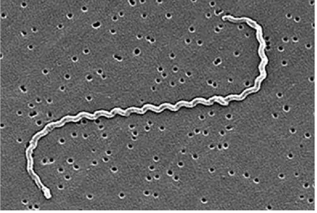
L. interrogans contains over 250 serovars grouped into >25 serogroups. The outer membrane harbors:
- Lipopolysaccharide (LPS) - antigenic, but with unusual structure that poorly activates innate immunity
- Cytotoxic glycolipoprotein (GLP) - directly toxic to tubular Na+/K+-ATPase
- Lipoproteins (especially LipL32, the most abundant outer membrane protein) - highly immunogenic, binds TLR-2, triggers NF-κB activation
The organism survives for days to weeks in water at pH >7.0 but is rapidly killed by acidic conditions and desiccation. - Goldman-Cecil Medicine, p. 3242
Transmission
Humans acquire infection through:
- Broken skin or mucous membranes (conjunctiva, nasal/oral mucosa) exposed to water or soil contaminated with urine from infected reservoir animals
- Ingestion through the upper alimentary tract mucosa
- The hooked ends and periplasmic flagella drive active burrowing through tissues
Primary reservoir: urban brown rat (Rattus norvegicus), which colonizes renal tubules persistently with L. interrogans serovar Icterohaemorrhagiae and sheds organisms lifelong in urine. - Comprehensive Clinical Nephrology, 7th Ed., p. 976
Pathophysiology
Step 1 - Entry and Bacteremia (Leptospiremic Phase, Days 1-7)
After penetrating skin/mucous membranes, leptospires disseminate hematogenously to all organs. They evade the innate immune system by multiple mechanisms:
| Mechanism | Effect |
|---|---|
| Outer membrane proteins bind LPS and peptidoglycan | Shields PAMPs from TLR recognition |
| Surface proteins bind complement regulators (Factor H) | Renders organisms resistant to complement-mediated killing |
| Disruption of endothelial cell junctions | Facilitates tissue invasion and organ dissemination |
Organisms reach the liver, kidney, lungs, meninges, eyes, and muscle. - Goldman-Cecil Medicine, p. 3243
Step 2 - Organ Injury Mechanisms
Hepatic injury:
- Leptospires in hepatic sinusoids cause direct hepatocyte disruption
- Bile leak → conjugated (direct) hyperbilirubinemia and jaundice
- Aminotransferase elevation is typically modest (contrasting with viral hepatitis)
Renal injury (almost universal):
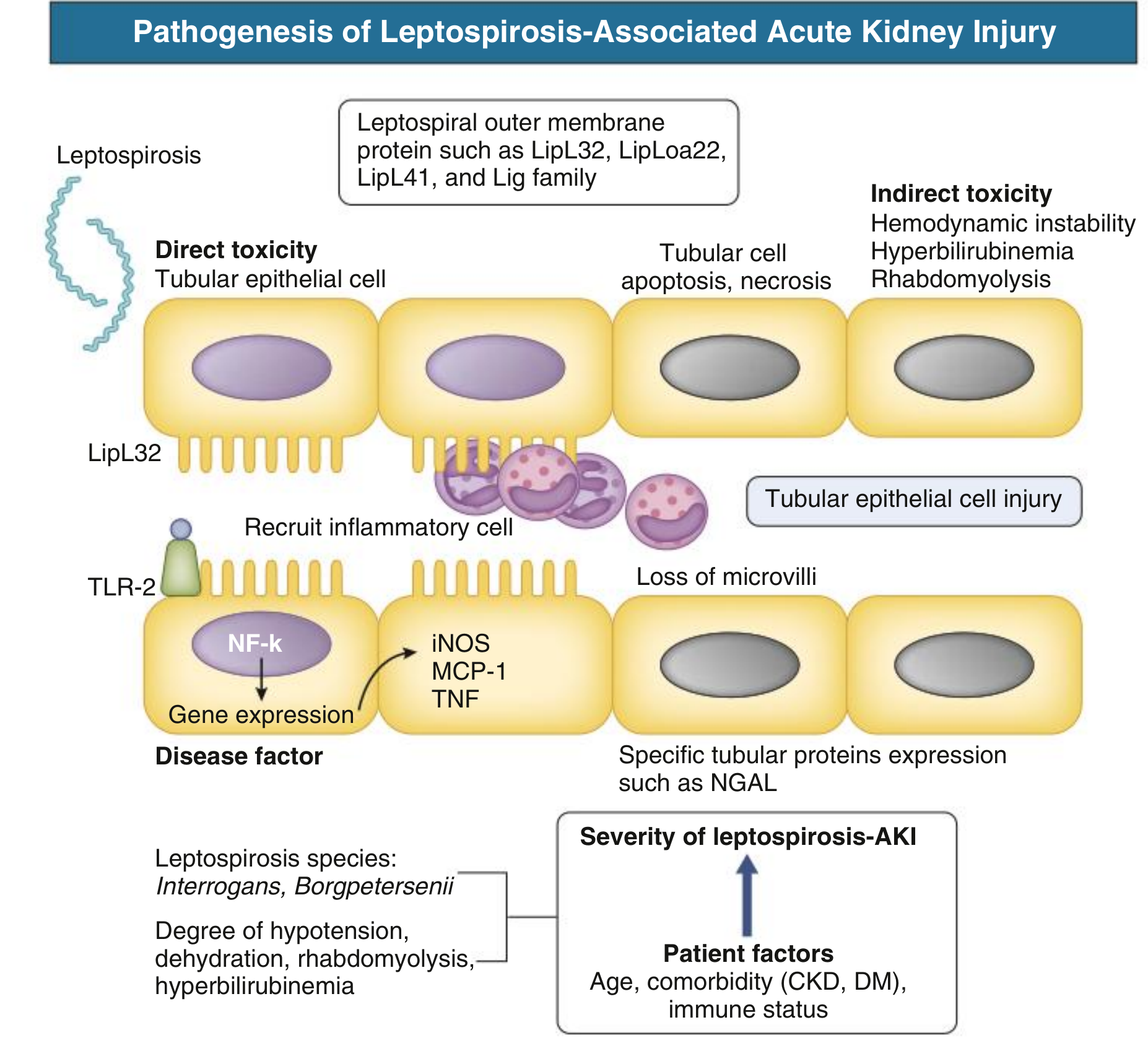
- Direct tubular toxicity: LipL32 binds TLR-2 → NF-κB activation → iNOS, MCP-1, TNF release → inflammatory cell recruitment → tubular cell apoptosis and necrosis
- Na+/K+-ATPase inhibition by GLP: impairs proximal tubular sodium and water reabsorption
- Result: non-oliguric renal failure with urinary Na+/K+ wasting → paradoxical hypokalemia (a hallmark)
- Tubular functional defects precede GFR reduction, explaining early hypokalemia
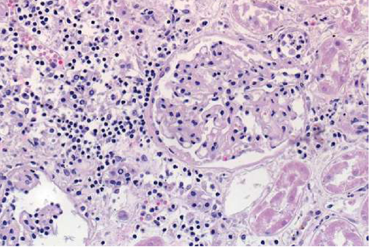
- Histopathology: tubulointerstitial nephritis with cellular infiltration and interstitial edema (glomeruli are typically spared)
Pulmonary injury:
- Disruption of endothelial junctions + thrombocytopenia + coagulopathy → diffuse alveolar hemorrhage
- Can progress to ARDS
- CT: diffuse ground-glass opacities and airspace nodules
Bleeding diathesis:
- Thrombocytopenia
- Coagulopathy (elevated INR, aPTT, D-dimer)
- Endothelial disruption
Muscle (myositis):
- Direct leptospiral invasion of myocytes
- Elevated CK and aldolase
- Rhabdomyolysis can contribute to AKI
Step 3 - Immune Phase (Days 7-14+)
- Clearance of bacteremia coincides with appearance of agglutinating IgM antibodies (typically week 1-2)
- However, the immune response itself drives disease - generalized immune activation may trigger a sepsis-like syndrome with multiorgan failure
- Organisms persist in the renal tubules despite bloodstream clearance (explaining urinary shedding into week 2-3)
- Aseptic meningitis during this phase is immune-mediated (organisms rarely recovered from CSF despite meningeal signs) - Sherris & Ryan's Medical Microbiology, 8th Ed., p. 1424
Clinical Features
Biphasic Course
Incubation period: 6-29 days (median ~15 days)
Phase 1: Leptospiremic (days 1-7)    →    Phase 2: Immune (days 7-14+)
[Bacteremia, initial flu-like illness]      [Antibody appearance, organ complications]
Frequency of Presenting Signs and Symptoms
The box plot below (data from 9 large case series) shows the range of frequency of key symptoms:
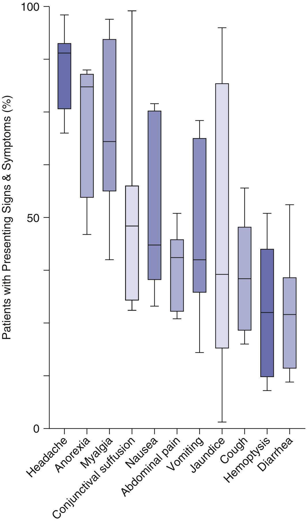
Phase 1 - Anicteric (Mild) Leptospirosis (~90% of cases)
| System | Features |
|---|---|
| Constitutional | Abrupt onset fever, chills, rigors, prostration |
| Head | Severe bitemporal/frontal throbbing headache, retro-orbital pain, photophobia |
| Musculoskeletal | Myalgia - characteristically calf muscles and lower back; exquisite calf tenderness on palpation |
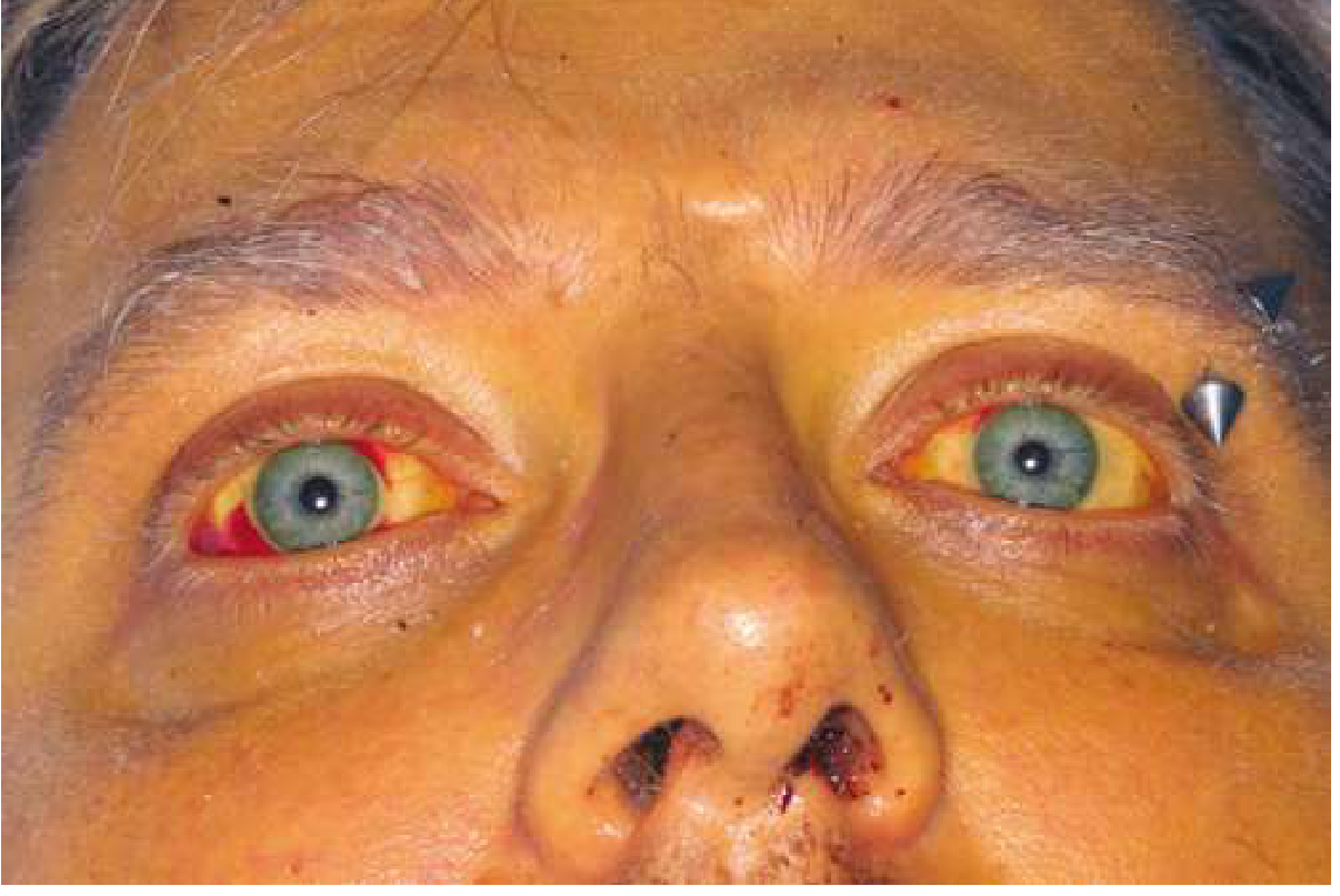
| Ocular | Conjunctival suffusion (pathognomonic - injection without exudate), subconjunctival hemorrhage |
| Respiratory | Nonproductive cough (~50% of cases) |
| GI | Nausea, vomiting, diarrhea, abdominal pain; poor fluid intake worsens dehydration |
| Skin | Maculopapular erythematous rash (especially pretibial in anicteric form - "Fort Bragg fever") |
| Renal | Early non-oliguric renal insufficiency, potassium wasting, hypokalemia, proteinuria |
| CNS | Aseptic meningitis (headache, neck stiffness, CSF: lymphocytes ↑, protein ↑, glucose normal) |
Phase 2 - Weil Disease (Icteric/Severe Leptospirosis, ~10% of cases)
The classic triad of Weil disease is: Jaundice + Acute renal failure + Bleeding
| Organ System | Features in Weil Disease |
|---|---|
| Liver | Deep jaundice (conjugated bilirubin), modest aminotransferase elevation (unlike viral hepatitis) |
| Kidney | Oliguric AKI, pyuria, hematuria, proteinuria, rising creatinine; hypokalemia; may need dialysis |
| Lungs | Pulmonary hemorrhage, hemoptysis, ARDS (most common cause of death today) |
| Bleeding | Petechiae, ecchymoses, GI hemorrhage, frank hemoptysis |
| Heart | Arrhythmias (bradycardia, heart block), myocarditis |
| Hematologic | Thrombocytopenia, hemolytic anemia (especially in G6PD deficiency), leukocytosis (>80% neutrophils) |
| Eyes | Uveitis (may occur weeks after acute illness) |
| Pancreas | Elevated amylase/lipase, pancreatitis |
| Mortality | 5-10% in Weil disease; higher with ARDS and multiorgan failure |
WHO Case Definitions (Table 298-1)
| Category | Criteria |
|---|---|
| Suspect | Fever ≥38.5°C + headache + myalgia/prostration/conjunctival suffusion + history of exposure |
| Probable (clinical) | Suspect + any 2 of: calf tenderness, cough ± hemoptysis, dyspnea, rash, jaundice, hemorrhage, meningeal signs, oliguria/proteinuria, arrhythmia |
| Probable (laboratory) | Suspect + positive rapid IgM OR any 3 of: urinary abnormalities, neutrophilia >80%, platelets <100,000/µL, elevated bilirubin |
| Confirmed | Suspect/probable + isolation, positive PCR, seroconversion, or MAT titer ≥400 |
Key Laboratory Findings
| Test | Typical Finding |
|---|---|
| WBC | Leukocytosis, neutrophilia >80% |
| Platelets | Thrombocytopenia (<100,000/µL in severe disease) |
| Coagulation | Elevated INR, aPTT, D-dimer |
| LFTs | Elevated direct bilirubin >> mild ALT/AST elevation |
| Renal | Elevated creatinine, BUN; pyuria, hematuria, proteinuria |
| Electrolytes | Hypokalemia (characteristic, due to tubular K+ wasting) |
| CK | Elevated (myositis, rhabdomyolysis) |
| CSF | Lymphocytic pleocytosis, elevated protein, normal glucose |
| Chest CT | Ground-glass opacities, airspace nodules (alveolar hemorrhage) |
| Serology | IgM detectable from day 5-7; MAT gold standard (reference labs only) |
| PCR | High sensitivity on blood (first week), urine (second week) |
Key Distinguishing Features from Differential Diagnoses
Leptospirosis mimics dengue, malaria, typhoid, and influenza. Features that favour leptospirosis:
- Conjunctival suffusion (not conjunctivitis - no exudate)
- Calf/lower limb myalgia (often dominant complaint)
- Hypokalemia with renal insufficiency (paradoxical)
- Modest transaminase elevation with deep jaundice (unlike viral hepatitis)
- Elevated CRP/procalcitonin helps distinguish from dengue
- Epidemiologic exposure (flooding, rodent contact, water sports)
Sources:
- Goldman-Cecil Medicine, International Edition, Chapter 298 (DAVID A. HAAKE), pp. 3242-3245
- Comprehensive Clinical Nephrology, 7th Edition, Chapter 71, pp. 976-977
- Sherris & Ryan's Medical Microbiology, 8th Edition, Chapter 5, pp. 1424-1425
- Andrews' Diseases of the Skin, Chapter on Leptospirosis, pp. 1399-1407
- Recent reviews: Nature Reviews Disease Primers 2025 (PMID 40316520); Clinical Medicine 2022 (PMID 35078790)
Clinical features in leptospirosis
I already have comprehensive, well-sourced content on leptospirosis clinical features from the previous response. Here is a focused, detailed breakdown:
Clinical Features of Leptospirosis
Overview
Leptospirosis is a biphasic illness. About 90% of cases are mild and self-limited (anicteric); ~10% progress to the severe icteric form (Weil disease). Most infections are subclinical and detected only serologically.
Incubation period: 6-29 days (median ~15 days)
Biphasic Course at a Glance
Phase 1: Leptospiremic Phase         Phase 2: Immune Phase
(Days 1-7)                           (Days 7-30)
─────────────────────────            ──────────────────────────────
Bacteremia present                   Antibody-mediated clearance
Flu-like illness                     Organ complications develop
Fever, myalgia, headache             Jaundice, AKI, hemorrhage
↓ (brief 1-3 day remission)          Aseptic meningitis, uveitis
Phase 1 - Leptospiremic Phase (Anicteric Leptospirosis)
This phase is indistinguishable from many acute febrile illnesses. Key features:
Constitutional
- Abrupt-onset high fever with chills and rigors
- Prostration and anorexia
Headache
- Severe, bitemporal or frontal, throbbing in character
- Accompanied by retro-orbital pain and photophobia
- One of the most prominent complaints
Musculoskeletal - Hallmark Feature
- Myalgia - characteristically involves the calves and lower back
- Calf tenderness on palpation is a characteristic and diagnostically important sign
- Severity can mimic a surgical emergency or deep vein thrombosis
Ocular - Pathognomonic Sign
- Conjunctival suffusion - bilateral conjunctival hyperemia/injection without exudate (distinguishes it from conjunctivitis)
- Subconjunctival hemorrhage
- Retro-orbital pain and photophobia
Respiratory
- Nonproductive cough in up to ~50% of cases
- Chest pain may occur
Gastrointestinal
- Nausea, vomiting, diarrhea
- Abdominal pain (can suggest pancreatitis)
- Poor oral intake worsens dehydration, which can precipitate oliguric renal failure
Skin
- Maculopapular erythematous rash, most marked on the shins (pretibial)
- In anicteric disease associated with serovar autumnalis - called "Fort Bragg fever" or pretibial fever
- Rash consists of 1-5 cm erythematous patches/plaques; resolves in 4-7 days spontaneously
- Petechiae and purpura in severe cases
Early Renal Involvement
- Non-oliguric renal insufficiency (early, often subtle)
- Hypokalemia - paradoxical and characteristic, due to renal tubular K+ wasting (Na+/K+-ATPase inhibition by GLP)
- Proteinuria, hematuria, pyuria
CNS
- Aseptic meningitis - headache, neck stiffness
- CSF: lymphocytic pleocytosis, elevated protein, normal glucose
- Organisms are rarely recovered from CSF despite meningeal signs (immune-mediated)
Frequency of Symptoms (Across 9 Large Case Series)
| Symptom/Sign | Approximate Frequency |
|---|---|
| Headache | ~80% |
| Myalgia | ~75% |
| Anorexia | ~65% |
| Nausea | ~50% |
| Conjunctival suffusion | ~45% |
| Vomiting | ~35% |
| Abdominal pain | ~35% |
| Jaundice | Variable (5-75%) |
| Cough | ~35% |
| Diarrhea | ~30% |
| Hemoptysis | ~10-15% |
Phase 2 - Immune Phase: Weil Disease (Icteric/Severe Leptospirosis, ~10%)
Weil disease is the severe form, caused most often by serovar Icterohaemorrhagiae. It is defined by the classic triad:
Jaundice + Acute Renal Failure + Bleeding
Hepatic Features
- Deep jaundice - predominantly conjugated (direct) hyperbilirubinemia
- Mild-to-moderate aminotransferase elevation only (unlike viral hepatitis where transaminases are very high)
- This dissociation - deep jaundice with modest transaminases - is a clue to leptospirosis
- Hepatomegaly and tenderness
Renal Features
- AKI develops in 10-80% depending on severity
- Initially non-oliguric with hypokalemia (tubular dysfunction)
- Can progress to oliguric AKI requiring dialysis
- Urinary findings: proteinuria, hematuria, pyuria, casts
- Long-term sequelae: CKD and persistent tubular dysfunction reported on follow-up
- Hypokalemia persists and may be profound
Pulmonary Features (Most Common Cause of Death)
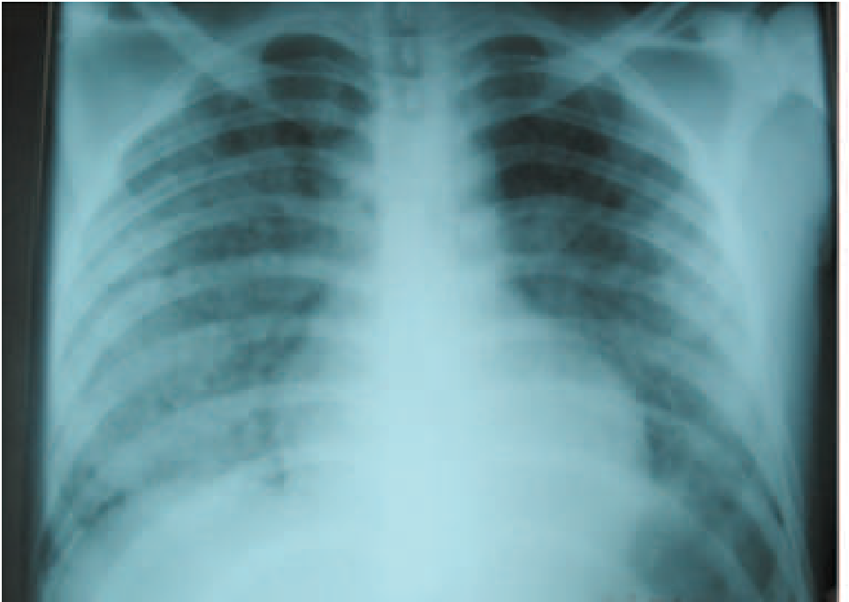
- Pulmonary hemorrhage - hemoptysis, frank alveolar bleeding
- ARDS - diffuse alveolar hemorrhage
- Chest CT: bilateral ground-glass opacities and airspace nodules
- Rapidly fatal if not recognized; can occur without jaundice
Hemorrhagic Features
- Petechiae and ecchymoses on skin and mucous membranes
- Severe GI hemorrhage
- Pulmonary hemorrhage (hemoptysis)
- Driven by: thrombocytopenia + coagulopathy + endothelial injury
Cardiac Features
- Arrhythmias (bradycardia, heart block, atrial fibrillation)
- Myocarditis
- Cardiac arrhythmia is an independent predictor of severity
Hematologic Features
- Leukocytosis (WBC 15,000-30,000/mm³, >80% neutrophils)
- Thrombocytopenia (<100,000/µL)
- Coagulopathy: elevated INR, aPTT, D-dimer
- Hemolytic anemia - particularly in G6PD-deficient patients; contributes to jaundice
Ocular
- Uveitis (anterior or panuveitis) - may develop weeks to months after acute illness; important to recognize as a delayed manifestation
Neurological
- Aseptic meningitis (continued from phase 1)
- Encephalitis in severe cases
- Peripheral neuropathy (rare)
Pancreatic
- Elevated serum amylase and lipase
- Frank pancreatitis (elevated amylase may also reflect reduced renal clearance)
WHO Clinical Case Definitions
| Category | Criteria |
|---|---|
| Suspect | Fever ≥38.5°C and/or severe headache + myalgia/prostration/conjunctival suffusion + history of exposure |
| Probable (clinical) | Suspect + any 2 of: calf tenderness, cough ± hemoptysis, dyspnea, rash, jaundice, hemorrhage, meningeal signs, oliguria/proteinuria, arrhythmia |
| Probable (laboratory) | Suspect + positive rapid IgM or any 3 of: urinary abnormalities, neutrophilia >80%, platelets <100,000/µL, elevated bilirubin |
| Confirmed | Positive culture / PCR / MAT seroconversion or single titer ≥1:400 |
Key Distinguishing Clinical Clues
| Feature | Why It Helps |
|---|---|
| Conjunctival suffusion (no exudate) | Pathognomonic; not seen in dengue/malaria |
| Calf tenderness | Highly characteristic |
| Hypokalemia + non-oliguric AKI | Paradoxical; reflects tubular Na+/K+-ATPase dysfunction |
| Deep jaundice + modest transaminases | Unlike viral hepatitis (transaminases very high) |
| Elevated CRP/procalcitonin | Helps distinguish from dengue (where they are low) |
| Exposure history | Flooding, rodents, water sports, occupational (farmers, veterinarians, sewer workers) |
Complications Summary
| Complication | Notes |
|---|---|
| Pulmonary hemorrhage / ARDS | Leading cause of death in modern series |
| Oliguric AKI | May need dialysis; usually reversible |
| Weil disease | Jaundice + AKI + bleeding triad |
| Aseptic meningitis | Immune-mediated, benign course usually |
| Uveitis | Delayed, weeks-months post-infection |
| Myocarditis / arrhythmia | Independent mortality predictor |
| Hemolytic anemia | Especially in G6PD deficiency |
| Mortality | 5-10% in Weil disease; higher with pulmonary involvement |
Sources:
- Goldman-Cecil Medicine, International Edition, Chapter 298, pp. 3242-3245
- Andrews' Diseases of the Skin, pp. 1399-1407
- Sherris & Ryan's Medical Microbiology, 8th Ed., pp. 1423-1425
- Comprehensive Clinical Nephrology, 7th Ed., pp. 976-977
Why in this 2 type of clinical features appear in one desease
Great question. The short answer: the two phases exist because the cause of illness shifts - Phase 1 is caused by the bacteria itself, Phase 2 is caused by your own immune system fighting it.
Searching sources...
Finding Sources
Finding Sources
Harrison's has a dedicated pathogenesis section. Let me read it:
Reading File
Excellent - Harrison's has rich pathogenesis content in the truncated section. Let me read the key pathogenesis and biphasic illness explanation portion:
Reading File
There's a critical timeline diagram image in Harrison's. Let me fetch it and continue reading:
Reading File
Loading Image
I now have excellent Harrison's content explaining exactly why the biphasic illness occurs. Let me fetch the timeline diagram:
Loading Image
Finding Sources
I now have all the material needed for a thorough, conceptual explanation. Here is the full answer:
Why Does Leptospirosis Produce Two Different Phases of Clinical Features?
The answer lies in a fundamental principle: the two phases have completely different causes. Phase 1 is caused by the bacteria itself; Phase 2 is caused by your own immune system's response to it. One disease, two different attackers.
The Core Concept
Phase 1 (Days 1-7)                    Phase 2 (Days 7-30)
─────────────────────────             ──────────────────────────────
CAUSE = Living bacteria               CAUSE = Immune system activation
in the bloodstream                    + bacteria hiding in organs

Bacteria → directly damage            Antibodies appear → clear blood
tissues and trigger                   But bacteria persist in kidneys,
non-specific inflammation             liver, lungs, brain →
                                      Immune response attacks tissues
This is why there is often a brief 1-3 day period of apparent improvement between the two phases - the bacteria are cleared from the blood, but before the immune response escalates fully. - Goldman-Cecil Medicine, p. 3243
Phase 1 in Detail: Direct Bacterial Damage (Leptospiremic Phase)
During Phase 1, living leptospires circulate freely in the bloodstream and spread to every organ. The symptoms come from:
1. Direct tissue invasion
- The hooked ends and periplasmic flagella physically burrow through tissues
- Leptospires invade liver sinusoids, renal tubules, muscle, meninges, and lungs
- Disruption of endothelial cell junctions increases vascular permeability
2. Bacterial toxins directly injure cells
- LipL32 (major outer membrane lipoprotein) binds TLR-2 on cells → triggers NF-κB → release of inflammatory cytokines (TNF, iNOS, MCP-1)
- Cytotoxic glycolipoprotein (GLP) directly inhibits Na+/K+-ATPase on renal tubular cells → explains the hypokalemia seen even in early disease
3. Non-specific febrile response
- Bacteremia itself activates innate immunity - fever, rigors, myalgia, headache, prostration
- This is similar to the leptospiremic phase of any spirochetal infection
Result: Flu-like syndrome with fever, severe headache, myalgia (especially calves), and conjunctival suffusion. The bacteremia can be detected in blood cultures and PCR.
The Turning Point: Why Does the Immune System Then Cause More Damage?
This is the key question. Here is the sequence, as described in Harrison's:
Step 1 - Bacteria escape the immune system (Phase 1)
Leptospires are remarkably good at evading the immune system during Phase 1:
| Evasion Mechanism | How It Works |
|---|---|
| Bind complement regulators (Factor H) | Resist complement-mediated killing |
| Mask LPS and peptidoglycan with outer membrane proteins | Avoid TLR4 recognition |
| Rapid intracellular spread | Avoid antibody neutralization |
| Invade "immune-privileged" sites (kidney tubules, eye) | Persist despite systemic immunity |
This is why early-phase illness is relatively mild despite active bacteremia. - Harrison's Principles of Internal Medicine, 22nd Ed., p. 1484
Step 2 - Antibodies appear (start of Phase 2)
Around Day 5-7, IgM antibodies form against leptospiral surface antigens. These antibodies:
- Clear leptospires from the bloodstream effectively
- But cannot reach bacteria hiding in kidney tubules, liver, lungs, brain, and aqueous humor of the eye
- This is why blood cultures and PCR on blood become negative in Phase 2
Step 3 - The immune response itself becomes the problem
This is where the damage escalates. Harrison's states clearly: "Earlier studies have highlighted the relation between an exaggerated proinflammatory immune response and mortality." - Harrison's, p. 1484
Multiple immune-mediated mechanisms fire simultaneously:
A. Consumptive Coagulopathy / DIC
- Elevated thrombin-antithrombin complexes, prothrombin fragments 1+2, D-dimer
- Diminished anticoagulant markers (antithrombin, protein C)
- Deregulated fibrinolysis
- Thrombocytopenia (platelet consumption on activated endothelium)
- Result: bleeding - petechiae, pulmonary hemorrhage, GI hemorrhage
B. Endothelial Activation
- Elevated soluble E-selectin and von Willebrand factor in blood = markers of endothelial cell activation
- These correlate directly with disease severity
- Activated endothelium → platelets aggregate on lung vasculature → pulmonary hemorrhage
- Increased vascular permeability → edema, ARDS
- Harrison's: "Experimental models show that pathogenic leptospires or leptospiral proteins are able to activate endothelial cells in vitro and to disrupt endothelial-cell barrier function, thus increasing permeability."
C. Persistent Organ Damage from Bacteria in Tissues
Even as blood is cleared, leptospires persist in organs. Histopathology shows:
- Kidney: Acute tubular damage → interstitial edema → interstitial nephritis → AKI
- Liver: Focal necrosis, hepatocyte apoptosis, bile canaliculi disruption → jaundice
- Lungs: Hemorrhage and alveolar destruction
- "Autopsy findings illustrate the involvement of multiple organ systems in severe disease." - Harrison's, p. 1485
D. Why Does Antibiotic Treatment Fail in Phase 2?
This is a critical clinical clue that confirms Phase 2 is immune-mediated, not bacterial:
- Antibiotics given during Phase 2 do not stop the immune cascade
- The sepsis-like syndrome continues even after bacteria are killed
- This parallels other post-infectious immune syndromes (e.g., rheumatic fever, reactive arthritis)
- The meningitis of Phase 2 is also immune-mediated - organisms are rarely recovered from CSF despite signs of meningeal irritation
Why Does Only ~10% Progress to Weil Disease?
Not everyone who gets leptospirosis develops the severe immune phase. The factors that push someone toward Weil disease:
| Factor | Effect |
|---|---|
| Infecting serovar | Icterohaemorrhagiae much more likely to cause Weil disease than autumnalis |
| Bacterial load (inoculum size) | Higher exposure → more organisms → stronger immune response |
| Host genetics | HLA-DQ6 haplotype is an independent risk factor |
| G6PD deficiency | Worsens hemolytic anemia component |
| Age and comorbidities | CKD, diabetes worsen AKI severity |
| Immune response magnitude | An exaggerated proinflammatory response = more organ damage |
Paradoxically, a stronger immune response correlates with worse outcomes. The immune system that should be clearing the infection is simultaneously destroying organs. This is the same mechanism seen in severe dengue, septic shock, and cytokine storm syndromes. - Harrison's, p. 1484; Comprehensive Clinical Nephrology, 7th Ed., p. 976
Summary: Why Two Phases in One Disease?
| Phase 1 (Leptospiremic) | Phase 2 (Immune) | |
|---|---|---|
| Cause | Live bacteria in blood | Immune response + tissue-trapped bacteria |
| Duration | Days 1-7 | Days 7-30+ |
| Dominant mechanism | Direct bacterial toxins, tissue invasion | Coagulopathy, endothelial activation, cytokines |
| Blood cultures/PCR | Positive | Negative |
| Antibodies | Absent | Present (IgM rising) |
| Antibiotic effect | Effective if given early | Limited benefit |
| Main symptoms | Fever, myalgia, headache, conjunctival suffusion | Jaundice, AKI, bleeding, ARDS |
The brief window of apparent improvement between phases corresponds to the moment antibodies clear the blood of bacteria - but before the full immune cascade and organ damage of Phase 2 has developed. This is the critical window for antibiotic intervention - treatment given in Phase 1 (or at the transition) is far more effective than treatment given deep into Phase 2.
Sources:
- Harrison's Principles of Internal Medicine, 22nd Ed., Chapter 189, pp. 1484-1485
- Goldman-Cecil Medicine, International Edition, Chapter 298, pp. 3242-3243
- Sherris & Ryan's Medical Microbiology, 8th Ed., pp. 1424-1425
- Comprehensive Clinical Nephrology, 7th Ed., pp. 976-977
This is a shared conversation. Sign in to Orris to start your own chat.