
Heme synthesis
I'll start with a quick overview, then pull from the medical library for a thorough answer.
Heme synthesis is an 8-step pathway that begins and ends in the mitochondria, with intermediate steps in the cytoplasm. The rate-limiting enzyme is ALA synthase (ALAS), which condenses succinyl-CoA + glycine to form delta-aminolevulinic acid (ALA). The final product, heme, is incorporated into hemoglobin, myoglobin, cytochromes, and other hemoproteins.
Searching sources...
Reading File
Now let me search the library for heme synthesis content:
Finding Sources
Finding Sources
I have great sources. Let me now read the key sections in parallel - Lippincott Biochemistry (most detailed), Basic Medical Biochemistry, and the Fitzpatrick's section:
Reading File
Reading File
Excellent content. Let me read more of Lippincott to get the full pathway and regulation details, and also fetch the key pathway diagram:
Reading File
Reading File
I now have comprehensive data. Let me fetch the key pathway diagrams from Lippincott:
Loading Image
Loading Image
I have excellent pathway diagrams. Let me also get the full pathway diagram (Figure 21.4) and the porphyria summary image, plus read the porphyria/clinical section:
Loading Image
Reading File
I now have everything needed for a thorough, well-illustrated answer. Let me compose it.
Heme Synthesis
Heme is an iron-containing porphyrin that serves as the prosthetic group for hemoglobin, myoglobin, cytochromes (including cytochrome P450), catalase, and peroxidases. Over 85% of all heme synthesis occurs in erythroid tissue (bone marrow); the liver accounts for most of the remainder.
Structure of Heme
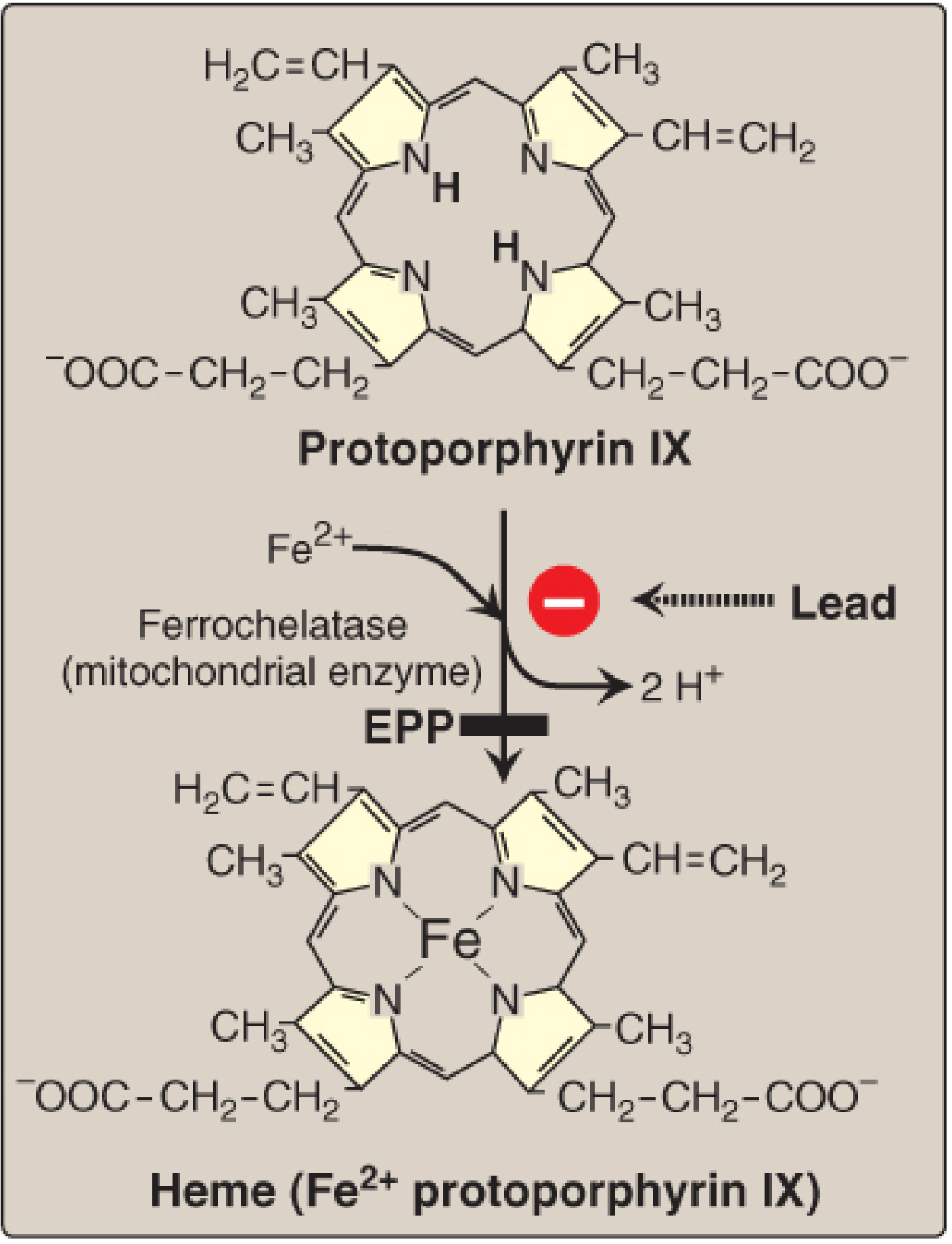
Heme is protoporphyrin IX chelated with a ferrous iron (Fe²+) atom. Four pyrrole rings joined by methenyl bridges (=CH-) form the porphyrin macrocycle. Eight side chains substitute the ring - two per pyrrole - in the order M-V / V-M / V-P / P-M (methyl-vinyl/vinyl-methyl/vinyl-propionate/propionate-methyl), the signature of the type III porphyrin series.
Cellular Compartmentalization
The pathway shuttles between mitochondria and cytosol:
| Location | Steps |
|---|---|
| Mitochondria | Steps 1 (ALA formation), 6-8 (coproporphyrinogen III → heme) |
| Cytosol | Steps 2-5 (PBG → coproporphyrinogen III) |
The 8 Steps
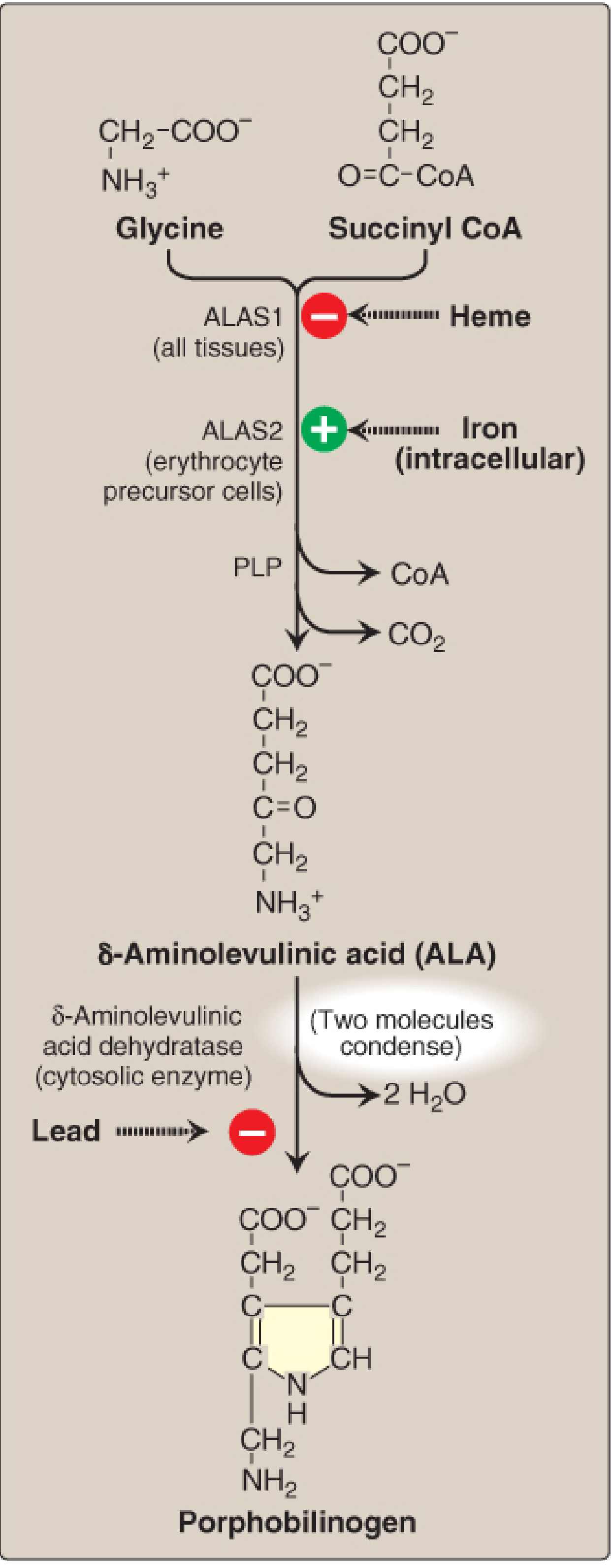
Step 1 - ALA Formation (Mitochondria) - Rate-Limiting Step
Substrates: Succinyl-CoA + Glycine
Enzyme: δ-Aminolevulinic acid synthase (ALAS) - requires pyridoxal phosphate (PLP, vitamin B6) as cofactor
Product: δ-Aminolevulinic acid (ALA); releases CO₂ and CoA
Enzyme: δ-Aminolevulinic acid synthase (ALAS) - requires pyridoxal phosphate (PLP, vitamin B6) as cofactor
Product: δ-Aminolevulinic acid (ALA); releases CO₂ and CoA
Two isoforms exist:
- ALAS1 - expressed in all tissues (especially liver); inhibited by heme (feedback)
- ALAS2 - expressed only in erythroid cells; regulated by intracellular iron availability (stimulated by iron)
Step 2 - Porphobilinogen (PBG) Formation (Cytosol)
Substrate: 2 × ALA
Enzyme: ALA dehydratase (PBG synthase) - a zinc-containing enzyme
Product: Porphobilinogen (PBG), a monopyrrole; releases 2 H₂O
Inhibited by: Lead (Pb²+) - replaces zinc, blocking this enzyme
Enzyme: ALA dehydratase (PBG synthase) - a zinc-containing enzyme
Product: Porphobilinogen (PBG), a monopyrrole; releases 2 H₂O
Inhibited by: Lead (Pb²+) - replaces zinc, blocking this enzyme
Step 3 - Hydroxymethylbilane Formation (Cytosol)
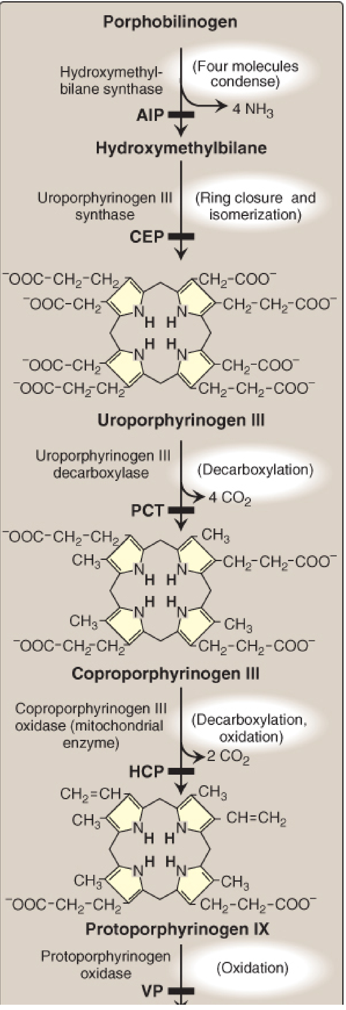
Substrate: 4 × PBG
Enzyme: Hydroxymethylbilane synthase (also called porphobilinogen deaminase or uroporphyrinogen I synthase)
Product: Hydroxymethylbilane (a linear tetrapyrrole); releases 4 NH₃
Deficiency: Acute Intermittent Porphyria (AIP) - the most common acute porphyria
Enzyme: Hydroxymethylbilane synthase (also called porphobilinogen deaminase or uroporphyrinogen I synthase)
Product: Hydroxymethylbilane (a linear tetrapyrrole); releases 4 NH₃
Deficiency: Acute Intermittent Porphyria (AIP) - the most common acute porphyria
Step 4 - Uroporphyrinogen III Formation (Cytosol)
Substrate: Hydroxymethylbilane
Enzyme: Uroporphyrinogen III synthase
Reaction: Ring closure + isomerization (reverses the D ring, giving the asymmetric type III series)
Product: Uroporphyrinogen III
Deficiency: Congenital Erythropoietic Porphyria (CEP) - without isomerization, type I porphyrins accumulate
Enzyme: Uroporphyrinogen III synthase
Reaction: Ring closure + isomerization (reverses the D ring, giving the asymmetric type III series)
Product: Uroporphyrinogen III
Deficiency: Congenital Erythropoietic Porphyria (CEP) - without isomerization, type I porphyrins accumulate
Step 5 - Coproporphyrinogen III Formation (Cytosol)
Substrate: Uroporphyrinogen III
Enzyme: Uroporphyrinogen III decarboxylase
Reaction: Decarboxylation of 4 acetate side chains → 4 methyl groups; releases 4 CO₂
Product: Coproporphyrinogen III
Deficiency: Porphyria Cutanea Tarda (PCT) - the most common porphyria overall
Enzyme: Uroporphyrinogen III decarboxylase
Reaction: Decarboxylation of 4 acetate side chains → 4 methyl groups; releases 4 CO₂
Product: Coproporphyrinogen III
Deficiency: Porphyria Cutanea Tarda (PCT) - the most common porphyria overall
Step 6 - Protoporphyrinogen IX Formation (Mitochondria)
Substrate: Coproporphyrinogen III (re-enters mitochondria)
Enzyme: Coproporphyrinogen III oxidase
Reaction: Decarboxylation + oxidation of 2 propionate side chains → vinyl groups; releases 2 CO₂
Product: Protoporphyrinogen IX
Deficiency: Hereditary Coproporphyria (HCP)
Enzyme: Coproporphyrinogen III oxidase
Reaction: Decarboxylation + oxidation of 2 propionate side chains → vinyl groups; releases 2 CO₂
Product: Protoporphyrinogen IX
Deficiency: Hereditary Coproporphyria (HCP)
Step 7 - Protoporphyrin IX Formation (Mitochondria)
Substrate: Protoporphyrinogen IX
Enzyme: Protoporphyrinogen oxidase
Reaction: Oxidation (removes 6 H); creates the fully conjugated, colored porphyrin ring
Product: Protoporphyrin IX
Deficiency: Variegate Porphyria (VP)
Enzyme: Protoporphyrinogen oxidase
Reaction: Oxidation (removes 6 H); creates the fully conjugated, colored porphyrin ring
Product: Protoporphyrin IX
Deficiency: Variegate Porphyria (VP)
Step 8 - Heme Formation (Mitochondria)
Substrate: Protoporphyrin IX + Fe²+
Enzyme: Ferrochelatase (also called heme synthase)
Reaction: Chelation of Fe²+ into the porphyrin ring; releases 2 H+
Product: Heme (Fe²+-protoporphyrin IX)
Inhibited by: Lead (Pb²+) - inhibits ferrochelatase
Deficiency: Erythropoietic Protoporphyria (EPP)
Enzyme: Ferrochelatase (also called heme synthase)
Reaction: Chelation of Fe²+ into the porphyrin ring; releases 2 H+
Product: Heme (Fe²+-protoporphyrin IX)
Inhibited by: Lead (Pb²+) - inhibits ferrochelatase
Deficiency: Erythropoietic Protoporphyria (EPP)
Pathway Summary Table
| Step | Location | Enzyme | Substrate → Product | Porphyria if Deficient |
|---|---|---|---|---|
| 1 | Mito | ALAS (PLP-dependent) | Succinyl-CoA + Gly → ALA | - (rate-limiting) |
| 2 | Cytosol | ALA dehydratase (Zn²+) | 2 ALA → PBG | - (Pb toxicity target) |
| 3 | Cytosol | HMB synthase | 4 PBG → HMB | AIP |
| 4 | Cytosol | Uroporphyrinogen III synthase | HMB → UPG III | CEP |
| 5 | Cytosol | Uroporphyrinogen III decarboxylase | UPG III → CPG III | PCT |
| 6 | Mito | Coproporphyrinogen III oxidase | CPG III → PPG IX | HCP |
| 7 | Mito | Protoporphyrinogen oxidase | PPG IX → PP IX | VP |
| 8 | Mito | Ferrochelatase | PP IX + Fe²+ → Heme | EPP |
ALA = aminolevulinic acid; PBG = porphobilinogen; HMB = hydroxymethylbilane; UPG = uroporphyrinogen; CPG = coproporphyrinogen; PPG = protoporphyrinogen; PP = protoporphyrin; Mito = mitochondria
Regulation
ALAS1 (Hepatic)
Heme acts as a negative feedback regulator on ALAS1 via three mechanisms:
- Represses transcription of the ALAS1 gene
- Increases degradation of ALAS1 mRNA
- Decreases mitochondrial import of the ALAS1 protein
When hepatic heme is consumed (e.g., by cytochrome P450 induction from drugs like barbiturates, phenytoin, rifampicin), free heme falls, ALAS1 is derepressed, and ALA production surges. This is the basis for drug-induced porphyria attacks.
ALAS2 (Erythroid)
- Stimulated by iron availability - when intracellular iron is adequate, ALAS2 activity increases to match globin synthesis
- Heme in erythroid cells also stimulates globin synthesis by maintaining the ribosomal initiation complex in an active state
Clinically Relevant Enzyme Inhibition by Lead
Lead (Pb²+) inhibits two enzymes:
- ALA dehydratase (step 2) - accumulation of ALA
- Ferrochelatase (step 8) - accumulation of protoporphyrin IX; zinc is substituted for iron → zinc protoporphyrin (ZPP) accumulates
Lab findings in lead poisoning: elevated urine ALA, elevated blood/urine ZPP, microcytic hypochromic anemia (resembles iron deficiency)
Porphyrias Overview
Porphyrias are inherited (usually autosomal dominant) or acquired defects in heme synthesis. They fall into two clinical categories:
| Type | Deficient Enzyme | Features |
|---|---|---|
| Acute Intermittent Porphyria (AIP) | HMB synthase (step 3) | Neuropsychiatric attacks, abdominal pain, autonomic dysfunction; NO photosensitivity; urine turns red/port-wine on standing |
| Congenital Erythropoietic Porphyria (CEP) | UPG III synthase (step 4) | Severe photosensitivity, hemolytic anemia, skin fragility |
| Porphyria Cutanea Tarda (PCT) | UPG decarboxylase (step 5) | Most common porphyria; skin blistering/fragility in light-exposed areas; associated with alcohol, estrogens, hepatitis C |
| Hereditary Coproporphyria (HCP) | CPG III oxidase (step 6) | Acute attacks + photosensitivity |
| Variegate Porphyria (VP) | Protoporphyrinogen oxidase (step 7) | Acute attacks + skin lesions; dangerous with barbiturates |
| Erythropoietic Protoporphyria (EPP) | Ferrochelatase (step 8) | Photosensitivity; no acute attacks |
Sources
- Biochemistry, 8th ed. Lippincott Illustrated Reviews, Chapter 21 (Porphyrin Metabolism)
- Basic Medical Biochemistry: A Clinical Approach, 6e, Chapter 42
Heme metabolism
Heme metabolism refers to the degradation of heme (primarily from senescent RBCs) into biliverdin, then bilirubin, which is conjugated in the liver and excreted in bile. Key steps: heme oxygenase cleaves the porphyrin ring releasing CO and biliverdin; biliverdin reductase converts biliverdin to unconjugated bilirubin; in the liver, UDP-glucuronosyltransferase conjugates bilirubin for excretion.
Searching sources...
Finding Sources
Finding Sources
Great sources found. Let me read the best sections in parallel - Lippincott's heme degradation section and Harper's catabolism of heme:
Reading File
Reading File
Excellent. Now let me read more from Lippincott (excretion, urobilinogen, clinical disorders) and get the key diagrams:
Reading File
Reading File
Loading Image
Loading Image
Now let me get the full catabolism summary diagram and the jaundice types section:
Loading Image
Reading File
I now have everything needed for a comprehensive answer. Let me also read the neonatal jaundice and hereditary hyperbilirubinemia sections:
Reading File
I have comprehensive content from all sections. Let me now compose the full answer.
Heme Metabolism (Degradation)
Heme metabolism refers to the catabolism of heme - primarily from hemoglobin in senescent red blood cells (RBCs) - through a series of steps that produce bilirubin, which is then conjugated and excreted. Adults destroy roughly 200 billion RBCs per day, yielding ~250-350 mg of bilirubin daily.
Overview: Sources of Heme for Degradation
| Source | Contribution |
|---|---|
| Senescent RBCs (120-day lifespan) | ~75-80% of daily bilirubin |
| Ineffective erythropoiesis | ~10-15% |
| Cytochromes, myoglobin, catalase | ~5-10% |
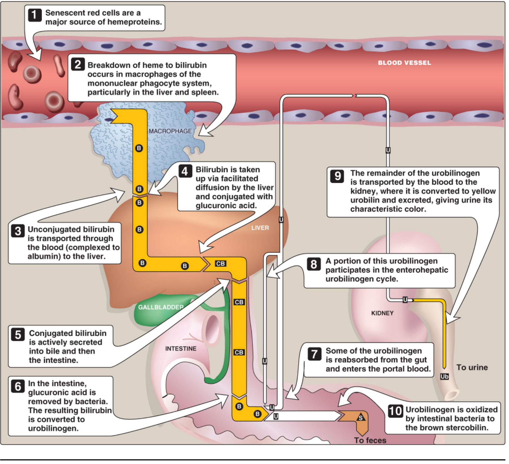
Mature RBCs are phagocytosed by macrophages of the mononuclear phagocyte system (MPS) - particularly in the liver (Kupffer cells), spleen, and bone marrow. Globin chains are hydrolyzed to amino acids; iron is recycled; the porphyrin ring of heme is degraded.
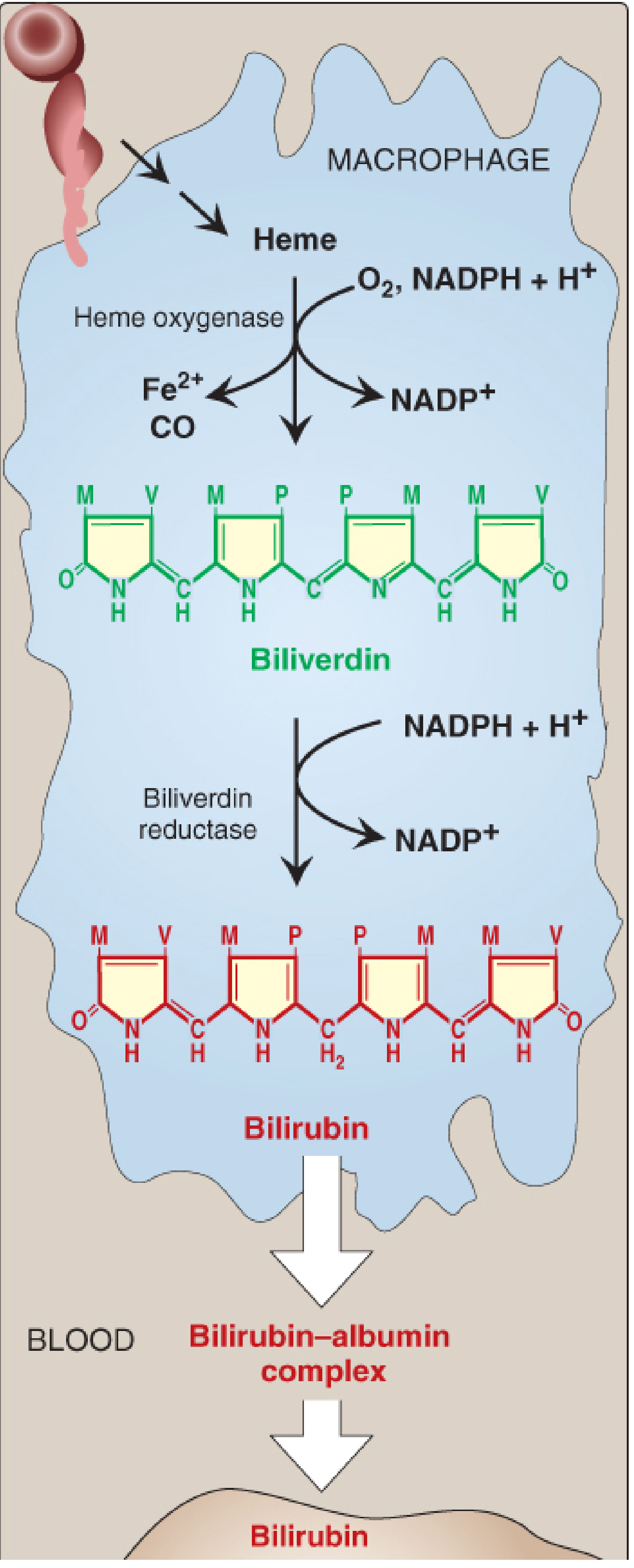
Step 1 - Heme → Biliverdin (Macrophages)
Enzyme: Heme oxygenase (microsomal; NADPH-dependent)
Cofactors: O₂, NADPH
Reaction: The α-methene bridge of the porphyrin ring is cleaved oxidatively
Heme-Fe³+ + 3 O₂ + 7 e⁻ → Biliverdin + CO + Fe²+ (or Fe³+)
Three products are released:
- Biliverdin - a linear, green tetrapyrrole
- Carbon monoxide (CO) - has signaling and anti-inflammatory roles; is why a bruise turns green-then-yellow
- Fe²+ - returned to the iron pool (ferritin/transferrin)
Note: Heme oxygenase is substrate-inducible - heme itself induces its own oxygenase.
Step 2 - Biliverdin → Bilirubin (Macrophages)
Enzyme: Biliverdin reductase
Cofactor: NADPH
Reaction: Reduction of the central methylene bridge of biliverdin
Biliverdin + NADPH + H⁺ → Bilirubin + NADP⁺
Bilirubin is a yellow-orange pigment. It is the end product of heme catabolism in mammals. (Birds and amphibians excrete biliverdin directly without this step.)
Bilirubin as antioxidant: At low concentrations, bilirubin functions as an antioxidant - it is oxidized back to biliverdin, then regenerated by biliverdin reductase, forming a redox cycle that scavenges reactive oxygen species.
Step 3 - Transport to the Liver (Blood)
Bilirubin is poorly water-soluble (unconjugated/indirect bilirubin, UCB). It is transported in blood bound noncovalently to albumin:
- Albumin has a high-affinity site (binds ~25 mg bilirubin/100 mL plasma) and a low-affinity site
- This form is called unconjugated bilirubin (UCB) or indirect bilirubin
- Certain drugs (salicylates, sulfonamides, some antibiotics) competitively displace bilirubin from albumin, risking CNS entry - especially dangerous in neonates (kernicterus)
Step 4 - Hepatic Uptake
At the sinusoidal surface of hepatocytes, bilirubin:
- Dissociates from albumin
- Enters via facilitated diffusion (saturable transport system)
- Binds to intracellular proteins, especially ligandin (glutathione S-transferase) - this prevents back-diffusion into blood
Step 5 - Conjugation in the Liver (Hepatocytes)
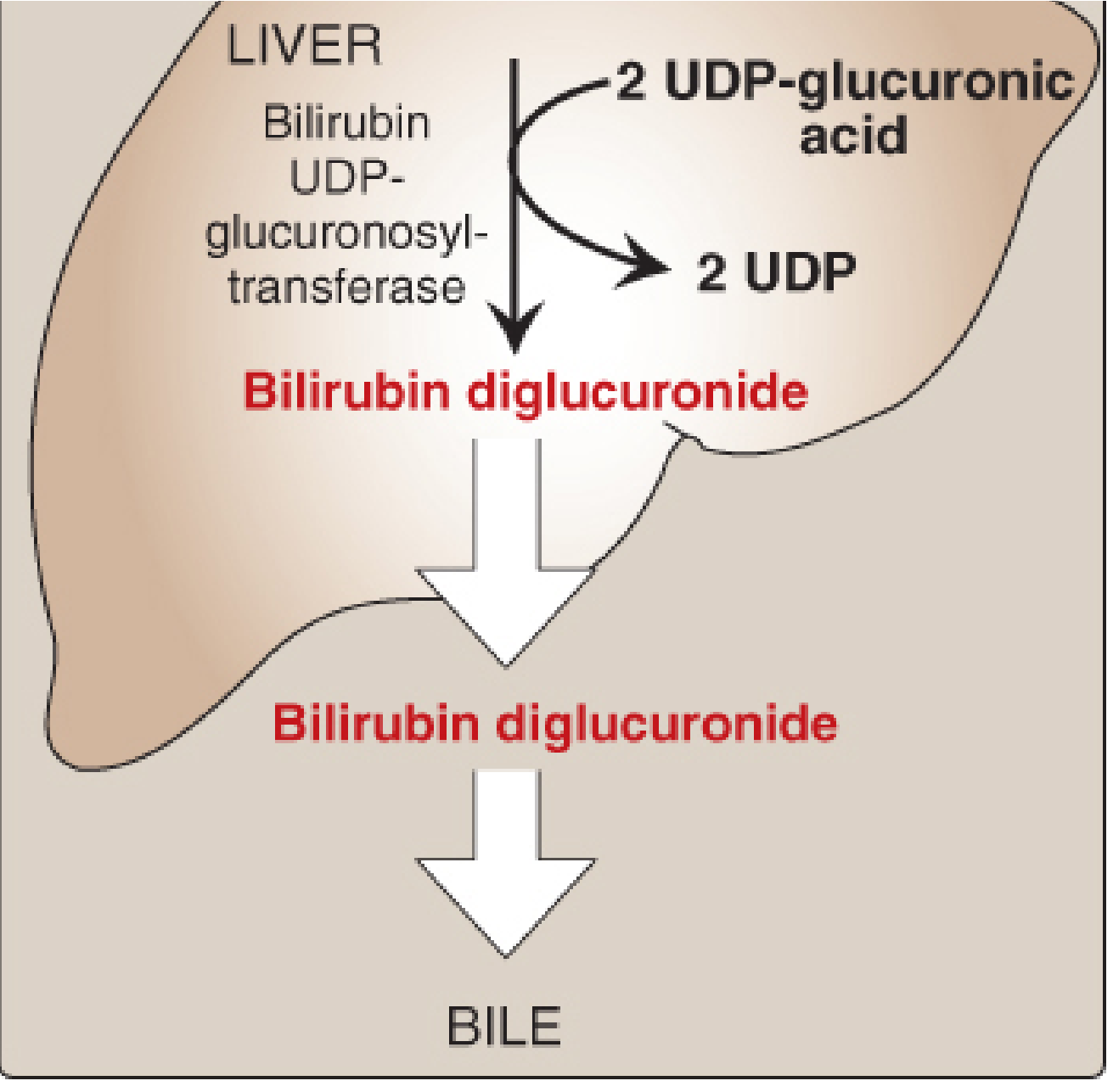
Enzyme: Bilirubin UDP-glucuronosyltransferase (bilirubin UGT) - located in the smooth ER
Donor: UDP-glucuronic acid (×2)
Reactions (sequential):
Bilirubin + UDP-glucuronate → Bilirubin monoglucuronide + UDP
Bilirubin monoglucuronide + UDP-glucuronate → Bilirubin diglucuronide + UDP
The product - bilirubin diglucuronide (conjugated bilirubin, CB / direct bilirubin) - is water-soluble. This is the predominant form secreted into bile. Note: In obstructive jaundice, the plasma form is predominantly monoglucuronide.
Induction: Phenobarbital and other drugs induce bilirubin UGT activity (clinically exploited in neonatal jaundice treatment).
Step 6 - Biliary Secretion
Transporter: MOAT (multispecific organic anion transporter), an ATP-binding cassette (ABC) transporter on bile canalicular membranes
Direction: Active transport of CB into bile canaliculi - this is the rate-limiting step of hepatic bilirubin metabolism
Conjugated bilirubin passes from bile canaliculi → bile ducts → gallbladder → duodenum.
Step 7 - Intestinal Processing: Urobilinogen & Stercobilin
In the distal ileum and colon, gut bacteria:
- Remove glucuronic acid (hydrolyze CB back to free bilirubin)
- Reduce bilirubin → urobilinogen (colorless)
- Oxidize most urobilinogen → stercobilin (brown) - gives feces their characteristic brown color
Fate of urobilinogen (enterohepatic circulation):
| Fraction | Destination |
|---|---|
| ~80-90% | Oxidized to stercobilin → excreted in feces |
| ~10-20% reabsorbed | Enters portal blood → liver (enterohepatic urobilinogen cycle) → re-excreted in bile |
| Small remainder | Reaches kidney → oxidized to urobilin (yellow) → excreted in urine (gives urine its color) |
Full Pathway at a Glance
Senescent RBC → Hemoglobin
                    ↓ (Macrophage/MPS)
              Heme (Fe²+-protoporphyrin IX)
                    ↓ Heme oxygenase (+O₂, NADPH)
                         → CO (gaseous signaling)
                         → Fe²+ (recycled)
              Biliverdin (green, linear tetrapyrrole)
                    ↓ Biliverdin reductase (+NADPH)
              Bilirubin (UCB) - yellow-orange
                    ↓ Bound to albumin → transported to LIVER
              Hepatocyte uptake → binds ligandin
                    ↓ Bilirubin UGT (+UDP-glucuronate ×2)
              Bilirubin diglucuronide (CB, "direct bilirubin")
                    ↓ Active secretion via MOAT → BILE
              Intestine: bacteria → Urobilinogen
                    ↓ (most)                ↓ (some reabsorbed)
              Stercobilin              Enterohepatic cycle / Kidney
              (feces, brown)           → Urobilin (urine, yellow)
Bilirubin Measurement: van den Bergh Reaction
| Fraction | Also called | Behavior | Significance |
|---|---|---|---|
| Direct bilirubin | Conjugated bilirubin (CB) | Reacts rapidly in aqueous solution | Water-soluble; appears in urine when elevated |
| Indirect bilirubin | Unconjugated bilirubin (UCB) | Reacts only in methanol | Lipid-soluble; crosses BBB; bound to albumin |
| Total bilirubin | - | Reacts in methanol | CB + UCB |
Normal total bilirubin: ≤1 mg/dL. Jaundice becomes visible at 2-3 mg/dL.
Jaundice - Types and Pathophysiology
1. Hemolytic (Pre-hepatic) Jaundice
- Cause: Excessive RBC destruction (sickle cell, G6PD deficiency, hereditary spherocytosis, malaria)
- Bilirubin: UCB (indirect) elevated; CB normal
- Urine: Urobilinogen increased; no bilirubin in urine (UCB not water-soluble, cannot be filtered)
- Stool: Dark (more stercobilin)
- Key: Liver conjugates maximally but is overwhelmed
2. Hepatocellular (Hepatic) Jaundice
- Cause: Hepatitis, cirrhosis, liver failure - impairs uptake, conjugation, and secretion
- Bilirubin: BOTH UCB and CB elevated (mixed picture)
- Urine: Dark urine (bilirubinuria from CB), elevated urobilinogen
- Stool: Pale (reduced stercobilin)
- Labs: ALT, AST elevated
3. Obstructive (Post-hepatic) Jaundice
- Cause: Bile duct obstruction by stone, tumor (pancreatic/cholangiocarcinoma), stricture
- Bilirubin: CB (direct) elevated - regurgitates into blood
- Urine: Dark (bilirubinuria); urobilinogen absent (CB cannot reach intestine)
- Stool: Pale/clay-colored (no stercobilin)
- Key feature: Absent urinary urobilinogen distinguishes obstruction from hepatocellular disease
Summary Table
| Feature | Hemolytic | Hepatocellular | Obstructive |
|---|---|---|---|
| Serum UCB | ↑↑ | ↑ | Normal |
| Serum CB | Normal/slight ↑ | ↑ | ↑↑ |
| Urine bilirubin | Absent | Present | Present |
| Urine urobilinogen | Increased | Increased | Absent |
| Stool color | Dark | Pale | Pale/clay |
| ALT/AST | Normal | ↑↑ | Mildly ↑ |
| ALP/GGT | Normal | Mildly ↑ | ↑↑ |
Hereditary Hyperbilirubinemias
| Disorder | Defect | Bilirubin Type | Severity |
|---|---|---|---|
| Gilbert syndrome | Mild UGT reduction (~30%); often combined with reduced uptake | UCB ↑ (mild) | Benign; triggered by fasting/stress |
| Crigler-Najjar type I | Complete absence of bilirubin UGT | UCB ↑↑↑ | Severe; fatal without liver transplant; phototherapy required continuously |
| Crigler-Najjar type II | Severely reduced UGT | UCB ↑↑ | Moderate; responds to phenobarbital |
| Dubin-Johnson syndrome | Defective MOAT (MRP2) - cannot secrete CB into bile | CB ↑ | Benign; black liver pigment |
| Rotor syndrome | Defective sinusoidal re-uptake of CB | CB ↑ | Benign; no liver pigment |
Neonatal Jaundice (Physiologic)
- Affects ~60% of full-term and ~80% of preterm newborns in the first week of life
- Cause: Bilirubin UGT activity is low at birth (reaches adult levels by ~4 weeks); high rate of fetal Hb turnover adds to the load
- Risk: UCB > 20-25 mg/dL can exceed albumin binding capacity, cross the blood-brain barrier, and deposit in the basal ganglia causing kernicterus (toxic encephalopathy)
- Treatment: Phototherapy with blue fluorescent light (wavelength ~460 nm) converts UCB to more polar, water-soluble photoisomers that can be excreted in bile without conjugation. Phenobarbital can also be used to induce UGT.
Sources
- Biochemistry, 8th ed. Lippincott Illustrated Reviews, Chapter 21 (Porphyrin & Bile Pigment Metabolism)
- Harper's Illustrated Biochemistry, 32nd ed., Chapter 31 (Catabolism of Heme)
This is a shared conversation. Sign in to Orris to start your own chat.