
Rheumatoid Arthritis
"rheumatoid arthritis"[MeSH Terms] AND treatment
rheumatoid arthritis joint pathology pannus formation histology
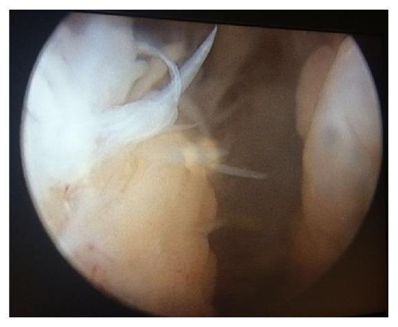
This diagnostic image shows an arthroscopic view within a joint space, specifically demonstrating significant synovial pathology. The central focus is on extensive pannus formation, characterized by a proliferation of hypertrophic synovial tissue. The pannus exhibits a variegated appearance, with areas of pale whitish-translucent fibrous strands and thicker, opaque yellowish-tan projections extending into the joint cavity. The tissue texture is notably irregular and friable, consistent with active synovial inflammation or inflammatory arthritis. In the background, deeper joint structures and synovial lining appear congested and brownish in hue. This visual evidence of synovial hypertrophy and pannus formation is a key clinical indicator of chronic inflammatory processes, such as rheumatoid arthritis or pigmented villonodular synovitis (PVNS), where such tissue can lead to articular cartilage erosion and joint degradation.
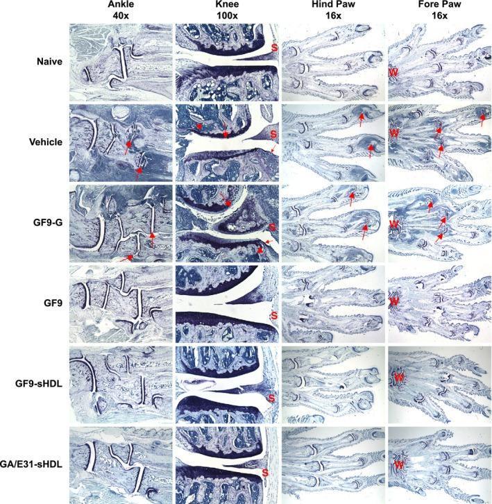
This composite of histological photomicrographs illustrates the joint pathology in a murine model of collagen-induced arthritis (CIA) across various treatment groups. The sections are stained with toluidine blue to visualize proteoglycan content and structural integrity in the ankle (40x), knee (100x), hind paw (16x), and fore paw (16x). Comparison between the 'Naive' and 'Vehicle' or 'GF9-G' control groups reveals marked inflammation, synovial thickening ('S'), pannus formation (small arrows), cartilage destruction (large arrows), and bone resorption (arrowheads) in the arthritic controls. In contrast, mice treated with TREM-1 inhibitory sequences—specifically free GF9, GF9-sHDL, and GA/E31-sHDL—demonstrate significant preservation of joint architecture. These therapeutic groups show increased staining intensity indicating better proteoglycan retention, minimal pannus formation, and reduced inflammatory cell infiltration. Labels 'W' and 'S' indicate the wrist and synovium, respectively. The images provide evidence that ligand-independent TREM-1 inhibition, particularly when delivered via synthetic high-density lipoprotein (sHDL) platforms, protects against the pathological hallmarks of rheumatoid arthritis-like disease.
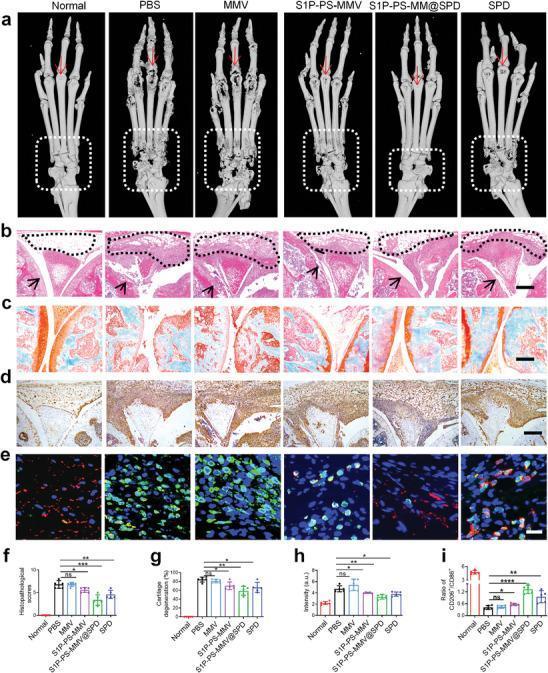
This composite educational image illustrates the therapeutic effects of various treatments on joint pathology in a mouse model of rheumatoid arthritis (RA). Row A presents Micro-CT reconstructions of mouse paws; the PBS and MMV groups show severe bone erosion and joint space narrowing in the phalanges (arrows) and tarsal bones (dashed boxes), while the S1P-PS-MMV@SPD group demonstrates significantly preserved bone architecture. Rows B through D show histological sections of joint tissue: H&E staining (B) highlights synovial hyperplasia (triangles) and pannus formation; Safranin-O/Fast Green staining (C) evaluates cartilage proteoglycan content (red staining), showing severe loss in PBS/MMV groups compared to the treatment groups; and immunohistochemistry (D) displays TNF-α expression (brown staining) as a marker of inflammation. Row E features immunofluorescence staining for macrophage polarization, using CD86 (green, M1) and CD206 (red, M2) markers. Panels F through I provide quantitative analysis for histopathological scores, cartilage degeneration, TNF-α intensity, and M2/M1 macrophage ratios, collectively demonstrating that S1P-PS-MMV@SPD reduces inflammation and promotes tissue repair in arthritic joints.
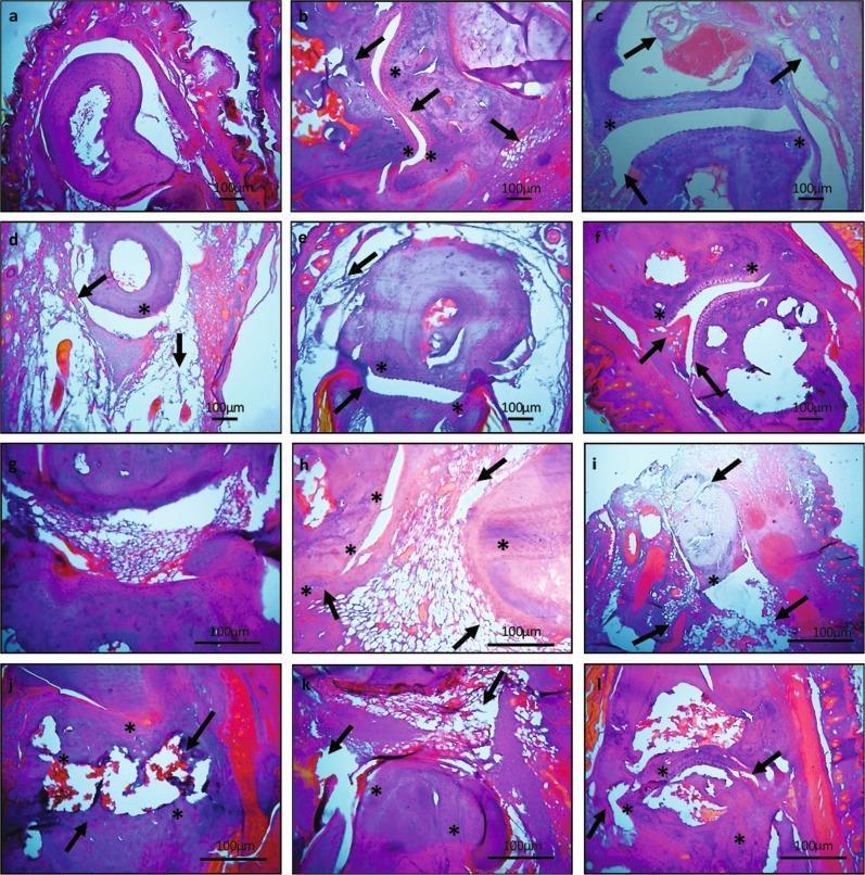
This composite diagnostic image consists of twelve light microscopy panels (a-l) showing hematoxylin and eosin (H&E) stained sections of mouse tarsal (a-f) and ankle (g-l) joints. The image serves as a comparison chart for rheumatoid arthritis pathology and treatment efficacy. Panels 'a' and 'g' represent normal controls, displaying intact articular cartilage, smooth synovial linings, and clear joint spaces. Panels 'b' and 'h' depict untreated arthritic joints, characterized by significant pannus formation, continuous cartilage degradation, bone erosion (indicated by arrows), and dense inflammatory cellular infiltration (marked by asterisks). Subsequent panels (c-f and i-l) illustrate the histological effects of various interventions, including hydroxychloroquine, leflunomide, and experimental plant extracts (aqueous and ethyl acetate). These panels demonstrate varying degrees of therapeutic response, ranging from persistent joint modification and bone loss in standard drug groups to preserved joint architecture and reduced synovial hyperplasia in extract-treated groups. The magnification (20x and 40x) allows for the visualization of key pathophysiological markers such as cellular infiltration, joint space narrowing, and osteoclast-mediated erosion.
"rheumatoid arthritis"[MeSH Terms] AND "biological therapy"[MeSH Terms]
rheumatoid arthritis hand deformity X-ray erosions
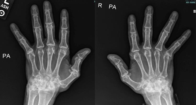
Posteroanterior (PA) view X-ray of both hands demonstrating chronic inflammatory arthritis, consistent with advanced rheumatoid arthritis. The bilateral images reveal significant involvement of the metacarpophalangeal (MCP) joints, characterized by marked joint space narrowing and subluxation where the proximal phalanges are partially displaced from the metacarpal heads. Classical ulnar deviation of the digits is present. The radiograph also shows cortical erosions and architectural distortion at the MCP and carpal joints, indicative of progressive erosive disease. The interphalangeal joints show milder changes compared to the MCP and wrist regions. These findings represent a late-stage manifestation of an inflammatory arthropathy, resulting in substantial joint deformity and malalignment. The image serves as a clinical teaching tool for recognizing radiographic hallmarks of chronic rheumatoid arthritis, including ulnar drift, subluxation, and symmetric joint space loss in the small joints of the hand.
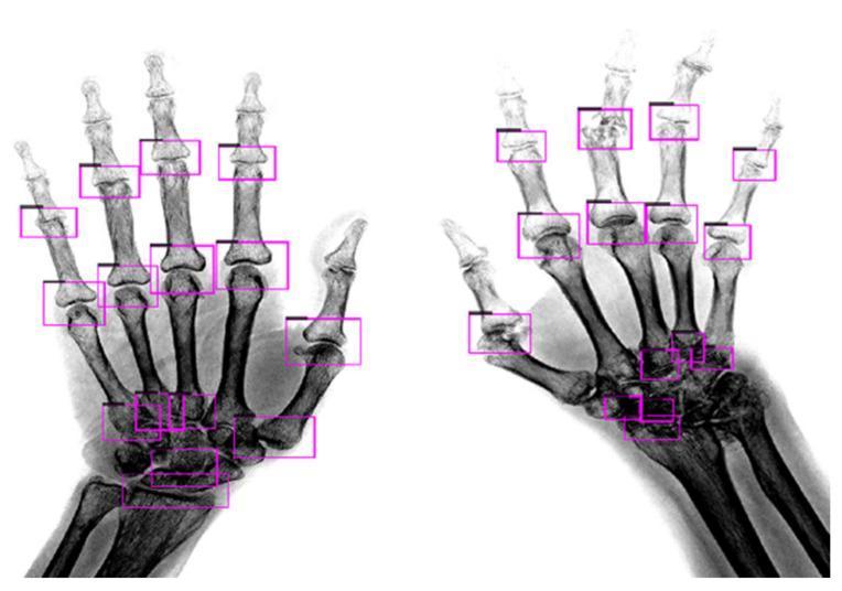
This diagnostic X-ray image (radiography) displays a comparison between two hands demonstrating different stages of Rheumatoid Arthritis (RA). The left hand represents mild RA, characterized by relatively preserved joint spaces and well-defined cortical margins. In contrast, the right hand illustrates severe RA, showing significant joint space narrowing (JSN), subchondral erosions, and architectural distortion of the bone structures. Both hands are overlaid with purple bounding boxes indicating the automated detection of regions of interest (ROIs) for clinical assessment. These ROIs target the proximal interphalangeal (PIP) joints, metacarpophalangeal (MCP) joints, and carpal bones. The image highlights the progression of RA from clear joint margins and identifiable anatomy to the blurred, eroded, and collapsed structures typical of advanced inflammatory arthritis. This visualization is intended for rheumatology and radiology education, specifically focusing on the application of deep learning algorithms for the detection and scoring of joint damage.
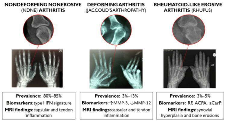
This infographic presents three radiographic patterns of musculoskeletal involvement in systemic lupus erythematosus (SLE). Each section includes a full hand X-ray, a magnified view of a metacarpophalangeal (MCP) or interphalangeal joint, and summary text on prevalence, biomarkers, and MRI findings. 1) Non-deforming Non-erosive (NDNE) Arthritis: Shows normal joint space and alignment without cortical irregularities. Prevalence is 80-85%, associated with a type I IFN signature and capsular/tendon inflammation on MRI. 2) Deforming Arthritis (Jaccoud’s Arthropathy): Demonstrates significant ulnar deviation and joint subluxation but lacks intrinsic bone erosions. Prevalence is 3-13%, associated with elevated MMP-3 and reduced MMP-12. 3) Rheumatoid-like Erosive Arthritis (Rhupus): Characterized by overt marginal bone erosions and joint space narrowing, similar to rheumatoid arthritis. Prevalence is 3-5%, associated with RF, ACPA, and aCarP biomarkers, with MRI showing synovial hyperplasia and bone erosions. This comparison illustrates the spectrum from reversible soft-tissue-mediated deformity to irreversible erosive bone damage in SLE patients.
Rheumatoid Arthritis (RA)
Overview
- Textbook of Family Medicine 9e, p. 877
- The Washington Manual of Medical Therapeutics, p. 943
Epidemiology
| Feature | Detail |
|---|---|
| Prevalence | 1-2% of adults |
| Sex ratio | Women : Men = 3:1 |
| Peak incidence | Ages 20-50 years |
| Genetic factor | HLA-DR4 association (MHC class II genes) |
| Twin concordance | Higher in monozygotic than dizygotic twins |
- Textbook of Family Medicine 9e, p. 877
Pathophysiology
- Synovial lining cells and subsynovial vessels proliferate, forming a pannus - a hypertrophic, invasive synovial tissue
- Leukocyte invasion follows, triggering an inflammatory cascade
- Proteases and cytokines (including TNF-alpha, IL-1, IL-6) mediate cartilage and bone destruction
- Rheumatoid Factor (RF) - an autoantibody against IgG Fc fragment - is produced; influenced by HLA-DR polymorphism and associated with more severe, extra-articular disease
- Anti-citrullinated protein antibodies (ACPA/anti-CCP) appear early in the disease course and are highly specific (>90%) for RA
Clinical Features
Onset
- Insidious in ~70% of cases (weeks to months)
- Acute abrupt onset in ~10% (best prognosis)
- Intermediate onset in ~20%
- Palindromic RA: brief episodic swelling of a large joint (knee, wrist, elbow) - may progress to classic RA
Articular Features
- Symmetric synovitis - the hallmark of RA
- Morning stiffness > 1 hour (results from joint immobilization, not time of day)
- Joints typically warm but not erythematous
- Small joints first: PIP, MCP (hands/feet), then larger joints
- Palpable pannus as a rubbery tissue mass
Classic Joint Deformities (late disease)
| Deformity | Description |
|---|---|
| Ulnar deviation | Fingers deviate ulnarly at MCPs |
| Swan neck | MCP flexion, PIP hyperextension, DIP flexion |
| Boutonniere | PIP flexion, DIP hyperextension |
| Cock-up deformity | Feet involvement |
| Atlantoaxial subluxation | C1-C2 involvement - can cause myelopathy |
Constitutional Symptoms
X-ray of RA Hands (advanced disease showing ulnar deviation and erosions):
Extra-articular Manifestations
| System | Manifestation |
|---|---|
| Skin | Rheumatoid nodules (extensor surfaces), cutaneous vasculitis |
| Lung | Interstitial lung disease (ILD), pleuritis, pleural effusion |
| Cardiovascular | Pericarditis, accelerated atherosclerosis |
| Eye | Scleritis, episcleritis, keratoconjunctivitis sicca (secondary Sjogren syndrome) |
| Neurological | Mononeuritis multiplex, CNS vasculitis, cervical myelopathy |
| Hematological | Felty syndrome (RA + splenomegaly + neutropenia, ~1%), anemia of chronic disease |
| Vascular | Raynaud phenomenon |
- The Washington Manual of Medical Therapeutics, p. 944
Diagnosis
1987 ACR Criteria (Traditional - requires 4 of 7, criteria 1-4 present ≥ 6 weeks)
| Criterion | Definition |
|---|---|
| 1. Morning stiffness | ≥ 1 hour before maximal improvement |
| 2. Arthritis of ≥ 3 joint areas | Simultaneous soft tissue swelling in 14 possible areas |
| 3. Arthritis of hand joints | At least one wrist, MCP, or PIP joint involved |
| 4. Symmetric arthritis | Same joint areas on both sides |
| 5. Rheumatoid nodules | Over bony prominences, observed by physician |
| 6. Serum rheumatoid factor | Positive RF |
| 7. Radiologic changes | Erosions or periarticular bony decalcification on hand/wrist X-ray |
Note: The 2010 ACR/EULAR criteria are now preferred for early RA detection, incorporating anti-CCP and acute-phase reactants.
Laboratory Tests
| Test | Sensitivity | Specificity | Notes |
|---|---|---|---|
| RF | ~80% | Lower | Positive in many other conditions (mnemonic: CHRONIC - Chronic disease, Rheumatoid arthritis, Other CTDs, Neoplasms, Infections, Cryoglobulinemia) |
| Anti-CCP (ACPA) | 50-60% | >90% | More specific; predictor of erosive disease |
| ESR / CRP | Non-specific | Non-specific | Markers of active inflammation |
| Synovial fluid | WBC > 2000/mm³ | Confirms inflammatory arthritis |
Imaging
- Plain X-ray: Periarticular osteopenia, joint space narrowing, erosions (appears months to 1 year after onset)
- MRI and Ultrasound: More sensitive; detect subclinical synovitis and early erosions
Treatment
Step 1 - Bridging Therapy (while DMARDs take effect, typically 1-3 months)
- NSAIDs - symptomatic relief only; do NOT modify disease course
- Glucocorticoids - short-term bridging; some disease-modifying activity but long-term toxicity limits use
Step 2 - Conventional Synthetic DMARDs (csDMARDs)
| Severity | First Choice |
|---|---|
| Mild RA | Hydroxychloroquine (Plaquenil) or Sulfasalazine |
| Moderate-Severe RA | Methotrexate 7.5-20 mg weekly (anchor drug) |
| Alternative | Leflunomide |
⚠ Methotrexate + Leflunomide combination carries additive hepatotoxicity risk - use cautiously.
Step 3 - Biologic and Targeted Synthetic DMARDs (if csDMARDs fail)
| Class | Drugs |
|---|---|
| Anti-TNF | Adalimumab, Etanercept, Infliximab, Golimumab, Certolizumab pegol |
| Anti-IL-6 | Tocilizumab, Sarilumab |
| Anti-IL-1 | Anakinra |
| Costimulation inhibitor | Abatacept |
| Anti-CD20 (B-cell depletion) | Rituximab |
| JAK inhibitors (targeted synthetic) | Tofacitinib, Baricitinib, Upadacitinib |
-
Methotrexate is commonly combined with TNF antagonists (additive efficacy, reduced anti-drug antibody formation)
-
Two biologic DMARDs should NEVER be combined - greatly increased infectious risk
-
Methotrexate is also commonly paired with rituximab or abatacept
-
The Washington Manual of Medical Therapeutics, p. 944-945
Monitoring & Prognosis
Poor Prognostic Factors
- RF positivity and high anti-CCP titers
- High disease activity at onset
- Early erosions on imaging
- Extra-articular manifestations
- Low functional status
Monitoring (methotrexate)
- CBC with platelet count, LFTs periodically
- Supplemental folic acid to reduce mucosal and hepatic toxicity
Complications
- Progressive joint destruction and disability
- Increased cardiovascular mortality (leading cause of death in RA)
- Secondary Sjogren syndrome, Felty syndrome
- Iatrogenic infections (especially with biologics)
- Cervical myelopathy from C1-C2 subluxation
Key Takeaways
- RA is a systemic autoimmune disease, not just an arthritis - extra-articular involvement is common and can be life-threatening
- Symmetric synovitis with morning stiffness >1 hour is the clinical hallmark
- Anti-CCP antibodies are more specific than RF for diagnosis and predict erosive disease
- Early DMARD therapy (within months, not years) prevents irreversible joint destruction
- Methotrexate remains the anchor drug for moderate-severe disease
- The treat-to-target approach (aim for remission or low disease activity) guides all management decisions
Sources: Textbook of Family Medicine 9e | The Washington Manual of Medical Therapeutics | Firestein & Kelley's Textbook of Rheumatology | Quick Compendium of Clinical Pathology 5e