
Clinical features and management of vestibulitis
vestibulitis vulvar vestibule treatment
nasal vestibulitis clinical features management 2024
nasal vestibulitis infection folliculitis furuncle nose
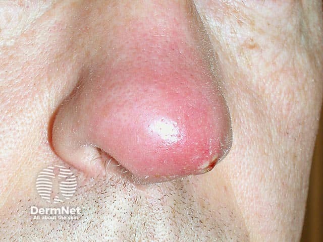
Clinical photography of a single nasal skin lesion showing an acute furuncle (boil) on the nasal vestibule/dorsum. The image depicts a solitary, erythematous, dome-shaped nodule with a shiny surface and surrounding mild edema. A palpable tenderness is suggested by the lesion’s conspicuous inflamed halo. A central pustule or crust may be present at the core, consistent with progression toward abscess formation within a pilosebaceous follicle. The lesion is located on the external nasal skin near the nostril, with frontal/anterior perspective offering clear visualization of the nasal bridge region. The clinical diagnosis is furunculosis of the nose, most often caused by Staphylococcus aureus; in this case PVL (Panton-Valentine leukocidin)–producing strains are implicated, which are associated with more aggressive skin and soft tissue infections and a higher risk of complications. Imaging is not radiologic; this is a dermatologic/clinical photograph used for documentation, teaching, and correlating with microbiology culture results. Management implications include incision and drainage if fluctuant, culture to guide antibiotics, and consideration of toxin-producing strains. The image serves educational purposes for dermatology, infectious disease, microbiology, and medical training in recognizing PVL-associated nasal furunculosis and differentiating it from impetigo, cellulitis, and Sty.
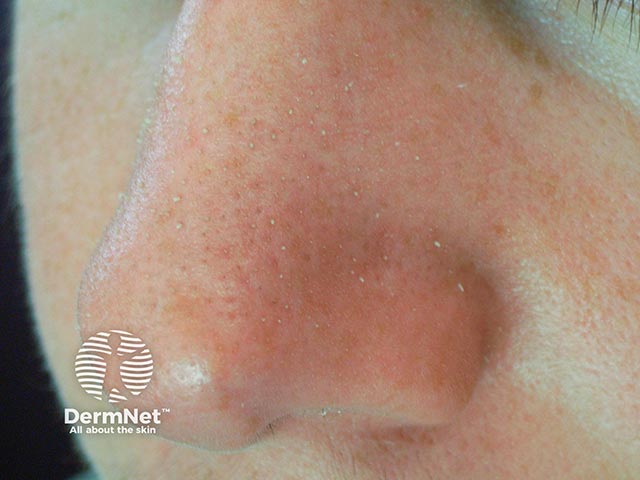
This is a clinical dermatology photograph of the nasal region illustrating Trichostasis spinulosa. Modality and technique: digital clinical photography, close-up macro view under standard white-light illumination. The image shows the nose with numerous tiny follicular papules; many dilated follicular openings contain bundles of fine vellus hairs that emerge from a single pore, producing characteristic hair tufts. Surrounding skin may show mild erythema or scale, but there is no overt inflammation. The distribution is localized to the nasal skin (dorsum and tip) and reflects a benign follicular disorder where multiple hair shafts are retained within a single follicle. Diagnostic significance lies in distinguishing TPS from acneiform lesions and folliculitis, guiding appropriate management rather than antimicrobial therapy. Clinically relevant use cases include dermatology education, image-based case discussions, and database curation for machine learning models aimed at recognizing hair-containing follicular disorders. This image captures the hallmark feature of TPS: multiple hair shafts within one follicular opening on nasal skin, contributing to its cosmetic appearance and patient counseling considerations.
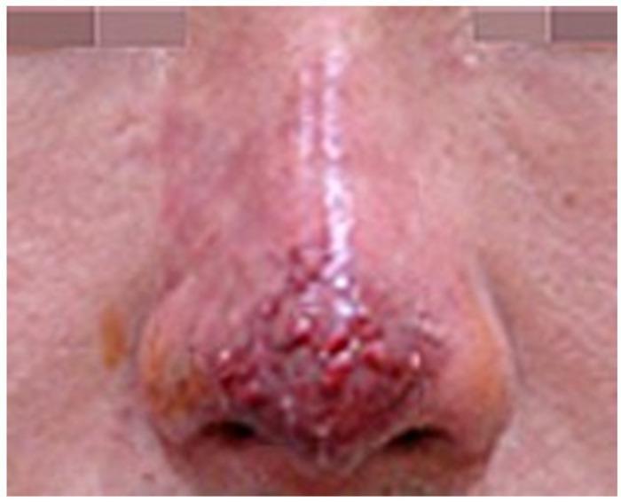
This clinical photograph shows a close-up view of the human nose exhibiting signs of vascular compromise and secondary infection following a dermatological procedure. The primary pathology is localized to the nasal tip and dorsum, where a cluster of small, raised, erythematous pustules is prominently visible. These lesions are characterized by a rounded morphology and a deep red to violaceous hue, suggesting intense underlying inflammation and possible infection. The skin of the nasal bridge appears edematous and displays a shiny texture with diffuse erythema extending towards the glabella. In contrast, the skin on the adjacent cheeks maintains a more normal tone and texture, highlighting the localized nature of the inflammatory process. The visual findings are consistent with an infection state and impending tissue necrosis, likely resulting from vascular occlusion post-filler injection. This image serves as a clinical reference for identifying early progression from ischemic stages to pustule formation and potential eschar development in aesthetic medicine complications.
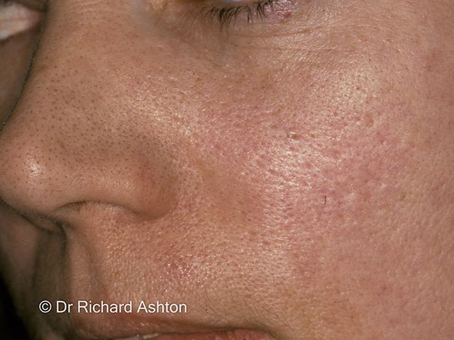
High-resolution clinical photograph of the left nasal ala and midface showing multiple skin-colored to slightly erythematous follicular papules with conspicuous comedo-like plugs along the nasal dorsum and alar rims. The image demonstrates numerous small keratotic plugs within dilated hair follicles, producing a pattern reminiscent of open comedones but clustered centrally on the nose. The underlying process is trichostasis spinulosa, a folliculocentric condition in which several vellus hairs are retained within a single follicular unit and obstructed by keratinous material. Lesion distribution is centrofacial, with highest density on the nose and mild textural coarsening of adjacent skin. The epidermis shows mild hyperkeratosis without overt pustulation or crusting; pigmentation is variable in some areas. Differential diagnoses include acne vulgaris with comedones, keratosis pilaris rubra faceii, and superficial folliculitis. Clinically, recognizing trichostasis spinulosa is important to avoid mislabeling as acne and to guide management toward follicular clearance. Treatment considerations include topical keratolytics (retinoids such as adapalene or tretinoin, benzoyl peroxide), gentle extraction, and adjunctive exfoliation; persistent cases may benefit from laser hair removal or chemical depilation. Image enhances educational awareness of centrofacial follicular occlusion disorders. This image provides a reference for clinicians diagnosing follicular occlusion disorders and tailoring patient education.
nasal vestibulitis treatment antibiotics
PMID: 36373460
For ent subject of mmbs
Nasal Vestibulitis - ENT (MBBS)
Definition
Anatomy Reminder
Etiology / Causative Organism
- Primary organism: Staphylococcus aureus (most common, both MSSA and MRSA)
- Less common: Streptococcal species, fungal infections
- Herpetic (HSV) infections - typically more severe and acute rather than chronic
Risk Factors (Common Exam Point)
| Category | Examples |
|---|---|
| Local trauma | Nose picking, nasal hair plucking, excessive nose blowing |
| Iatrogenic | Nasal steroid sprays (though no confirmed association), nasal surgeries, cauterization |
| Chemotherapy | Taxanes (paclitaxel, nab-paclitaxel), bevacizumab - >75% develop nasal symptoms |
| Systemic | Diabetes mellitus, immunosuppression, obesity |
| Miscellaneous | Nasal piercing, nasal foreign body, poor hygiene |
Clinical Features
Symptoms
- Anterior nasal pain (often the presenting complaint)
- Swelling and tenderness at the nasal tip / ala
- Nasal crusting
- Bleeding (epistaxis - minor)
- Pustule formation just inside the nostril
- Chronic irritation with recurrent symptoms in some patients
Signs on Examination
- Erythema, cracking, and yellow crusting just inside the nostril
- Mild erythema of the vestibular skin
- Tenderness to palpation or movement of the nose
- Nasal septum is typically spared (disease limited to vestibular skin/hair follicles)
- May appear fairly normal in low-grade chronic cases
Spectrum of Disease (Important Classification)
1. Simple Vestibulitis (Folliculitis)
- Superficial infection of hair follicles
- Mild erythema, crusting, tenderness
- Localized to vestibule
2. Furunculosis (Furuncle / Boil)
- Deep variety of vestibulitis
- Perifollicular erythema + edema
- Possible abscess formation (pustule with fluctuation)
- More severe pain and swelling
3. Carbuncle
- A group/cluster of furuncles coalescing together
- More extensive and severe
Non-infective Vestibulitis
- Caused by physical/chemical irritation
- No pus or organisms
- Dryness, cracking, crusting without frank infection
Complications (High-Yield for Exams)
Nasal Vestibulitis
        ↓
  Untreated / Severe
        ↓
  Facial Cellulitis
        ↓
  Cavernous Sinus Thrombosis  ← MOST DANGEROUS
        ↓
  Meningitis / Brain Abscess
- Nasal tip cellulitis
- Periorbital cellulitis
- Septicemia (in immunocompromised)
Diagnosis
- Clinical diagnosis - based on history and examination
- No special tests usually needed
- Nasal swab culture - indicated when:
- Symptoms recur or persist
- MRSA suspected
- Systemic involvement
- Immunocompromised patient
- Cultures from abscess yield positive results in ~83% of cases (predominantly MSSA)
Management
A. Non-infective Vestibulitis
- Vaseline (petroleum jelly) applied to vestibule - moisturizes and protects
- Mild topical corticosteroid to reduce inflammation
- Avoid precipitating factors (nose picking, excessive blowing)
B. Infective Vestibulitis - Mild/Moderate (Outpatient)
| Agent | Notes |
|---|---|
| Mupirocin (Bactroban nasal) | Drug of choice; also used for MRSA decolonization |
| Chlorhexidine + Neomycin (Naseptin) | First-line combination; contraindicated in peanut allergy (contains arachis oil); contraindicated in pregnancy |
| Bacitracin, Polymyxin | Alternative topical agents |
| Retapamulin | Alternative topical |
- Applied 3 times a day for 5 days
- Warm compresses help reduce inflammation
- Amoxicillin-clavulanate - most commonly used
- Clindamycin - for penicillin allergy or as second-line
- Anti-staphylococcal agents (flucloxacillin, dicloxacillin)
C. Severe / Complicated Cases (Inpatient)
- Treatment failure in outpatient setting
- Presence of cellulitis or abscess
- Immunocompromised patient
- Signs of spreading infection
- IV antibiotics (IV amoxicillin-clavulanate is most common)
- Incision and drainage (I&D) - for fluctuant abscess
- Add antiviral (valacyclovir) if herpetic infection suspected
D. MRSA Eradication Protocol
- Mupirocin is the second-line agent for MRSA
- Applied 3 times daily for 5 days
- A swab is taken 2 days after completion to confirm MRSA eradication
E. Chemotherapy-associated Nasal Vestibulitis
- Saline nasal sprays - basic supportive care
- Rose geranium in sesame oil nasal spray - evidence supports superiority over saline
Clinical Image
High-Yield Summary Table for MBBS Exams
| Feature | Detail |
|---|---|
| Organism | S. aureus (MSSA > MRSA) |
| Risk factors | Nose picking, hair plucking, DM, immunosuppression |
| Classic symptom | Anterior nasal pain + crusting + tenderness |
| Septum involvement | Usually spared |
| Deep form | Furunculosis (abscess) |
| Multiple furuncles | Carbuncle |
| Dangerous complication | Cavernous sinus thrombosis |
| First-line topical Rx | Mupirocin OR Naseptin |
| MRSA treatment | Mupirocin |
| Naseptin contraindication | Peanut allergy, pregnancy |
| Oral antibiotic | Amoxicillin-clavulanate |
| I&D indication | Fluctuant abscess |
Sources
- K.J. Lee's Essential Otolaryngology - Nasal Vestibulitis section
- Scott-Brown's Otorhinolaryngology Head & Neck Surgery Vol. 1 - Vestibulitis section
- Harrison's Principles of Internal Medicine 22E (2025) - Nasal Vestibulitis
- Textbook of Family Medicine 9e - Nasal Vestibulitis
- Marra P et al. Nasal Vestibulitis and Vestibular Furunculosis: a systematic review. Clin Ter. 2022 (PMID: 36373460)
Clinical features and management of chronic rhinosinusitis for ent subject with deep from standard textbooks
"chronic rhinosinusitis" diagnosis management guidelines
chronic rhinosinusitis nasal polyps endoscopy CT scan sinusitis
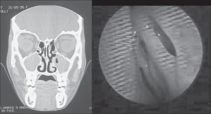
This composite clinical image presents a side-by-side comparison of diagnostic modalities for chronic rhinosinusitis (CRS). On the left, a coronal CT scan of the paranasal sinuses shows significant soft tissue opacification in the bilateral maxillary and ethmoid sinuses, indicating a positive diagnosis for sinusitis according to Lund-Mackay staging criteria. The nasal septum is centered, and inferior turbinates are visible. On the right, a corresponding nasofibroscopy (endoscopy) frame provides a direct view of the nasal passage and mucosa. In this specific case, the endoscopic view appears negative for classic signs of CRS, demonstrating smooth mucosa without evident purulent secretion, polyps, or significant edema in the visible meatus. The pairing illustrates a clinical 'disagreement' where radiologic evidence suggests disease that is not immediately visible via endoscopy, highlighting the diagnostic complexity of sinonasal pathology in otolaryngology.
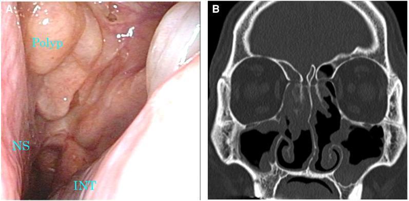
This composite figure displays diagnostic findings of chronic rhinosinusitis with nasal polyps (CRSwNP). Image A is a nasal endoscopy showing a large, pale-pink, fleshy nasal polyp occupying the middle meatus and partially obstructing the nasal passage. Key landmarks include the nasal septum (NS) on the medial side and the inferior nasal turbinate (INT) at the inferior aspect. Image B is a coronal non-contrast CT scan of the paranasal sinuses. It reveals partial opacification and mucosal thickening within the bilateral maxillary, ethmoid, and frontal sinuses. The soft tissue density lesions signify inflammatory polyposis and retained secretions, though significant aeration is visible compared to total opacification. The nasal septum is midline, and the turbinate structures are clearly delineated. These images demonstrate the clinical and radiological presentation of eosinophilic chronic rhinosinusitis (ECRS) and are used to assess the Total Polyp Score (TPS) and Lund-Mackay Score (LMS) in response to treatment, such as biologics like dupilumab.
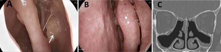
This composite figure illustrates the clinical and radiological response to treatment for eosinophilic chronic rhinosinusitis (ECRS) in an adult patient. Panel A displays a right-sided nasal endoscopy view showing a persistent, smooth, pale yellow-colored polypoid mass located within the olfactory fissure (marked by a white arrow). Panel B presents a left-sided nasal endoscopy view showing resolution of previously documented nasal polyps, with visible nasal mucosa and patent passages. Panel C is a coronal paranasal sinus CT scan following four months of therapy, demonstrating significant improvement in sinus aeration. The black, air-filled spaces within the maxillary, ethmoid, and frontal sinuses indicate reduced mucosal thickening and clearance of soft-tissue opacities, although residual mucosal thickening remains visible in the floor of the maxillary sinuses. Collectively, the images provide a comparison of endoscopically visible localized disease versus global sinus improvement monitored via diagnostic imaging.
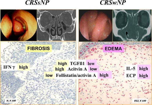
A multi-modal comparison of Chronic Rhinosinusitis without Nasal Polyps (CRSsNP) and with Nasal Polyps (CRSwNP). The image is divided into two clinical panels, each containing endoscopy, CT imaging, and histopathology. The CRSsNP panel (left) shows endoscopic nasal mucosal thickening, a coronal CT scan with ethmoid sinus opacification, and a light microscopy image (H&E, x100) labeled 'FIBROSIS'. This state is characterized by high levels of IFN̳, TGF̲1, and Activin A, and a low Follistatin/activin A ratio. The CRSwNP panel (right) displays endoscopic visualization of translucent nasal polyps, a coronal CT scan showing extensive sinus opacification, and a histopathological image (EG2, x100) labeled 'EDEMA'. It highlights eosinophilic infiltration (red staining) with high levels of IL-5 and ECP, but low TGF̲1 and Activin A. This comparison illustrates the different tissue remodeling patterns in CRS: predominant fibrosis and Th1/pro-fibrotic markers in CRSsNP versus predominant edema and Th2 markers in CRSwNP.
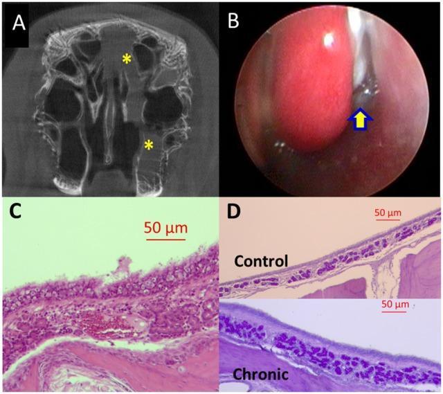
Educational visual panel depicting a rabbit model of chronic rhinosinusitis (CRS) at 14 weeks. (A) Coronal micro-CT scan of the paranasal sinuses demonstrating partial opacification of the left maxillary and ethmoid sinuses (yellow asterisks), indicating fluid or mucosal thickening. (B) Nasal endoscopy image showing the left middle meatus with purulent secretions (yellow arrow) positioned between the middle turbinate and the lateral nasal wall. (C) H&E stained histopathology slide showing significant infiltration of chronic inflammatory cells (plasma cells and lymphocytes), epithelial hyperplasia, and submucosal edema. (D) Periodic Acid-Schiff (PAS) stained comparison between control (upper) and chronic sinusitis (lower) tissue. The chronic group exhibits marked hypertrophy and increased density of submucous glands, staining deep purple, which characterizes mucus hyperplasia. This composite illustrates diagnostic imaging, clinical findings, and microscopic cellular changes associated with long-term sinus inflammation.
Chronic Rhinosinusitis (CRS) - ENT (MBBS)
Definition
"CRS is diagnosed when symptoms last more than 12 consecutive weeks." - Cummings Otolaryngology
Classification (High-Yield)
| Feature | CRS without Nasal Polyps (CRSsNP) | CRS with Nasal Polyps (CRSwNP) |
|---|---|---|
| Sex predominance | More common in women | More common in men |
| Age of onset | Childhood / young adulthood | Adulthood |
| Inflammation type | TH1-mediated, IFN-γ dominant | TH2-mediated, eosinophilic |
| Histology | Fibrosis, BM thickening, goblet cell hyperplasia, mononuclear infiltrate | Edematous stroma, albumin deposition, pseudocyst formation, eosinophilic infiltrate |
| Key cytokines | IFN-γ high; TGF-β1 high | IL-5 high; ECP (eosinophil cationic protein) high |
| Presenting symptom | Facial pain/pressure dominant | Decreased/loss of smell dominant |
| Associations | Bacterial infection/colonization, deviated septum | Asthma, aspirin sensitivity (Samter triad), AFRS |
Subtypes of CRS:
- CRSsNP - without polyps
- CRSwNP - with nasal polyps (most common polypoidal form)
- Allergic Fungal Rhinosinusitis (AFRS) - associated with polyps; IgE-mediated; characteristic "peanut butter" greenish-brown mucus
- Pediatric CRS
Pathophysiology
OMC Obstruction
      ↓
Impaired mucociliary clearance + Reduced ventilation
      ↓
Mucus stasis + Bacterial colonization
      ↓
Persistent mucosal inflammation
      ↓
Chronic Rhinosinusitis
- Bacterial superantigen response (Staphylococcal enterotoxins)
- Fungal sensitization
- Atopy and allergens
- Biofilm formation (polymicrobial - S. aureus, P. aeruginosa)
- Osteitis (bone remodeling)
- Unified airway disease (association with lower airway disease)
CRSsNP: TH1 milieu with increased IFN-γ in inflamed mucosa and fibrotic remodeling
Endoscopy and CT Imaging Comparison: CRSsNP vs CRSwNP
Clinical Features
Cardinal Symptoms (EPOS Diagnostic Criteria)
- Nasal obstruction / congestion (blockage)
- Nasal discharge - anterior rhinorrhea or postnasal drip (may be mucopurulent)
- Facial pain / pressure / fullness
- Reduction or loss of smell (hyposmia/anosmia) - more prominent in CRSwNP
"Cardinal symptoms: facial pain or pressure, nasal discharge or postnasal drip, congestion, and hyposmia or anosmia." - Harrison's Principles 22E
Additional/Associated Symptoms:
- Fatigue and malaise
- Ear pressure / fullness
- Hoarseness (from postnasal drip)
- Chronic cough
- Headache
- Sleep disturbance
Sinus-Specific Pain Locations:
| Sinus | Pain Location |
|---|---|
| Maxillary | Cheek, upper teeth, infraorbital region |
| Frontal | Forehead, supraorbital area |
| Ethmoid | Between eyes, medial canthal region, bridge of nose |
| Sphenoid | Vertex of skull, occiput, retro-orbital ("worst headache") |
Signs on Examination:
- Mucosal erythema and edema
- Mucopurulent discharge in the middle meatus
- Nasal polyps: pale/grey/yellow translucent growths in middle meatus (in CRSwNP)
- Nasal septal deviation (predisposing factor)
- Mucosal disease in middle meatus and ethmoid bulla
- Purulent secretions from sinus ostia
- Polyps visualized
- Posterior disease (sphenoid/posterior ethmoid) not visible on anterior rhinoscopy
Nasal Polyps: Key Features
- Appear as white, grey, tan, or yellow translucent growths
- Arise from the middle meatus (bilateral and multiple in CRSwNP)
- Pale and insensate (do not bleed on probing - differentiates from turbinate hypertrophy)
- Unilateral polyp = always suspect malignancy (inverted papilloma, SCC, especially in older patients / wood-dust exposure)
- Nasal polyposis + Bronchial asthma + Aspirin/NSAID sensitivity
Investigations
1. Nasal Endoscopy
- Best for confirming mucosal inflammation in middle meatus and posterior areas invisible on anterior rhinoscopy
- Visualizes polyps, purulent secretions, anatomical variants
2. CT Paranasal Sinuses (Non-contrast) - Imaging of Choice
- Coronal cuts are most informative (shows OMC anatomy clearly)
- Findings in CRS:
- Diffuse or polypoid mucosal thickening
- Partial or complete opacification of sinuses
- Bone remodeling/thickening (osteitis)
- Intrasinus calcifications
- OMU obstruction (infundibular, OMC, or sphenoethmoidal recess patterns)
- Chronic secretions: increased density (30-60 HU) vs acute (10-25 HU)
-
40% of patients with symptom-based CRS have normal CT - objective confirmation is mandatory
- Each sinus scored 0 (clear), 1 (partial opacification), 2 (complete opacification)
- OMC scored 0 or 2
- Maximum total score: 24
3. Nasal Swab / Culture
- To identify organisms and antibiotic sensitivity
- Important in recurrent/recalcitrant cases
4. Allergy Testing
- Skin prick test or RAST - important especially in CRSwNP
5. MRI
- Not routine; used when malignancy, fungal disease, or intracranial extension suspected
- Superior to CT for soft tissue differentiation
6. Special Tests (when indicated):
- Saccharin test / Ciliary biopsy - if primary ciliary dyskinesia suspected
- Sweat chloride test / CFTR mutation - if cystic fibrosis suspected
- Immunoglobulin levels - if immunodeficiency suspected
- ANCA, ACE levels - if granulomatous disease (Wegener's, sarcoid) suspected
Management
Step 1 - General/First-Line Medical Management
A. Saline Nasal Irrigation
- High-volume saline washes (e.g., Neil Med, Neti pot) - more effective than simple sprays
- Clears mucus, promotes drainage, reduces mucosal edema
- Safe, well-tolerated, used in all subtypes
B. Intranasal Corticosteroids (INCS) - Mainstay of Treatment
- Mometasone, Fluticasone, Budesonide (sprays or rinses)
- Reduce mucosal edema and polyp size
- High-volume budesonide rinses have superior evidence vs standard sprays for CRSwNP
- Should be tried for at least 4 weeks by primary care before ENT referral (EPOS guidelines)
- Excellent safety profile; minimal systemic absorption
C. Antibiotics
- Role is limited in CRS; not first-line
- Macrolides (e.g., clarithromycin, azithromycin) for 3 months - modest quality evidence supports use in CRSsNP without polyps (anti-inflammatory + antibacterial mechanism)
- Short-course antibiotics (2-4 weeks) frequently prescribed but little evidence of benefit
- Biofilm-forming organisms (S. aureus, P. aeruginosa) lead to resistance
- Antifungal agents: NOT recommended for any subtype of CRS
D. Oral Corticosteroids
- Short course (2-3 weeks) for CRS unresponsive to intranasal steroids
- Especially effective for CRSwNP (shrinks polyps temporarily)
- Used as "rescue" or bridging therapy
E. Antihistamines
- For patients with allergic component (IgE-mediated disease)
- Intranasal or systemic
F. Leukotriene Antagonists
- Montelukast - particularly helpful when allergic component present
- Useful adjunct in AERD/aspirin sensitivity
G. Decongestants
- Used sparingly and briefly only (risk of rhinitis medicamentosa with prolonged use)
Step 2 - Second-Line / Escalated Medical Management
Biologic Therapies (for Severe, Recalcitrant CRSwNP)
| Biologic | Mechanism | Target |
|---|---|---|
| Dupilumab (FDA approved) | Anti-IL-4Rα (blocks IL-4 + IL-13) | Type 2 inflammation |
| Mepolizumab | Anti-IL-5 | Eosinophil reduction |
| Benralizumab | Anti-IL-5Rα | Eosinophil depletion |
| Omalizumab | Anti-IgE | Allergic pathway |
- Meta-analysis (PMID: 39729039) confirms budesonide nasal irrigation benefits for CRS
- Biologics reduce polyp size and symptom burden, improving QOL
Step 3 - Surgical Management
Indications for Surgery:
- Failure of appropriate maximal medical therapy (most common indication)
- CT-confirmed residual disease after medical treatment
- Complications of CRS
- Suspected malignancy
- Antrochoanal polyp (relative indication)
"CRS that is recalcitrant to medical therapy is the most common indication for surgery." - Cummings Otolaryngology
Functional Endoscopic Sinus Surgery (FESS) - Surgery of Choice
- Mucosal preservation (Messerklinger technique)
- Restoration of ventilation and drainage
- Removal of obstructing disease while preserving normal structures
- Provides wide passages for topical drug delivery post-operatively
- Relieve obstruction at OMC
- Restore mucociliary clearance
- May be curative in some subtypes (e.g., odontogenic sinusitis)
- Remove polyps and debris
- Complete ethmoidectomy (most critical step)
- Create wide sinus openings for topical medication delivery
- Surgery adjunctive - long-term medical therapy still required post-FESS
- Uncinectomy (removal of uncinate process)
- Middle meatal antrostomy (maxillary sinus opening)
- Anterior ethmoidectomy
- Posterior ethmoidectomy
- Sphenoidotomy
- Frontal sinusotomy (Draf procedures for frontal sinus)
Other Surgical Options:
- Polypectomy - simple removal; often combined with FESS
- Caldwell-Luc operation - now largely obsolete; reserved for specific indications
- Adenoidectomy - effective in pediatric CRS
Pre-surgical Requirements (Scott-Brown's):
- CT scan must be obtained to:
- Assess residual disease
- Identify anatomical risk factors
- Plan surgical approach
- Informed consent including:
- Natural history of disease
- Surgical complications (orbital, intracranial injury)
- Risk of recurrence
- Need for ongoing medical management
Special Associations (High-Yield)
Samter's Triad (AERD):
- Nasal polyposis + Asthma + Aspirin/NSAID sensitivity
- Treatment: salicylate desensitization therapy
Allergic Fungal Rhinosinusitis (AFRS):
- Occurs in 20s-30s; warm, humid regions; atopic patients
- Characteristic mucus: thick, greenish-brown, peanut butter consistency with fungal hyphae (Aspergillus)
- CT: double-density sign, often unilateral, can expand sinus walls
- Resistant to medical therapy; surgery + perioperative steroids is treatment of choice
Cystic Fibrosis:
- Nearly universal CRS and nasal polyps
- Young patients with bilateral polyps should be tested
Primary Ciliary Dyskinesia (Kartagener's syndrome):
- CRS + bronchiectasis + situs inversus
- Defective cilia - impaired mucociliary clearance
Granulomatous Disease:
- Granulomatosis with polyangiitis (Wegener's): ANCA positive; saddle nose deformity
- Sarcoidosis: lupus pernio, non-caseating granulomas
Complications of CRS
| Category | Complications |
|---|---|
| Orbital (most common) | Periorbital edema, orbital cellulitis, subperiosteal abscess, orbital abscess, cavernous sinus thrombosis |
| Intracranial | Meningitis, epidural abscess, subdural empyema, brain abscess |
| Osseous | Osteomyelitis, Pott's puffy tumour (frontal bone osteomyelitis) |
| Local | Mucocele, mucopyocele |
Key High-Yield Summary Table
| Feature | Detail |
|---|---|
| Definition | >12 weeks of rhinosinusitis symptoms |
| Cardinal symptoms | Obstruction, discharge, facial pain/pressure, hyposmia/anosmia (≥2 needed) |
| Imaging of choice | Non-contrast CT PNS (coronal cuts) |
| Best for diagnosis | Nasal endoscopy + CT (combined) |
| CRSsNP inflammation | TH1, IFN-γ, fibrosis |
| CRSwNP inflammation | TH2, IL-5, ECP, eosinophilic edema |
| Polyp appearance | Pale, grey, translucent; insensate; bilateral (middle meatus) |
| Unilateral polyp | Suspect malignancy / inverted papilloma |
| Mainstay medical Rx | Intranasal corticosteroids + saline irrigation |
| Antibiotics | Macrolides 3 months for CRSsNP; limited evidence overall |
| Antifungals | NOT indicated in standard CRS |
| Surgery | FESS (after failed medical therapy) |
| Samter's Triad | Polyps + Asthma + Aspirin sensitivity |
| AFRS mucus | Thick, peanut butter, greenish-brown |
| Pediatric CRS surgery | Adenoidectomy first-line |
| CT scoring | Lund-Mackay system |
| Biologic for refractory CRSwNP | Dupilumab (anti-IL-4Rα) |
Sources
- Cummings Otolaryngology Head and Neck Surgery - Chapters 38, 43, 44
- Scott-Brown's Otorhinolaryngology Head & Neck Surgery Vol.1 - Chapter 94
- Harrison's Principles of Internal Medicine 22E (2025) - Chronic Sinusitis
- K.J. Lee's Essential Otolaryngology
- Goldman-Cecil Medicine International Edition
- Payne SC et al. Executive Summary - Clinical Practice Guideline on Adult Sinusitis Update. Otolaryngol Head Neck Surg. Aug 2025
- Shin JJ et al. Clinical Practice Guideline: Surgical Management of CRS. Otolaryngol Head Neck Surg. Jun 2025
- Magboul N et al. Budesonide nasal irrigation for CRS: meta-analysis. Rhinology. 2025
Investigation and surgical management of chronic rhinosinusitis
"chronic rhinosinusitis" surgical management endoscopic sinus surgery
FESS endoscopic sinus surgery procedure nasal endoscopy CT paranasal sinuses
coronal CT paranasal sinuses ostiomeatal complex opacification chronic sinusitis
Investigation and Surgical Management of Chronic Rhinosinusitis
PART A: INVESTIGATIONS
Overview
"Almost all symptoms [of CRS] are nonspecific and without objective demonstration of mucosal inflammation; therefore the symptom-based diagnosis is unreliable. More than 40% of patients who fulfil the symptom-based diagnosis of CRS may have normal CT and endoscopy results." - Cummings Otolaryngology
1. Nasal Endoscopy
- Anterior rhinoscopy is insufficient - only allows anterior nasal cavity visualization
- Rigid nasal endoscopy (0°, 30°, 45° telescopes) visualizes:
- Middle meatus and ethmoid bulla (key area for CRS)
- Mucopurulent secretions from sinus ostia
- Mucosal edema, polypoid change
- Posterior nasal cavity and nasopharynx
- Sphenoethmoidal recess (posterior ethmoid/sphenoid area)
- Mucosal edema and erythema in the middle meatus
- Mucopurulent/purulent discharge from the middle meatus
- Nasal polyps (pale, grey, translucent - bilateral in CRSwNP)
- Loss of normal anatomical landmarks (turbinate hypertrophy, septal deviation)
- Crust formation in post-surgical cases
2. CT Paranasal Sinuses (Non-Contrast) - Imaging of Choice
CT Findings in CRS (Cummings):
- Diffuse or polypoid mucosal thickening (most common finding)
- Partial or complete opacification of one or more sinuses
- Bone remodeling and thickening (osteitis) - secondary reaction to chronic mucosal inflammation
- Intrasinus calcifications (especially in fungal disease)
- OMU (ostiomeatal unit) obstruction - blockage of infundibulum
- Chronic secretions have increased attenuation: 30-60 HU (vs. acute: 10-25 HU)
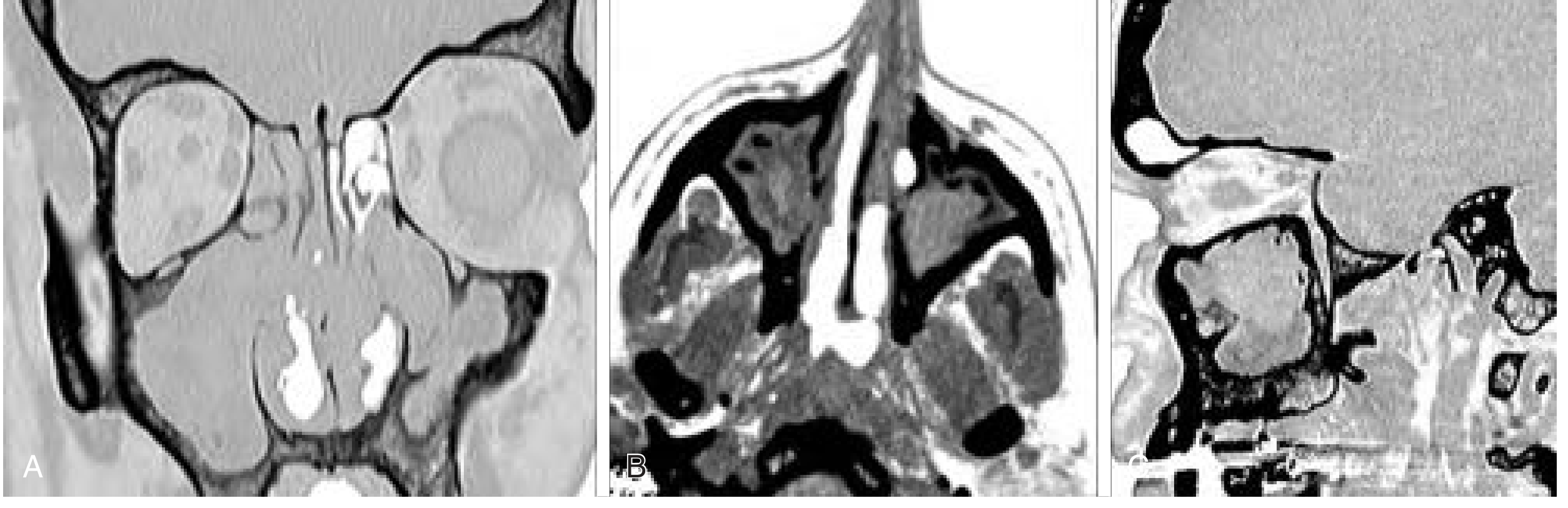
Five CT Patterns of Sinusitis (Babbel Classification):
| Pattern | Frequency | Description |
|---|---|---|
| Infundibular | 26% | Obstruction at maxillary ostium/infundibulum → maxillary disease |
| OMU pattern | 25% | Middle meatus obstruction → ipsilateral maxillary + frontal + anterior ethmoid disease |
| Sphenoethmoidal recess | 6% | Sphenoid and/or posterior ethmoid disease |
| Sinonasal polyposis | Wide extent | Diffuse bilateral disease |
| Sporadic/unclassifiable | Remainder | No clear pattern |
- OMU pattern is the most clinically relevant - obstruction of the middle meatus causes simultaneous disease in maxillary, frontal, and anterior ethmoid sinuses
- 72% of CRS patients have middle meatus opacification
- 100% of frontal sinus inflammatory disease has opacification of the frontoethmoidal recess
Lund-Mackay CT Scoring System (Most Widely Used):
| Sinus Subsite | Score |
|---|---|
| Each sinus (anterior ethmoid, posterior ethmoid, frontal, maxillary, sphenoid) | 0 = clear; 1 = partial opacification; 2 = total opacification |
| Each ostiomeatal complex (OMC) | 0 = patent; 2 = obstructed |
- 5 sinuses × 2 sides = 10 subsites (max score 20)
- 1 OMC × 2 sides = 2 subsites (max score 4)
- Maximum total Lund-Mackay score = 24
- Higher scores correlate with greater disease burden and guide surgical planning
CT's Role in Surgical Planning:
- Identifies anatomical variants that increase surgical risk (see below)
- Maps the extent of disease and affected sinuses
- CT must be available to the surgeon throughout the operation (Scott-Brown's)
- Image-guided navigation systems (IGS) use CT data for intraoperative orientation
Important Anatomical Variants on CT:
| Variant | Significance |
|---|---|
| Concha bullosa | Pneumatized middle turbinate; can narrow OMC |
| Paradoxical middle turbinate | Medially curved → narrows middle meatus |
| Haller cells (infraorbital cells) | Ethmoid cells over maxillary roof; narrow infundibulum |
| Onodi cells (sphenoethmoidal cells) | Posterior ethmoid cells surrounding the optic nerve - high surgical risk |
| Deviated nasal septum | Contributes to OMC obstruction |
| Atelectatic uncinate process | Apposed to orbital floor; obstructs infundibulum |
| Low-lying skull base / asymmetry | Increased risk of CSF leak during ethmoidectomy |
3. MRI of Paranasal Sinuses
- Malignancy suspected (soft tissue differentiation far superior to CT)
- Intracranial extension suspected (meningitis, abscess)
- Fungal sinusitis (characteristic MRI signal patterns)
- Orbital complications
- Watery/acute secretions: T1 hypointense, T2 hyperintense
- Chronic thickened secretions: T1 variable (high in inspissated/proteinaceous secretions), T2 hypointense
- Fungal concretions (mycetoma): T2 signal void (characteristic)
4. Nasal Swab / Endoscopically-Directed Culture
- Directed culture from the middle meatus (endoscopically guided)
- Organisms in CRS: Mixed flora; Staphylococcus aureus, Pseudomonas aeruginosa (especially post-surgery), Streptococcus pneumoniae, H. influenzae, Moraxella catarrhalis, anaerobes, MRSA
- Important for recalcitrant, post-surgical, and immunocompromised cases
- Guides targeted antibiotic therapy
5. Allergy Testing
- Skin prick testing or specific IgE (RAST) - for atopic CRS
- Particularly important in CRSwNP (TH2-dominant, eosinophilic disease)
- Positive in ~50% of CRSwNP patients
6. Histopathology
- Tissue from polyps or biopsies at FESS
- Confirms eosinophilic vs non-eosinophilic CRS (affects prognosis and treatment)
- Rules out malignancy in atypical presentations
- Identifies granulomatous disease (sarcoidosis, GPA)
7. Special Investigations (Selected Cases)
| Test | Indication |
|---|---|
| Saccharin transit time test | Mucociliary clearance screening |
| Ciliary biopsy + electron microscopy | Primary ciliary dyskinesia (PCD/Kartagener's syndrome) |
| Sweat chloride test / CFTR genotyping | Cystic fibrosis |
| Serum immunoglobulins (IgG, IgA, IgM, IgE) | Immunodeficiency |
| ANCA (c-ANCA / PR3-ANCA) | Granulomatosis with polyangiitis (Wegener's) |
| ACE level, serum calcium | Sarcoidosis |
| Nasal cytology | Eosinophilia, fungal elements |
| Total serum IgE + Aspergillus-specific IgE | Allergic fungal rhinosinusitis (AFRS) |
| Skin prick test for Aspergillus | AFRS |
Investigation Algorithm
SYMPTOM-BASED DIAGNOSIS OF CRS
(≥2 cardinal symptoms for >12 weeks)
             ↓
   Anterior Rhinoscopy / Nasal Endoscopy
   (confirm mucosal disease)
             ↓
   ┌─────────────────────────────┐
   │ Trial of Medical Therapy    │
   │ (INCS + Saline for 4 weeks) │
   └────────────┬────────────────┘
                ↓
        Persistent Symptoms?
                ↓
    CT PARANASAL SINUSES (Non-contrast)
    → Confirm disease, Lund-Mackay score
    → Identify anatomical variants
    → Surgical planning
                ↓
     Selectively: MRI, Allergy testing,
     Cultures, Special investigations
PART B: SURGICAL MANAGEMENT
Indications for Surgery
- CRS recalcitrant to appropriate medical therapy - most common indication
- CT-confirmed persistent mucosal disease after maximal medical treatment
- Complications of CRS (orbital, intracranial)
- Suspected sinonasal malignancy
- Mucocele / mucopyocele
- Fungal sinusitis (mycetoma, AFRS, invasive fungal)
- Odontogenic sinusitis (may be curative)
- Antrochoanal polyp (relative indication)
- Frontal: barotrauma, osteoma causing obstruction, osteomyelitis (Pott's puffy tumor)
"CRS that is recalcitrant to medical therapy is the most common indication for surgery. Surgery is adjunctive to medical therapy." - Cummings Otolaryngology
Pre-operative Assessment
- Adequate trial of medical therapy (minimum 4-8 weeks of INCS + saline)
- CT scan obtained and available (mandatory - must be displayed throughout surgery)
- Patient fitness - cardiorespiratory status, anticoagulation, comorbidities
- Informed consent including:
- Natural history and recurrence risk
- Surgical complications (orbital, intracranial, vascular, nasal)
- Need for ongoing post-operative medical treatment (especially in CRSwNP)
- Option to continue non-surgical management
Functional Endoscopic Sinus Surgery (FESS)
Principles of FESS (Messerklinger/Stammberger Technique):
- Mucosal preservation - remove disease while preserving functional mucosa
- Restoration of ventilation and drainage via the natural ostia
- Restoration of mucociliary clearance
- Minimum necessary surgery - address the specific disease
- For CRSwNP: creates wide passages for post-operative topical drug delivery - critical, as topical agents virtually cannot penetrate unoperated sinus cavities
Equipment:
- 0°, 30°, 45° rigid endoscopes (Karl Storz or similar)
- Microdebrider (powered shaver) - mucosal-preserving tissue removal
- Through-biting forceps (preferred over Blakesley-Wells to avoid tearing mucosa)
- Back-biting forceps (for uncinate incision)
- Freer's elevator (to identify uncinate)
- Kerrison's punch, Kuhn curettes (frontal recess)
- CT scan displayed on screen throughout surgery
- Image guidance / navigation system (optional but increasingly used)
Anaesthesia:
- General anaesthesia preferred in most Western centres
- Laryngeal mask (LMA) preferred over ET tube unless aspiration risk
- Total IV anaesthesia (TIVA) increasingly used (Remifentanil) - BIS monitoring recommended to prevent awareness
- Local infiltration with vasoconstrictor (1:80,000 adrenaline ± cocaine) for haemostasis
FESS Surgical Steps (Stepwise)
Step 1: Uncinectomy
- Identify the uncinate process (boomerang-shaped structure bordering the infundibulum)
- Use Freer's elevator to identify the free edge
- Use back-biting forceps for inferior incision (avoids orbital penetration - do NOT use sickle knife inferiorly)
- Sickle knife safe superiorly (frontal process of maxilla protects the orbit)
- Remove the uncinate to expose the ethmoid infundibulum and maxillary ostium
Step 2: Middle Meatal Antrostomy (Maxillary Sinusotomy)
- After uncinectomy, the maxillary sinus natural ostium is identified
- The ostium is enlarged posteriorly and inferiorly using back-biters or the microdebrider
- Creates a wide window for drainage and access
- Avoids anterior and inferior extensions (lacrimal duct anteriorly, inferior turbinate inferiorly)
Step 3: Anterior Ethmoidectomy
- The ethmoid bulla is the largest anterior ethmoid cell - it is opened first
- Use through-biting instruments (preferred over Blakesley-Wells)
- Remove anterior ethmoid partitions progressively, cell by cell
- Anterior ethmoidal artery lies in the suprabulllar recess (occasionally within bulla wall) - avoid microdebrider in this area
Step 4: Posterior Ethmoidectomy
- The ground lamella of the middle turbinate marks the boundary between anterior and posterior ethmoid
- Perforate the ground lamella in the infero-medial quadrant (avoids skull base and lamina papyracea injury)
- Use roof of maxillary sinus as a floor guide (always below the skull base)
- Caution: Onodi cell may contain the optic nerve - always know your position
Step 5: Sphenoidotomy
- Natural sphenoid ostium is located in the sphenoethmoidal recess, medial to the superior turbinate, at the level of the antral roof
- Identify by passing endoscope medial to superior turbinate (may need to resect inferior third of superior turbinate for access)
- If ostium cannot be found: create artificial opening through posterior ethmoid, then extend medially to incorporate natural ostium
- Always perforate in the infero-medial portion to avoid skull base injury
Step 6: Frontal Sinusotomy (Draf Procedures)
- The Agger nasi cell is the key landmark for all frontal recess approaches
- Remove posterior wall and roof of Agger nasi cell to expose the frontal recess
- Remove Kuhn cells (frontal recess cells) using Kuhn curettes or Rosemann punch
- Critical: Avoid circumferential mucosal injury in the frontal recess - causes scarring and stenosis
- Extended frontal procedures (Draf classification):
| Draf Procedure | Extent | Indication |
|---|---|---|
| Draf I | Removal of cells below frontal ostium | Mild frontal disease |
| Draf IIa | Unilateral frontal sinusotomy (between middle turbinate and lamina papyracea) | Standard frontal disease |
| Draf IIb | Unilateral - includes frontal sinus floor from septum to lamina papyracea | Recurrent frontal disease |
| Draf III (EMLP) | Bilateral - creates common frontal-nasal cavity opening; removes intersinus septum + nasal septum segment | Severe/recurrent frontal disease, mucocele, osteoma |
Additional Surgical Procedures
Polypectomy
- Simple avulsion (microdebrider or Tilley-Henkel forceps)
- Always combined with FESS for CRSwNP - polypectomy alone has high recurrence
- Histology mandatory on all polyp specimens
Antrochoanal Polyp Removal
- Requires removal of both the nasal + antral portions
- Middle meatal antrostomy approach (FESS) to remove antral origin
- Sublabial/transcanine or inferior meatal approach as supplementary access in some cases
- Traditional Caldwell-Luc with mucosal stripping NOT necessary
- Low recurrence if completely removed including antral component
Caldwell-Luc Operation (Largely Obsolete)
- External approach to maxillary sinus via sublabial incision through canine fossa
- Creates bone window into the anterior maxillary sinus wall
- Largely replaced by FESS - inferior results, more morbidity
- Still used in: odontogenic sinusitis, severely contracted maxillary sinus, failed FESS, tumour resection
Balloon Sinuplasty (Balloon Catheter Dilation)
- Balloon dilation of maxillary, frontal, and/or sphenoid ostia
- Controversial - does not remove disease, only dilates ostia
- Evidence shows 80.5% ostial patency maintained
- Suitable for: mild CRSsNP, frontal ostia dilation, elderly/unfit patients
- Not appropriate for CRSwNP, extensive disease, or anatomical correction
Image-Guided Surgery (Navigation)
- Uses preoperative CT fused with real-time intraoperative position tracking
- Increases safety for complex/revision cases, near skull base, optic nerve
- Not a substitute for CT on screen; both should be used
Adenoidectomy
- First-line surgery in pediatric CRS (done before FESS in children)
- Effective in some children by removing reservoir of infection in the nasopharynx
Post-operative Management
- Nasal packing (various materials - Merocel, Rapid Rhino, Bismuth iodoform paraffin paste)
- Topical vasoconstrictors (1:80,000 adrenaline ± cocaine) for bleeding
- Antibiotics: only if mucopus found at surgery (swab taken intraoperatively); no evidence for routine prophylactic antibiotics
- Oral prednisolone: 25 mg daily for 10 days (CRSwNP) - eosinophils persist up to 3 weeks in tissues; may need extended 3-week course for extensive eosinophilic disease (taper to avoid Addisonian crisis)
- Nasal saline irrigation (high-volume) - debrides crust, promotes mucociliary function
- Topical intranasal corticosteroids (spray or budesonide rinse)
- Review at 7-10 days (endoscopic debridement of crusts, clots, synechiae)
- Weekly review if concerns; fortnightly if stable
- Sinus cavity fully healed by 6 weeks
- Routine INCS sprays + less frequent irrigations continued
- 3-month review post-operatively
- Patients with eosinophilic CRS / CRSwNP: may need indefinite topical steroids to prevent polyp recurrence
- Biologic therapy (dupilumab, mepolizumab) for severe recalcitrant CRSwNP post-FESS
Surgical Complications of FESS
"In the hands of experienced clinicians, reported complications are rare. Analysis of the literature reveals a range of significant complications between 0.3 and 22.4% (median 7.0%)." - Scott-Brown's
Minor Complications:
- Bleeding/epistaxis (most common)
- Nasal adhesions / synechiae (middle turbinate to lateral wall)
- Crusting, infection
- Failure to improve / disease recurrence
Major Complications (Serious - Must Know for Exams):
| Complication | Cause | Management |
|---|---|---|
| Orbital haematoma | Breach of lamina papyracea | Recognize immediately; decompress orbit (remove LP, incise periorbita ± lateral canthotomy/cantholysis) |
| Medial rectus injury | Orbital fat exposed + continued manipulation; microdebrider risk | Ophthalmology review; late oculoplastics |
| Optic nerve injury | Intraorbital haematoma; Onodi cell dissection | IV steroids immediately; urgent ophthalmology consult |
| CSF leak | Skull base breach (cribriform plate area) | Identify and repair intraoperatively (fat plug, mucosal graft, fibrin glue) |
| Meningitis / intracranial abscess | Unrecognized CSF leak or skull base breach | Neurosurgical consult; IV antibiotics |
| Internal carotid artery injury | Sphenoid surgery with bony dehiscence | Pack sphenoid; anaesthetic resuscitation; endovascular stent |
| Anosmia | Olfactory filaments / cribriform area injury | Counselled pre-operatively |
| Epiphora | Nasolacrimal duct injury (during anterior antrostomy) | Dacryocystorhinostomy (DCR) if needed |
Outcomes of FESS
- 3,000+ patients analysed
- CRSwNP patients had better outcomes than CRSsNP following surgery
- Revision surgery rates:
- 3.6% at 12 months
- 11.8% at 36 months
- 19% by 5 years (21% for CRSwNP; 15% for CRSsNP)
- Eosinophilic CRS: greater clinical severity + higher recurrence post-operatively
- "Full-house" FESS + post-operative steroid irrigations reduces recurrence risk in CRSwNP
- FESS produces significant symptom improvement and QOL improvement for both CRSwNP and CRSsNP despite limited RCT evidence
High-Yield Summary for MBBS Exams
| Topic | Key Point |
|---|---|
| Imaging of choice | Non-contrast CT PNS, coronal cuts |
| CT scoring system | Lund-Mackay (max 24) |
| Key CT finding | Mucosal thickening, OMU obstruction, osteitis |
| CT attenuation (chronic) | 30-60 HU (vs acute 10-25 HU) |
| CT-endoscopy correlation | 70-80% |
| Endoscopy finding | Mucosal edema, purulent discharge from middle meatus, polyps |
| Surgery of choice | FESS (Functional Endoscopic Sinus Surgery) |
| Indication for surgery | Failed maximal medical therapy |
| First FESS step | Uncinectomy |
| Key landmark for frontal | Agger nasi cell |
| Ground lamella entry | Infero-medial quadrant (safe zone) |
| Risk in posterior ethmoid | Onodi cell → optic nerve |
| Risk in sphenoid | ICA dehiscence |
| Obsolete operation | Caldwell-Luc |
| Post-op oral steroid | Prednisolone 25 mg × 10 days (CRSwNP) |
| Routine prophylactic Abx | NOT recommended post-FESS |
| Most feared complication | ICA injury (catastrophic) / Optic nerve injury |
| CSF leak site | Cribriform plate vertical lamella |
| 5-year revision rate | 19% overall |
| Biologic for refractory CRSwNP | Dupilumab (anti-IL-4Rα) |
Sources
- Cummings Otolaryngology Head and Neck Surgery (2019) - Chapters 38 & 44
- Scott-Brown's Otorhinolaryngology Head & Neck Surgery Vol.1 - Chapter 98 (FESS)
- K.J. Lee's Essential Otolaryngology - CRS section
- Harrison's Principles of Internal Medicine 22E (2025)
- Shin JJ et al. Clinical Practice Guideline: Surgical Management of CRS. Otolaryngol Head Neck Surg. Jun 2025 (PMID: 40424072)
- Tran T et al. Extent of ESS in CRS: Systematic Review & Meta-Analysis. Curr Allergy Asthma Rep. 2024 (PMID: 39249643)
Etiology and treatment of maxillary sinusitis with explanation at the end of answer
maxillary sinusitis etiology management treatment
maxillary sinus anatomy teeth roots odontogenic sinusitis dental infection
maxillary sinus anatomy cross section coronal nasal cavity
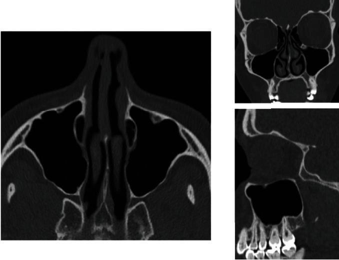
This diagnostic image set consists of three non-contrast Computed Tomography (CT) scans of the midface and paranasal sinuses, presented in axial, coronal, and sagittal planes. The axial view (left) displays the bilateral maxillary sinuses as large, air-filled (radiolucent) cavities lateral to the nasal cavity and posterior to the maxilla. The coronal view (top right) highlights the vertical relationship between the orbits superiorly, the nasal conchae centrally, and the maxillary sinuses. Inferiorly, the relationship between the sinus floor and the alveolar process is visible. The sagittal view (bottom right) provides a detailed cross-section of the maxilla, specifically illustrating the anatomical proximity of the maxillary molar and premolar tooth roots to the inferior cortical wall (floor) of the maxillary sinus. This anatomical relationship is critical for understanding risks of iatrogenic sinus effraction or oroantral communication during dental extractions. The images serve as an educational reference for normal sinus anatomy and the evaluation of the sinus floor thickness in relation to the dental apparatus.
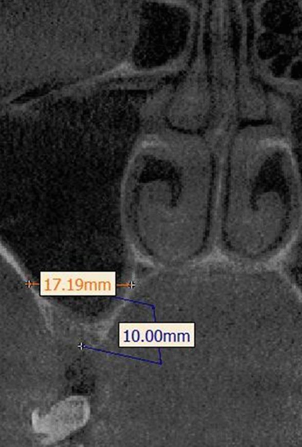
This diagnostic image is a coronal cross-section from a Cone Beam Computed Tomography (CBCT) scan, focusing on the maxillary sinus and midface anatomy. The image demonstrates a standardized methodology for measuring sinus width for implantology or oral surgery planning. An orange horizontal linear measurement of 17.19 mm indicates the sinus width, spanning from the buccal wall to the palatal wall. This measurement is positioned at a specific vertical reference height of 10.00 mm, marked by a blue vertical line originating from the alveolar crest. Key anatomical landmarks visible include the nasal cavity, inferior nasal turbinates, the floor of the maxillary sinus, and the underlying alveolar process. The image serves as a clinical guide for evaluating pneumatization and available bone volume in the posterior maxilla, highlighting the use of fixed reference points (10 mm above the crest) to ensure reproducible radiographic measurements in preoperative assessments.
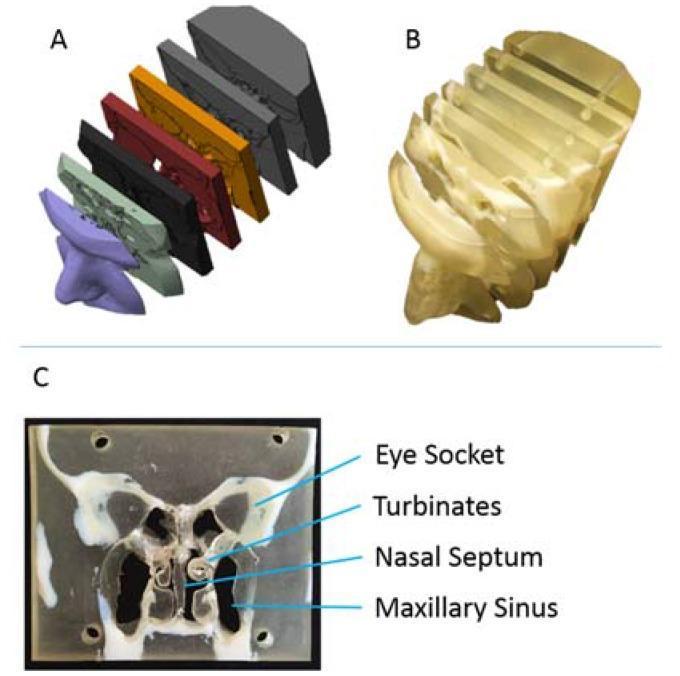
This composite image illustrates the workflow for creating a multi-material, 3D-printed educational model of the human nasal cavity and paranasal sinuses derived from CT data. Panel A shows a 3D digital rendering of seven sequential coronal slabs, color-coded to differentiate anatomical segments from the external nose to the posterior nasopharynx. Panel B displays the physical 3D-printed model, fabricated using dual polymers: a translucent, flexible material for soft tissues and a rigid, opaque material for bony structures. Panel C provides a detailed view of a single coronal cross-section of the physical model. Key anatomical landmarks are labeled, including the bilateral eye sockets (orbits), the intricate scrolls of the nasal turbinates, the vertical nasal septum dividing the airway, and the large maxillary sinuses situated inferior to the orbits. This model is designed for otolaryngology (ENT) education to demonstrate complex sinus anatomy, airflow pathways, and surgical considerations for conditions such as sinusitis or septal deviation.
Etiology and Treatment of Maxillary Sinusitis
Anatomy Recall (Foundation)
- Drainage ostium located high on the medial wall (superomedially) - gravity-dependent drainage is poor
- Ostium opens into the infundibulum → middle meatus (ostiomeatal complex)
- Floor of the maxillary sinus lies in close proximity to the roots of the upper premolars (4th, 5th teeth) and first and second molars (6th, 7th teeth) - basis of odontogenic sinusitis
- Supplied by the maxillary artery branches; innervated by V2 (maxillary division of trigeminal nerve)
Classification
| Category | Type | Duration |
|---|---|---|
| By duration | Acute | < 4 weeks |
| Subacute | 4-12 weeks | |
| Chronic | > 12 weeks | |
| Recurrent acute | ≥4 episodes/year with symptom-free intervals | |
| By etiology | Viral (AVRS) | Most common overall |
| Bacterial (ABRS) | Superimposed on viral | |
| Fungal | Mycetoma, AFRS, invasive | |
| Odontogenic | Dental source | |
| Non-infective | Allergic, vasomotor, irritant |
ETIOLOGY
1. Viral Causes (Most Common - >90% of Acute Cases)
- Rhinovirus (most common)
- Coronavirus, Adenovirus, Influenza virus, Parainfluenza virus, RSV
- Acute viral rhinosinusitis (AVRS) is the most common form
- Viral infection → mucosal edema → obstruction of the sinus ostium → impaired mucociliary clearance → stasis of secretions
- Self-limiting: resolves in 7-10 days
- Only <2% of viral sinusitis cases progress to bacterial superinfection (Harrison's)
2. Bacterial Causes (ABRS)
| Organism | Frequency |
|---|---|
| Streptococcus pneumoniae | Most common (~30-40%) |
| Haemophilus influenzae | Common (~30%) |
| Moraxella catarrhalis | Common (especially children) |
| Streptococcus pyogenes (Group A) | Less common |
| Staphylococcus aureus | Less common; important in chronic/post-surgical |
- Gram-negative organisms (Pseudomonas, Enterobacter, Klebsiella)
- Occur in ICU patients, nasotracheal intubation, immunocompromised hosts
- Often polymicrobial
- Staphylococcus aureus (including MRSA), Pseudomonas aeruginosa, Fusobacterium, Prevotella, Bacteroides (anaerobes), Corynebacterium, Proteus mirabilis, E. coli
- Biofilm formation by S. aureus and P. aeruginosa → antibiotic resistance → recalcitrant disease
3. Odontogenic Causes (Unique to Maxillary Sinus)
- Exclusively affects the maxillary sinus (due to proximity of upper tooth roots to the sinus floor)
- Causes:
- Periapical abscesses of upper teeth (2nd premolar, 1st and 2nd molars most commonly)
- Oroantral fistula (after dental extraction)
- Dental implants
- Periodontal disease
- Post-oral surgery infections
- Usually unilateral - key distinguishing feature
- Anaerobic organisms predominate
- Resolves with treatment of the causative tooth + sinus
"Odontogenic bacterial infections often result in unilateral maxillary sinusitis. Caused by tooth root/periapical abscesses, oroantral fistula, post-oral surgery infections (e.g., dental implants). Generally resolve with treatment of the affected tooth and sinus." - K.J. Lee's Essential Otolaryngology
4. Fungal Causes
- Non-invasive accumulation of fungal hyphae (usually Aspergillus fumigatus) in the maxillary sinus
- Occurs in immunocompetent individuals
- Unilateral; CT shows hyperdense material with calcifications (double density sign)
- Treatment: FESS removal (no antifungals needed for non-invasive type)
- Young atopic patients (20s-30s)
- Type I IgE-mediated hypersensitivity to fungal antigens
- Bent-Kuhn criteria: nasal polyps + type I hypersensitivity + eosinophilic mucin (peanut butter-consistency) + characteristic CT + fungal stain/culture positive
- Common fungi: Aspergillus, Bipolaris, Curvularia, Alternaria
- Seen in immunocompromised patients (uncontrolled diabetes mellitus, neutropenia, transplant recipients, HIV)
- Organisms: Mucorales (Mucor, Rhizopus) - causes rhinocerebral mucormycosis; Aspergillus
- Medical emergency - rapidly progressive; can invade orbit, intracranial structures
- Presentation: facial pain, headache, cranial nerve involvement, proptosis, chemosis, black necrotic eschar
5. Anatomical / Structural Causes
- Deviated nasal septum - displaces middle turbinate, narrows middle meatus
- Concha bullosa - pneumatized middle turbinate; narrows OMC
- Haller cells (infraorbital ethmoid cells) - narrow the maxillary infundibulum directly
- Turbinate hypertrophy - inferior or middle
- Nasal polyps - obstruct drainage
- Paradoxical middle turbinate
- Sinonasal tumours - cause secondary obstruction
6. Mucociliary Dysfunction
- Cystic fibrosis - altered Cl⁻/water transport → thick viscid mucus → poor clearance
- Primary Ciliary Dyskinesia (PCD / Kartagener's syndrome) - immotile cilia
- Cigarette smoking - impairs ciliary function
- Decreased hydration - inspissated secretions
7. Allergic / Immunological Causes
- Allergic rhinitis - type I hypersensitivity; eosinophilic mucosal inflammation; mucosal edema obstructs OMC
- Aspirin-Exacerbated Respiratory Disease (AERD) - arachidonic acid pathway dysregulation
- Immunodeficiency - hypogammaglobulinemia, IgA deficiency, HIV
8. Environmental / Miscellaneous Causes
- Barotrauma - scuba diving, air travel (pressure changes cause mucosal edema and haemorrhage)
- Atmospheric pollutants, tobacco smoke
- Nosocomial - nasotracheal intubation, nasogastric tube (direct obstruction of OMC or local trauma)
- Cold and damp weather
- Systemic disease - connective tissue disorders, malnutrition, Wegener's granulomatosis (GPA), sarcoidosis
Pathogenic Mechanism: Common Pathway
Precipitating Factor (viral URTI, allergy, structural, dental)
               ↓
   Mucosal edema of nasal cavity / OMC
               ↓
   Obstruction of maxillary sinus ostium (high on medial wall)
               ↓
   Impaired ventilation + Mucociliary stasis
               ↓
   Hypoxia in sinus → O2 ↓, pH ↓
               ↓
   Bacterial colonization → Infection
               ↓
   Acute Maxillary Sinusitis
               ↓ (if untreated / recurrent)
   Chronic Maxillary Sinusitis
CLINICAL FEATURES (Brief Recap)
- Facial pain/pressure over the cheeks (malar area) - worsens on bending forward
- Maxillary toothache (upper premolars/molars) - referred via V2
- Purulent nasal discharge (anterior or posterior)
- Nasal obstruction/congestion
- Fever, malaise, headache
- Unilateral pain = suspect odontogenic or fungal
- Tenderness on pressure/percussion over the malar area
- Purulent exudate in the middle meatus
- Erythema/swelling of overlying cheek (in severe cases)
TREATMENT
A. Acute Viral Rhinosinusitis (AVRS) - Symptomatic Only
"All patients with acute sinusitis should be counselled about symptom-based treatments." - Harrison's 22E
| Treatment | Drug/Dose | Notes |
|---|---|---|
| Analgesics/Antipyretics | Paracetamol or Ibuprofen | First-line for pain, fever |
| Nasal saline sprays/washes | High-volume isotonic saline | Clears mucus, reduces edema, promotes drainage |
| Oral decongestants | Pseudoephedrine 120 mg 12-hourly | Shrinks ostial mucosa → promotes drainage |
| Topical decongestants | Oxymetazoline (2 sprays BD) | Maximum 3-5 days only (risk of rhinitis medicamentosa) |
| Intranasal corticosteroids | Mometasone, Fluticasone | Helpful in allergic component or chronic rhinitis history |
| Mucolytics | Guaifenesin | Thins secretions |
| Facial hot packs | Warm compress over cheek | Symptomatic relief |
Antihistamines should be AVOIDED in acute sinusitis - their drying effect thickens secretions and impairs drainage.
B. Antibiotic Therapy - When to Prescribe
| Indication | Definition |
|---|---|
| Persistent | Symptoms lasting ≥10 days without improvement |
| Severe | Fever >102°F (38.9°C) + purulent nasal discharge or facial pain for ≥3-4 consecutive days |
| Worsening ("double sickening") | New fever/headache/increased discharge after initial improvement of a viral URI lasting 5-6 days |
- Comorbidities: diabetes mellitus, chronic respiratory disease, congestive heart failure, immunodeficiency
- High fever with systemic toxicity
- Possible orbital or intracranial involvement
- Unilateral acute maxillary tooth pain with purulent discharge
C. Antibiotic Regimens
First-Line:
| Drug | Dose | Duration |
|---|---|---|
| Amoxicillin/Clavulanate (preferred) | 875/125 mg BD | 5-7 days (adults) |
| Amoxicillin (alternative) | 875 mg PO BD or 1000 mg TDS | 5-10 days |
"Amoxicillin remains the treatment of choice. It is inexpensive, penetrates sinus tissues well, and can be changed if symptoms have not improved in 48-72 hours." - Berek & Novak's
Second-Line (or if beta-lactam resistant suspected):
| Drug | Dose | Notes |
|---|---|---|
| Amoxicillin/Clavulanate high-dose | 2000/125 mg BD × 7 days | For resistant organisms |
| Levofloxacin | 500 mg OD × 5-10 days | Fluoroquinolone (risk: dysglycemia, neuropathy, tendon/aortic rupture) |
| Moxifloxacin | 400 mg OD | Fluoroquinolone |
Penicillin Allergy:
| Allergy Type | Drug |
|---|---|
| Mild allergy | Cefuroxime (2nd gen cephalosporin) |
| Severe allergy | Doxycycline 200 mg OD × 5-10 days |
| Children, mild allergy | Cefdinir, cefuroxime, cefpodoxime (3rd gen cephalosporins) |
Macrolides (azithromycin, clarithromycin) are specifically NOT recommended for sinusitis due to high rates of macrolide-resistant S. pneumoniae. Trimethoprim/sulphamethoxazole also not recommended due to resistance. (Harrison's)
Children (American Academy of Pediatrics guidelines):
- Observation for 72 hours is acceptable for non-severe cases
- Antibiotics indicated for: severe onset, worsening symptoms, or failure to improve in 72 hours
- First-line: Amoxicillin ± clavulanate for minimum 10 days
D. Chronic Maxillary Sinusitis Treatment
- Nasal saline irrigation (high-volume) - first-line
- Intranasal corticosteroids - mainstay (mometasone, fluticasone, budesonide)
- Macrolides (long-term, low-dose) - 12 weeks (clarithromycin/erythromycin) for CRSsNP (anti-inflammatory + antibacterial effect)
- Oral corticosteroids - short course for acute exacerbations especially with polyps
- Allergy management - antihistamines, leukotriene antagonists (montelukast) for allergic component
- FESS (Functional Endoscopic Sinus Surgery) - for medically recalcitrant cases
E. Odontogenic Maxillary Sinusitis
- Treat the causative dental pathology first - extraction/root canal of offending tooth, closure of oroantral fistula
- Antibiotics - covering oral anaerobes (amoxicillin-clavulanate, metronidazole + amoxicillin, or clindamycin)
- FESS/antrostomy - to drain and wash out the maxillary sinus of infected material
- Most cases resolve after dental treatment + sinus washout
F. Fungal Maxillary Sinusitis
| Type | Treatment |
|---|---|
| Mycetoma (non-invasive) | FESS removal of fungal ball; NO antifungals |
| AFRS | FESS + perioperative steroids; topical/oral steroids long-term; NO routine antifungals |
| Invasive fungal | IV antifungals (Amphotericin B for Mucor; Voriconazole for Aspergillus) + urgent surgical debridement; treat underlying immunosuppression |
G. Nosocomial / ICU Sinusitis
- Remove precipitating factor (nasotracheal tube, NG tube)
- Broad-spectrum IV antibiotics (gram-negative coverage) until culture results available
- Consider antral washout/drainage
H. Surgical Management - Antral Washout (Proof and Drain)
- Failure of medical therapy
- Intractable pain
- Suspected complications
- Fungal sinusitis
- Odontogenic sinusitis with abscess
- Immunocompromised patient
- Antral washout (Proof puncture) - trocar inserted through inferior meatus to puncture medial wall; sinus irrigated with saline
- FESS with middle meatal antrostomy - endoscopic widening of the natural ostium; procedure of choice
- Caldwell-Luc operation - external approach via sublabial incision (largely obsolete, reserved for specific indications)
- Intranasal antrostomy via inferior meatus - creates dependent drainage (less preferred now)
Complications of Untreated Maxillary Sinusitis
| Complication | Notes |
|---|---|
| Orbital cellulitis / abscess | Via ethmoid extension; most common complication |
| Subperiosteal abscess | Facial bones |
| Cavernous sinus thrombosis | Via retrograde venous spread (danger triangle) |
| Meningitis | Via skull base |
| Brain abscess / Epidural abscess | Rare but serious |
| Osteomyelitis | Of facial bones |
| Mucocele / Mucopyocele | Chronic obstruction leading to cystic distension |
Summary Table - Treatment at a Glance
| Scenario | Treatment |
|---|---|
| Viral sinusitis | Saline, decongestants (≤3-5 days topical), analgesics, INCS |
| ABRS (mild, no criteria) | Watchful waiting + symptomatic |
| ABRS (criteria met) | Amoxicillin/clavulanate 875/125 mg BD × 5-7 days |
| Penicillin allergy (mild) | Cefuroxime |
| Penicillin allergy (severe) | Doxycycline |
| NOT recommended | Macrolides, TMP-SMX (resistance) |
| Antihistamines in acute | AVOID (drying effect worsens drainage) |
| Chronic (medical) | INCS + saline irrigation ± macrolides |
| Odontogenic | Dental treatment + amoxicillin-clavulanate + FESS |
| Fungal (mycetoma) | FESS removal only |
| Invasive fungal | IV amphotericin B + urgent surgery |
| Complicated/refractory | FESS / antral washout |
EXPLANATION (Simple Language)
Why Does Maxillary Sinusitis Happen?
Why Is Treatment Mostly Symptomatic?
- Pain relief (paracetamol/ibuprofen) for the cheek and tooth pain
- Saline washes to clear out the blocked passages
- Decongestant sprays (for maximum 3-5 days only) to shrink the swollen window/ostium and let the sinus drain
When Do Antibiotics Help?
The Danger of Untreated Sinusitis
Sources
- K.J. Lee's Essential Otolaryngology - Rhinosinusitis (Pathogenesis, Bacteriology, Treatment)
- Harrison's Principles of Internal Medicine 22E (2025) - Acute and Chronic Sinusitis
- Berek & Novak's Gynecology - Sinusitis section
- Scott-Brown's Otorhinolaryngology Head & Neck Surgery Vol.1 & 2
- Vitali FC et al. Global Prevalence of Maxillary Sinusitis of Odontogenic Origin. J Endod. 2023 (PMID: 36754253)
- Allevi F et al. Treatment of Sinusitis Following Dental Implantation: Meta-Analysis. Am J Rhinol Allergy. 2022 (PMID: 35244478)