
Explain axillary artery. Uh... Relations. ...in a stromosis with the scapula. Anastomosis. With scapula and its clinicals. Uh, give handwritten-- handmade diagrams. Which easy to draw. And explain. All these things, point to point. Short, short me samjha.
Reading File
Now let me search the library for axillary artery content in parallel.
Finding Sources
Finding Sources
Good results. Let me now read the key anatomy sections in parallel.
Reading File
Reading File
Reading File
Now let me read more from Gray's for relations, and the scapular anastomosis section from THIEME:
Reading File
Reading File
Excellent! Now let me fetch the key diagrams and also get the relations of the axillary artery:
Loading Image
Loading Image
Finding Sources
I now have all the key information. Let me also fetch the branches diagram for completeness:
Loading Image
Now I have all the information needed. Let me compile a clear, point-by-point explanation with diagrams.
Axillary Artery - Complete Notes
(Gray's Anatomy for Students; THIEME Atlas of Anatomy)
1. OVERVIEW
- Begins: Lateral margin of rib I (continuation of subclavian artery)
- Ends: Lower border of teres major muscle (continues as brachial artery)
- Location: Inside the axilla, enclosed in the axillary sheath (derived from prevertebral fascia of the neck)
2. THREE PARTS (divided by Pectoralis Minor)
HANDMADE DIAGRAM - Three Parts:
Rib I ──────────────────────────────── Teres Major
| |
[1st part]──[Pect. Minor]──[2nd part]──[3rd part]
↑ behind it
| Part | Position | No. of Branches |
|---|---|---|
| 1st | Proximal to pect. minor | 1 branch |
| 2nd | Behind / posterior to pect. minor | 2 branches |
| 3rd | Distal to pect. minor | 3 branches |
Memory trick: 1-2-3 rule (part 1 = 1 branch, part 2 = 2 branches, part 3 = 3 branches)
3. SIX BRANCHES
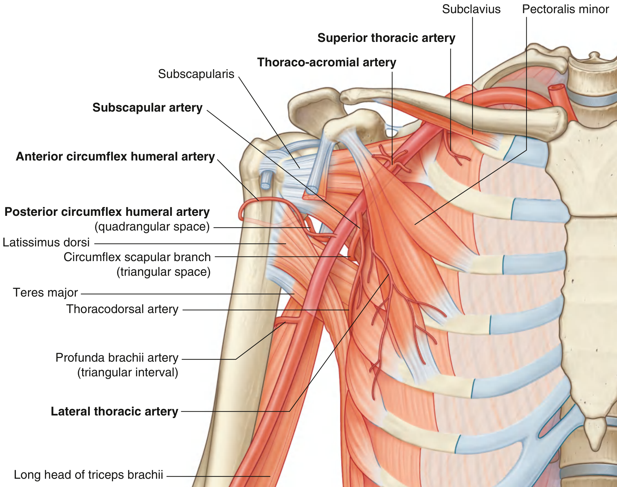
HANDMADE DIAGRAM - Branches:
AXILLARY ARTERY
|
┌────────────┴────────────┐
│ 1st Part │
└──► Superior Thoracic │
│
┌────────────────────────┐│
│ 2nd Part ││
└──► Thoraco-acromial ││
└──► Lateral Thoracic ││
│
┌────────────────────────┘│
│ 3rd Part │
└──► Subscapular ─┬─► Circumflex Scapular
└─► Thoracodorsal
└──► Anterior Circumflex Humeral (ACHA)
└──► Posterior Circumflex Humeral (PCHA)
| Branch | Supply |
|---|---|
| Superior thoracic | Upper axillary wall |
| Thoraco-acromial | Pectoral, deltoid, clavicular, acromial regions |
| Lateral thoracic | Medial axillary wall, breast (in women) |
| Subscapular | Posterior axillary wall + scapular region |
| ACHA | Anterior surgical neck of humerus, glenohumeral joint |
| PCHA | Passes through quadrangular space with axillary nerve; surgical neck posteriorly |
4. RELATIONS
The axillary artery is surrounded by the cords of the brachial plexus, which are named by their relation to it:
HANDMADE DIAGRAM - Cross-section Relations:
Medial cord
|
Lateral ─── AA ─── Axillary Vein (anteromedial)
cord |
Posterior cord
(AA = Axillary Artery)
Key relations:
- Medial: Axillary vein (lies anteromedial to artery)
- Lateral: Lateral cord of brachial plexus
- Posterior: Posterior cord of brachial plexus
- Medial side: Medial cord
- Anterior: Pectoralis minor crosses the 2nd part
- Whole vessel is enclosed in the axillary sheath
5. ANASTOMOSIS WITH SCAPULA - "Scapular Arcade"
This is the most clinically important part.
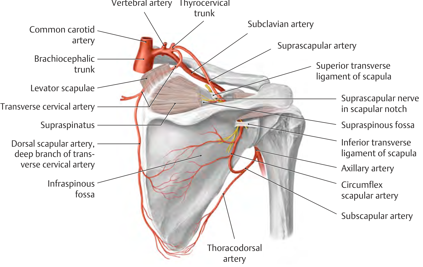
HANDMADE DIAGRAM - Scapular Anastomosis:
Subclavian artery
│
Thyrocervical trunk ──► Suprascapular artery
│ passes OVER
│ Superior transverse ligament
▼
Supraspinous fossa
│
│ passes UNDER
│ Inferior transverse ligament
▼
Infraspinous fossa
│
◄──────────┘ ← ANASTOMOSIS here
│
Axillary artery ──► Subscapular artery
│
Circumflex Scapular artery
(enters via Triangular Space)
│
Infraspinous fossa ◄────── Also joins:
Dorsal Scapular artery
(deep br. of transverse cervical a.)
Three arteries meet in the infraspinous fossa:
- Suprascapular artery (from thyrocervical trunk of subclavian)
- Circumflex scapular artery (from subscapular → axillary)
- Dorsal scapular artery (deep branch of transverse cervical artery)
6. CLINICAL IMPORTANCE
a) Collateral Circulation after Axillary Artery Ligation/Occlusion
- If axillary artery is ligated proximal to the subscapular artery, the scapular arcade takes over via:
- Suprascapular → Circumflex scapular (retrograde) → re-enters subscapular → distal axillary
- Blood can reach the arm even if the axillary artery is blocked!
- This is why ligation above the subscapular is safer than below it
b) Safe vs. Dangerous Ligation Sites
Proximal ligation (above subscapular):
✅ Collateral via scapular arcade INTACT → limb survives
Distal ligation (below subscapular + circumflex humeral):
❌ Collateral inadequate → limb ischemia risk
c) Shoulder Dislocation
- Anterior shoulder dislocation (commonest) can damage the axillary artery
- Also risks injury to axillary nerve (PCHA travels with it through quadrangular space)
- Clinical sign: loss of deltoid sensation (regimental badge area) + weakened abduction
d) Quadrangular Space Syndrome
- PCHA and axillary nerve pass through quadrangular space
- Compression here → shoulder pain + deltoid weakness
e) Axillary Artery Aneurysm
- Can present as pulsatile axillary mass
- Risk of distal embolism to upper limb
QUICK REVISION SUMMARY
HANDMADE CHEAT SHEET:
Subclavian a. → [Lat margin Rib I] → AXILLARY A. → [Below teres major] → Brachial a.
Parts: 1──[Pect.minor]──2──[Pect.minor]──3
↓ ↓↓ ↓↓↓
1 branch 2 branches 3 branches
3rd Part branches = S-A-P (Subscapular, Ant.CHA, Post.CHA)
Scapular Arcade = Suprascapular + Circumflex Scapular + Dorsal Scapular
↑ Subclavian side ↑ Axillary side
Clinical: Ligation above subscapular = SAFE (arcade kicks in)
Sources: Gray's Anatomy for Students, p.853-855 | THIEME Atlas of General Anatomy, p.404, p.410
This is a shared conversation. Sign in to Orris to start your own chat.