
10 mark answer for juvenile ossifying fibroma
juvenile ossifying fibroma histology microscopy jaw
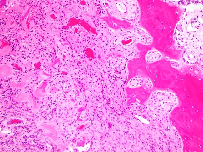
Histopathology of a gingival peripheral ossifying fibroma. Light microscopy using Hematoxylin and Eosin staining shows a dense fibrous stroma with proliferating spindle-shaped fibroblasts arranged in fascicles and whorls, with relatively low mitotic activity and mild chronic inflammatory infiltrate at the periphery. The stroma is intimately associated with scattered mineralized deposits representing a mineralized matrix. Notable mineralization includes dystrophic calcifications, cementum-like material, and small bony trabeculae or lamellar bone, consistent with ossifying changes. In older or healed areas, well-formed lamellar bone or cementum-like tissue is seen within the fibrous matrix. The lesion is typically confined to gingival tissue and may cause surface erosion or ulceration; histology demonstrates a reactive, non-neoplastic process with a fibrous proliferative component and focal calcified masses. The diagnostic significance is that presence of calcified material within a fibrous gingival lesion supports POF diagnosis, distinguishing it from fibroma or pyogenic granuloma. Differential considerations include irritational fibroma, cemento-ossifying fibroma, peripheral odontogenic fibroma, and pyogenic granuloma with calcifications. Clinically, this histology aligns with a gingival mass in the anterior maxilla or mandible, excised as part of periodontal therapy. Therapeutic implications include conservative excision with close follow-up due to potential recurrence. The specimen's micrograph underscores the biphasic pattern: fibrous stroma and mineralized nodules.
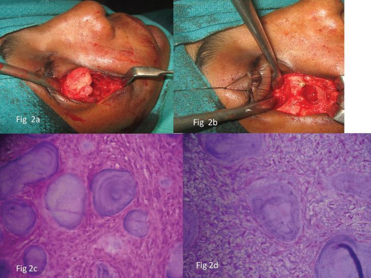
This composite image includes intraoperative clinical photographs and histopathological photomicrographs detailing the surgical management and cellular features of a psammomatoid juvenile ossifying fibroma (PsJOF) in a 15-year-old patient. Panels 2a and 2b show an infraorbital surgical approach. Panel 2a demonstrates the exposure of a pale, well-circumscribed, lobulated mass within the infraorbital rim, while panel 2b shows the site post-excision and peripheral ostectomy, revealing the underlying bony anatomy of the orbit. Panels 2c (10x magnification) and 2d (40x magnification) are H&E-stained photomicrographs showing the pathognomonic features of PsJOF. The histology displays a cellular fibrous stroma containing numerous small, spherical, mineralized ossicles. These ossicles resemble psammoma bodies, characterized by a basophilic appearance and distinct concentric lamellation. The combination of clinical findings and characteristic psammomatoid ossicles is essential for differentiating this aggressive fibro-osseous lesion from conventional ossifying fibromas in pediatric oral and maxillofacial pathology.
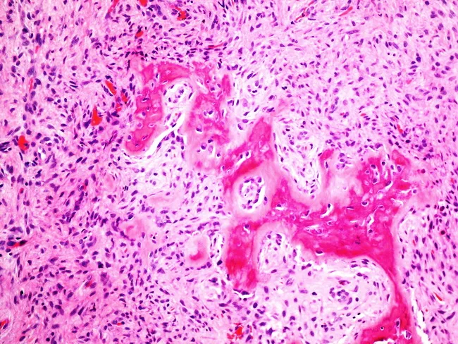
High-power light microscopy of an oral soft tissue biopsy stained with Hematoxylin and Eosin reveals a lesion consistent with peripheral ossifying fibroma. The overall architecture shows a relatively well-circumscribed, hypocellular to mildly cellular fibrous stroma populated by bland spindle-shaped and ovoid fibroblasts, with sparse mitotic activity and minimal atypia. Intermixed within the fibrous matrix are mineralized bony trabeculae, some of which are woven bone, indicating osteogenic differentiation within the lesion. The lamellar mineralization forms irregular islands that disrupt the uniform fibrous background, aligning with characteristic ossification seen in this entity. Inflammatory cells are limited, and the lesion lacks marked necrosis or malignant features. Vascular channels are present but not prominent, contributing to a vascular but non-aggressive appearance. The combination of a fibrous proliferative process with discrete osseous formation in the gingival/periodontal region is classic for a peripheral ossifying fibroma, a reactive, gingival overgrowth linked to irritation or trauma. Clinically, this histology supports conservative surgical excision with careful curettage to minimize recurrence. Differential considerations include fibroma, pyogenic granuloma, and other ossifying/subosseous lesions, but the presence of bone trabeculae within a fibrous lesion strongly favors peripheral ossifying fibroma. Correlation with clinical findings and radiographic imaging aids definitive diagnosis. Clinical correlation advised.
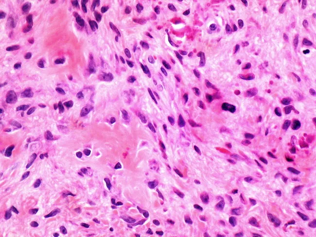
Imaging modality: Light microscopy of a hematoxylin and eosin (H&E) stained histology section. An oral gingival lesion, consistent with peripheral ossifying fibroma, shows a fibrous connective tissue stroma with osteoid matrix formation. The primary tissue features include bland, spindle-shaped fibroblasts arranged in sweeping fascicles and septa, with minimal atypia and low mitotic activity. Lakes of eosinophilic osteoid or bone-like material are identifiable within the fibrous matrix, sometimes accompanied by calcifications. The lesion exhibits mild chronic inflammatory infiltrate and increased vascularity perithreshold to the stroma. The osteoid matrix indicates osteogenic differentiation and is the key diagnostic feature distinguishing this entity from a simple fibroma. The overall architecture blends reactive fibrous tissue with calcified deposits, supporting a diagnosis of peripheral ossifying fibroma rather than other gingival lesions. Differential considerations include fibroma with calcifications, cemento-ossifying fibroma, pyogenic granuloma, and ossifying fibroma; clinical correlation and radiographic context help refine the impression. Clinically, these lesions arise from the interdental gingiva, predominantly in younger individuals, and are treated by complete surgical excision with removal of the lesion base; recurrence is possible. This image is valuable for dental pathology education and differential diagnostic training, illustrating characteristic fibroblastic proliferation coupled with osteoid formation. For clinical teaching and research.
Juvenile Ossifying Fibroma (JOF)
Definition and Classification
- Ossifying fibroma of odontogenic origin - affects tooth-bearing bone (mandible > maxilla) in adults
- Juvenile Trabecular Ossifying Fibroma (JTOF) - mostly in the alveolar process of the maxilla in children
- Juvenile Psammomatoid Ossifying Fibroma (JPOF) - mostly fronto-orbital and ethmoid bones
Epidemiology
- Occurs predominantly in children and adolescents (first and second decades of life)
- JTOF: mean age ~8-12 years; JPOF: mean age ~16-33 years (slightly older)
- Both sexes affected; some series show slight female predominance
- JTOF affects maxilla predominantly; JPOF affects the paranasal sinuses (ethmoid and fronto-orbital bones)
Pathogenesis
- Is well-capsulated and can be shelled out at surgery (in conventional OF)
- Has active osteoblastic rimming of trabeculae (differentiating it from fibrous dysplasia)
- Juvenile variants show a highly cellular stroma indicating more active proliferative behavior
Clinical Features
- Painless, progressive swelling of the jaw or sinonasal region - the most common presentation
- Lesions are slowly expansile but the juvenile aggressive variant grows faster
- Sinonasal symptoms: nasal obstruction, epistaxis, proptosis, diplopia, epiphora, facial swelling, headache
- Endoscopically: smooth, mucosa-covered mass
- Malignant transformation has not been reported
- JTOF: expansile swelling of maxilla, displacing teeth
- JPOF: symptoms related to orbit and paranasal sinuses
Radiological Features
- Well-defined, expansile, radiolucent lesion with variable calcification (unilocular or multilocular)
- "Eggshell rim" with central radiolucency in conventional OF
- Juvenile aggressive variants (JPOF/JTOF): thicker bony rim with central ground-glass appearance
- Cortical thinning and possible cortical perforation (especially JPOF - explaining high recurrence)
- Roots of adjacent teeth may be displaced
- Low to intermediate signal on T1-weighted imaging
- Variable signal intensity on T2-weighted imaging
- Similar in appearance to fibrous dysplasia on MRI
Histopathology
- Cellular fibrous stroma of proliferating spindle-shaped fibroblasts
- Juvenile variants show highly cellular stroma with plump cells
- JTOF: Trabeculae of woven bone with prominent osteoblastic rimming (plump osteoblasts lining the bony trabeculae) - this osteoblastic activity distinguishes it from fibrous dysplasia
- JPOF: Compact, rounded, concentrically laminated calcifications resembling psammoma bodies (acellular, EMA-positive, similar to those in meningiomas)
- Both types may have dystrophic calcifications, cementicles, and cementum-like material
| Feature | JOF | Fibrous Dysplasia |
|---|---|---|
| Capsule | Present (may be incomplete in JOF) | Absent |
| Osteoblastic rimming | Present | Absent |
| Stroma | Cellular, fibroblastic | Less cellular, "C-shaped" trabeculae |
| Nature | Neoplasm | Developmental anomaly (GNAS mutation) |
| Recurrence | Yes (juvenile aggressive) | No (ceases with skeletal maturation) |
Differential Diagnosis
- Fibrous dysplasia - no capsule, no osteoblastic rimming, GNAS mutation, "Chinese letters/C-shaped" bony trabeculae
- Conventional ossifying fibroma (OF) - older patients (20-40 years), mandible, less aggressive
- Cementifying fibroma / Cemento-ossifying fibroma (COF) - cementum-like spherules throughout the lesion
- Giant cell granuloma - multinucleated giant cells, no mineralised matrix
- Osteoblastoma - similar histology but lacks fibrous stroma; posterior mandible
- Aneurysmal bone cyst - blood-filled spaces, secondary change possible in JOF
- Osteosarcoma - malignant cytology, permeative growth, no well-defined border
Treatment
- Surgical excision is the treatment of choice
- Conventional OF: conservative enucleation and curettage - the fibrous capsule allows shelling-out
- Juvenile aggressive OF (JTOF/JPOF): requires more radical excision including peripheral ostectomy because of high recurrence rates
- Recurrence rates: 30-50% for juvenile/psammomatoid variants (due to tendency for cortical perforation and incomplete excision)
- Conventional OF: recurrence rates are low
- Endoscopic approach is increasingly used for sinonasal disease; extensive disease may need combined craniofacial resection
- No malignant transformation has been reported
- Follow-up is essential due to recurrence risk
Summary Table: JOF Subtypes
| Feature | JTOF | JPOF |
|---|---|---|
| Age | Younger children (~8-12 yrs) | Older children/young adults (~16-33 yrs) |
| Site | Maxillary alveolar process | Fronto-orbital, ethmoid bones |
| Histology | Woven bone trabeculae with osteoblastic rimming | Psammomatoid ossicles (EMA+) |
| Behavior | Moderately aggressive | More aggressive |
| Recurrence | Moderate | High (30-50%) |
- JOF = true neoplasm (not developmental anomaly like fibrous dysplasia)
- Two subtypes: trabecular (maxilla, younger children) and psammomatoid (orbit/ethmoid, slightly older)
- Histological hallmarks: highly cellular fibrous stroma + characteristic mineralized component (woven bone with osteoblastic rimming in JTOF; psammoma-like ossicles in JPOF)
- Psammoma bodies are EMA-positive, resembling extracranial meningioma ossicles
- Recurrence rate 30-50% - higher than conventional OF
- Malignant transformation not reported
- Treatment: radical surgical excision with long-term follow-up