
Type 1 hypersensitivity reactin mechanism
Reading File
Now let me search the medical library for Type 1 hypersensitivity, in parallel with a web search.
Finding Sources
Searching the Web
Type 1 hypersensitivity reaction mechanism IgE mast cells
Good - I found a dedicated section. Let me read it, and also search for more detailed content on the mechanism.
Reading File
Finding Sources
Excellent - I have a great hit from Cellular and Molecular Immunology (Abbas). Let me read the detailed mechanism section from that textbook, and also get the Katzung pharmacology section which had a diagram.
Reading File
Reading File
I have great content. Let me now read more of the Cellular and Molecular Immunology section for the detailed step-by-step mechanism, and also fetch the diagram image.
Reading File
Loading Image
Let me get the full mechanism diagram and read more detail on mast cell activation and mediators.
Loading Image
Reading File
I now have comprehensive information to give a thorough, well-structured answer. Let me compile everything.
Type I Hypersensitivity Reaction - Mechanism
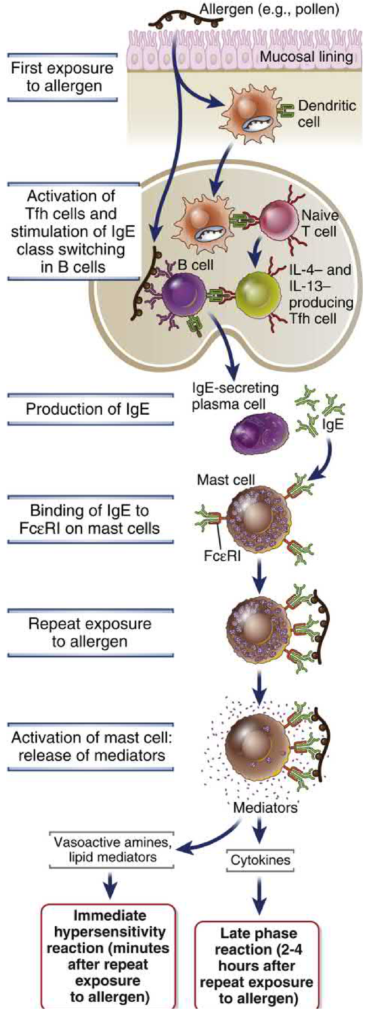
Type I hypersensitivity (immediate hypersensitivity) is an IgE-mediated allergic reaction that occurs within minutes of re-exposure to an antigen (allergen). It involves two key phases: sensitization and the effector (elicitation) phase.
Phase 1: Sensitization (First Exposure)
This phase primes the immune system - no symptoms occur yet.
-
Allergen entry: Allergens (pollen, house dust mite proteins, food proteins, drugs) enter via mucosal surfaces and are captured by dendritic cells (DCs).
-
T cell polarization: DCs present allergen peptides to naive T cells, which differentiate into Th2 cells (and T follicular helper [Tfh] cells). Th2/Tfh cells produce IL-4 and IL-13.
-
IgE class switching: IL-4 and IL-13 act on allergen-specific B cells, promoting class-switch recombination from IgM/IgG to IgE production. Plasma cells then secrete large amounts of IgE.
-
Mast cell sensitization: IgE binds with extremely high affinity to FcεRI (Fc epsilon receptor type I) on tissue mast cells and circulating basophils. The mast cells are now "armed" - this is sensitization.
At this point the person is sensitized but asymptomatic.
Phase 2: Effector Phase (Re-exposure / Elicitation)
Re-exposure to the same allergen triggers the reaction.
-
Allergen cross-linking: The allergen binds to and cross-links two or more adjacent IgE molecules on the mast cell surface. This aggregates the FcεRI receptors.
-
Mast cell activation: Cross-linking of FcεRI activates intracellular signaling (Lyn kinase → Syk kinase → downstream phospholipase C pathway), leading to:
- Rise in intracellular Ca²+
- Degranulation - preformed granule contents released within seconds to minutes
Mediators Released
| Category | Mediator | Effect |
|---|---|---|
| Preformed (granules) | Histamine | Vasodilation, increased vascular permeability, smooth muscle contraction |
| Heparin | Anticoagulant | |
| Tryptase, chymase | Tissue remodeling, inflammation marker | |
| Serotonin | Vasoconstriction/dilation depending on site | |
| Newly synthesized lipid mediators | Prostaglandin D2 (PGD2) | Vasodilation, bronchoconstriction |
| Leukotrienes C4, D4, E4 | Prolonged bronchoconstriction, mucus secretion, increased vascular permeability | |
| PAF (platelet-activating factor) | Vasodilation, leukocyte chemotaxis | |
| Cytokines (produced on activation) | IL-4, IL-13 | Sustain Th2 responses, mucus secretion |
| IL-5 | Eosinophil recruitment and activation | |
| TNF, IL-3, MIP-1α | Late-phase inflammation |
- Cellular and Molecular Immunology, Table 20.2
Immediate vs. Late-Phase Reactions
-
Immediate reaction (within minutes): Driven by vasoactive amines (histamine) and lipid mediators - causes rapid vasodilation, bronchoconstriction, and mucus secretion.
-
Late-phase reaction (2-4 hours after re-exposure): Driven by cytokines (especially IL-4, IL-5, TNF) released by mast cells. These recruit eosinophils, neutrophils, and more Th2 cells, causing a prolonged inflammatory response.
Clinical Manifestations
Depending on the tissue involved:
- Systemic - Anaphylaxis (most severe - airway restriction + cardiovascular collapse)
- Respiratory - Allergic asthma, allergic rhinitis (hay fever)
- Skin - Urticaria (hives), angioedema, atopic dermatitis
- GI - Food allergy symptoms (abdominal pain, diarrhea)
- Eyes - Allergic conjunctivitis
Key diagnostic markers:
- Serum tryptase - elevated in anaphylaxis (peaks at ~1 hour; also chronically elevated in systemic mastocytosis)
- Plasma histamine - peaks rapidly (~10 minutes), returns to normal within 1 hour
- Urinary histamine - may remain elevated for 24 hours
- Katzung's Basic and Clinical Pharmacology, 16th Ed., pp. 1524-1525
- Cellular and Molecular Immunology (Abbas), 10th Ed., pp. 1302-1308
- Quick Compendium of Clinical Pathology, 5th Ed., p. 4360
Why NOT Type I: Important Distinctions
- Anaphylactoid reactions - clinically similar to anaphylaxis but NOT IgE-mediated (direct mast cell degranulation by drugs like morphine, radiocontrast media) - not true Type I hypersensitivity
- Hereditary angioedema - complement-mediated (C1 inhibitor deficiency) - not Type I
- Type II - antibody-mediated cytotoxicity (IgG/IgM on cell surfaces)
- Type III - immune complex-mediated (IgG + antigen complexes + complement)
- Type IV - delayed-type (T cell and macrophage mediated, 48-72 hrs)
Haptens
Haptens are small molecules that can bind antibodies but cannot trigger an immune response on their own - they need to couple with a larger carrier protein to become immunogenic. Classic examples include penicillin, poison ivy (urushiol), and DNCB.
Searching sources...
Finding Sources
Searching the Web
hapten immunology mechanism carrier protein immune response
Excellent hits. Let me read both textbook sections in parallel.
Reading File
Reading File
Let me now read the detailed hapten-carrier text and also search for more context on hapten definition and properties.
Reading File
Finding Sources
Let me quickly get the Roitt's section and the figure for hapten-carrier.
Reading File
Loading Image
Let me also fetch the Roitt's hapten-carrier diagram for visual clarity.
Loading Image
I now have everything needed for a thorough, well-illustrated answer on haptens.
Haptens
Definition
A hapten is a small chemical molecule (typically <1 kDa) that is antigenic (can bind to a specific antibody) but not immunogenic by itself (cannot elicit an immune response alone). To become immunogenic, a hapten must be covalently coupled to a large carrier protein - this complex is called the hapten-carrier conjugate or immunogen.
The term comes from the Greek haptein ("to fasten"). The concept was first worked out using small molecules like m-aminobenzene sulfonate and dinitrophenol (DNP).
"Immunization with free hapten produces no antibodies to the hapten. However, immunization with hapten groups linked to a protein carrier generates antibodies that react with high affinity to hapten alone or linked to a molecule other than the carrier."
- Roitt's Essential Immunology
The Classic Demonstration
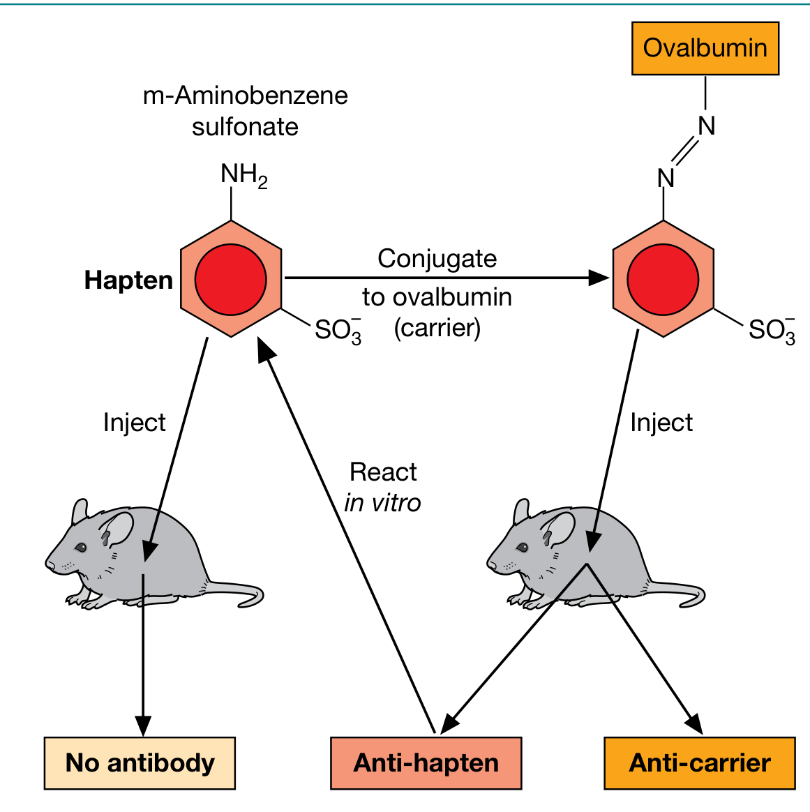
As shown above:
- Injecting a free hapten (e.g., m-aminobenzene sulfonate) alone → no antibody produced
- Injecting hapten conjugated to a carrier protein (e.g., ovalbumin) → produces anti-hapten AND anti-carrier antibodies
- The antibodies produced can then react with the free hapten in vitro
Why Haptens Alone Cannot Induce an Immune Response
The reason lies in the T cell-B cell collaboration required for a full humoral immune response:
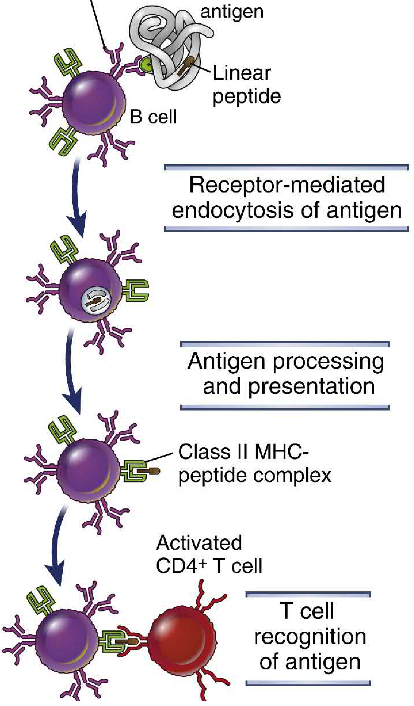
A T cell-dependent B cell response requires two distinct epitopes on the same molecule:
| Role | Recognized by | Epitope type |
|---|---|---|
| Hapten | B cell (via BCR/membrane Ig) | Native conformational epitope |
| Carrier protein | CD4+ T helper cell (via class II MHC) | Linear peptide epitope |
A hapten alone is too small to be processed into peptides and presented on MHC class II - so it cannot activate T helper cells. Without T cell help, B cells cannot undergo full activation, class switching, affinity maturation, or plasma cell differentiation.
Three Key Rules of the Hapten-Carrier Effect (Abbas, Cellular & Molecular Immunology)
- Both hapten-specific B cells AND carrier-specific T helper cells are required - the response is T cell-dependent
- Hapten and carrier must be physically linked - administering them separately cannot induce an anti-hapten response. The hapten is responsible for the efficient internalization of the carrier protein into the B cell
- The interaction is MHC class II-restricted - helper T cells cooperate only with B cells that express the same class II MHC molecules involved in the initial T cell activation by dendritic cells
Mechanism Step-by-Step
- Hapten-carrier conjugate enters the body
- Hapten-specific B cell binds the conjugate via its BCR (recognizing the hapten epitope)
- The B cell endocytoses the entire hapten-carrier conjugate
- The carrier protein is proteolytically processed inside the B cell into linear peptides
- These peptides are loaded onto class II MHC molecules and displayed on the B cell surface
- A carrier-specific CD4+ T cell (Th cell) recognizes the class II MHC-peptide complex
- T cell delivers help via CD40L-CD40 interaction + cytokines (IL-4, IL-21)
- B cell activates, proliferates, undergoes isotype switching, and differentiates into plasma cells secreting anti-hapten antibodies
Clinical Relevance
Drug allergies are the most important clinical application of hapten biology:
| Drug | Mechanism |
|---|---|
| Penicillin | The reactive β-lactam ring covalently binds to carrier proteins, forming a hapten. This is why ~10% of penicillin-allergic patients also react to cephalosporins (cross-reactive haptens) |
| Cephalosporins | Similar β-lactam haptenization |
| Tetracyclines, Tolbutamide | Hapten/drug adsorption mechanism |
Drug-induced immune hemolytic anemia uses the hapten mechanism:
- Drugs like penicillin bind to RBC membranes (haptenize them)
- Anti-drug IgG antibodies then coat the RBCs → complement activation → hemolysis (Type II hypersensitivity)
Application: Conjugate Vaccines
The hapten-carrier concept is the basis for conjugate vaccines against encapsulated bacteria:
- Polysaccharide capsule antigens (equivalent to haptens - recognized by B cells but T cell-independent and weak in infants) are covalently linked to carrier proteins (e.g., tetanus toxoid, CRM197)
- This converts the weak T-independent polysaccharide response into a strong T-dependent response with isotype switching, affinity maturation, and immunologic memory
- Examples: Hib (H. influenzae type b), PCV (pneumococcal), MCV (meningococcal) vaccines
- Cellular and Molecular Immunology (Abbas), 10th Ed., pp. 769-771
- Roitt's Essential Immunology, p. 163
- The Washington Manual of Medical Therapeutics, p. 1215
This is a shared conversation. Sign in to Orris to start your own chat.