
Seizure disorder classify and explain
seizure classification types diagram epilepsy
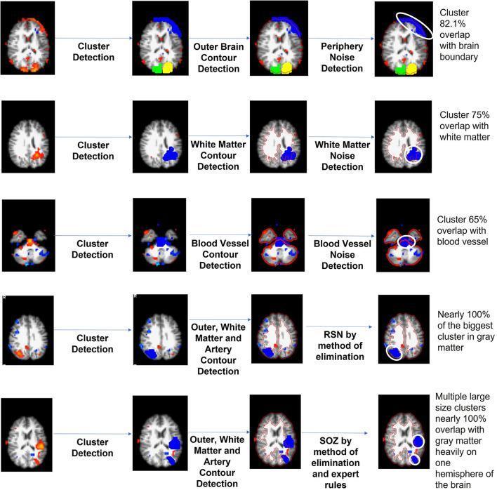
This diagnostic diagram illustrates an unsupervised independent component (IC) sorting mechanism for resting-state functional MRI (rs-fMRI), specifically designed for identifying Seizure Onset Zones (SOZ). The image is organized into a five-row flow-process chart demonstrating the automated classification of voxel clusters. The first three rows depict the 'waterfall' technique for noise purging: periphery noise (82.1% brain boundary overlap), white matter noise (75% white matter overlap), and blood vessel noise (65% vascular overlap). These are identified through sequential steps of cluster detection followed by specific contour detection (outer brain, white matter, or artery). The fourth row demonstrates the identification of Resting State Networks (RSN), characterized by the largest cluster showing nearly 100% overlap with gray matter. The final row identifies the Seizure Onset Zone (SOZ) based on multiple large-size clusters overlapping nearly 100% with gray matter, predominantly localized to one hemisphere. This algorithm aims to reduce manual IC sorting time for neurosurgeons evaluating children with drug-resistant epilepsy.
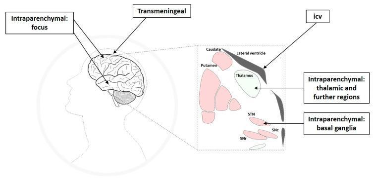
This medical illustration outlines intracranial drug delivery routes for pharmacological seizure control in treatment-resistant epilepsy. The diagram consists of a sagittal profile of a human head and an inset expanded view of subcortical structures. Four primary delivery pathways are identified: (1) Intraparenchymal targeting the cortical seizure focus directly; (2) Transmeningeal, where drugs diffuse through the meninges to reach neocortical areas; (3) Intracerebroventricular (icv), targeting regions adjacent to the lateral ventricle; and (4) Intraparenchymal targeting remote epileptic network structures. The expanded subcortical view highlights key anatomical targets for deep-brain delivery, including the thalamus, caudate, and putamen, as well as specific components of the basal ganglia: the subthalamic nucleus (STN), substantia nigra pars compacta (SNc), and substantia nigra pars reticulata (SNr). The illustration emphasizes the shift from systemic administration to network-specific targeting to modulate seizure propagation and initiation while minimizing functional loss in eloquent brain areas.
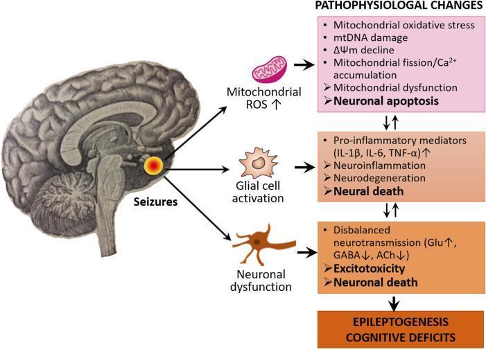
This pathophysiology diagram illustrates the interconnected pathways of epileptogenesis and cognitive deficits following seizure activity in the brain. The visual flow begins with a sagittal brain illustration highlighting a focal point of seizures, which triggers three primary cellular events: mitochondrial ROS increase, glial cell activation (microglia and astrocytes), and neuronal dysfunction. These events lead into three distinct pathophysiological cascades. The first cascade involves mitochondrial oxidative stress, mtDNA damage, membrane potential (ΔΨm) decline, and Ca2+ accumulation, resulting in mitochondrial dysfunction and neuronal apoptosis. The second cascade details neuroinflammation driven by pro-inflammatory mediators (IL-1β, IL-6, TNF-α), leading to neurodegeneration and neural death. The third cascade focuses on disbalanced neurotransmission, characterized by increased glutamate (Glu) and decreased GABA and acetylcholine (ACh), leading to excitotoxicity. These three pathways are shown to be bidirectional and mutually reinforcing, ultimately converging to drive the clinical outcomes of epileptogenesis and cognitive impairment. The diagram is intended for medical education on the molecular and cellular mechanisms of chronic epilepsy.
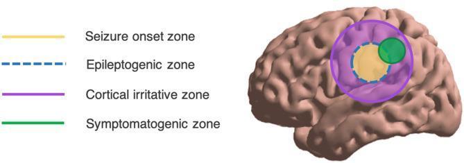
This medical illustration presents an anatomical diagram of the human brain, specifically focusing on the spatial localization and overlap of different zones involved in epilepsy. The diagram uses a lateral view of the cerebral cortex with four distinct color-coded regions to demonstrate the conceptual schema of epileptic phenomena. The Seizure Onset Zone (SOZ) is depicted as a central yellow solid circle, representing the area where clinical seizures originate. Encapsulating the SOZ is the Epileptogenic Zone (EZ), marked by a blue dashed circular line, defined as the minimum area of brain tissue necessary for resection to achieve seizure freedom. A larger purple circle represents the Cortical Irritative Zone, indicating the region generating interictal spikes, which typically encompasses both the EZ and SOZ. Finally, the Symptomatogenic Zone is shown as a green solid circle on the periphery; it partially overlaps with the other zones, representing the cortical area that, when activated, produces the initial clinical symptoms. This graphic illustrates the clinical challenge of surgical localization, where these theoretical zones may not perfectly align.
"seizure" AND "classification" AND "epilepsy"
Seizure Disorder: Classification and Explanation
- Harrison's Principles of Internal Medicine (22e), p. 3452
ILAE 2017 Classification Framework
| Onset | Definition |
|---|---|
| Focal | Originating within networks limited to one hemisphere |
| Generalized | Originating within and rapidly engaging bilaterally distributed networks |
| Unknown | Onset cannot be determined |
Important terminology change: The 2017 classification replaced "partial" with "focal," eliminated "simple partial" and "complex partial" as terms, and renamed secondarily generalized seizures as "focal-to-bilateral tonic-clonic."
I. FOCAL ONSET SEIZURES
A. By Awareness Status
- Awareness (the ability to know the seizure is happening and to respond to the environment) is fully preserved throughout
- Subjective experiences described are called auras
- Types of manifestations:
- Motor: tonic, clonic, or myoclonic movements of a contralateral limb; involuntary movements may spread in a "Jacksonian march" (progressive spread up the limb as seizure activity spreads across motor cortex)
- Sensory: paresthesias, flashing lights, formed visual hallucinations, vertigo
- Autonomic: flushing, sweating, piloerection, GI disturbance
- Psychic/experiential: fear, déjà vu, depersonalization, micropsia/macropsia, acrid odors (temporal lobe origin)
- Any alteration of awareness during any part of the seizure qualifies
- Often feature automatisms - semi-purposeful, repetitive behaviors (lip-smacking, picking at clothes, chewing, walking in circles)
- Most commonly arise from the temporal lobe (mesial temporal lobe epilepsy is the single most common focal epilepsy syndrome)
- Typically last 1-3 minutes, followed by a postictal state (confusion, fatigue, headache)
B. By Motor/Nonmotor Onset
- Motor onset: tonic, clonic, myoclonic, epileptic spasm, hyperkinetic, atonic, automatism
- Nonmotor onset: autonomic, behavior arrest, cognitive, emotional, sensory
C. Focal-to-Bilateral Tonic-Clonic (FBTC)
- Formerly called "secondary generalization" or "secondarily generalized tonic-clonic"
- A focal seizure (often brief, missed, or unwitnessed) that spreads to engage both hemispheres and produce a full convulsion
- Implies a focal brain lesion and carries localizing diagnostic significance
- Todd's paralysis: localized paresis lasting minutes to hours after a focal motor seizure
- Epilepsia partialis continua: a rare, often refractory state where focal motor seizures continue for hours or days
II. GENERALIZED ONSET SEIZURES
A. Generalized Motor Seizures
| Phase | Duration | Features |
|---|---|---|
| Tonic | 10-20 s | Sudden loss of consciousness; rigid extension of back, neck, arms, legs; cry from forced air through closed cords; cyanosis; pupil dilation |
| Clonic | ~30 s | Rhythmic, violent flexor spasms (8/s → 4/s); autonomic surge (tachycardia, HTN, hypersalivation) |
| Postictal | Minutes to hours | Deep coma → confusion → fatigue; Todd's paralysis possible; no memory of event |
- Sustained increase in muscle contraction, stiffening of limbs without clonic phase
- Seen in Lennox-Gastaut syndrome
- Rhythmic jerking movements without a preceding tonic phase
- Sudden, brief, shock-like involuntary muscle jerks
- Can be focal, multifocal, or generalized
- Classic of juvenile myoclonic epilepsy (JME): myoclonic jerks on awakening that may herald a GTC seizure later in the day
- Also seen in Dravet syndrome, progressive myoclonic epilepsies
- Sudden loss of muscle tone causing falls and injury
- Seen in Lennox-Gastaut syndrome
- Previously called "astatic" seizures
- Brief, symmetric flexion or extension of trunk and limbs
- Hallmark of West syndrome (infantile spasms)
- Occur in clusters, typically in the first year of life
B. Generalized Nonmotor Seizures (Absence)
- Abrupt, brief (5-30 s) loss of awareness without falling or convulsing
- Classic EEG: 3 Hz spike-and-wave discharges
- The child "blanks out" mid-sentence and resumes immediately after - no postictal confusion
- Multiple episodes per day are common
- Associated with childhood absence epilepsy; often resolves by adolescence
- More gradual onset/offset, longer duration, more pronounced tone changes
- Associated with epileptic encephalopathies (Lennox-Gastaut syndrome)
- EEG: slow (< 2.5 Hz) spike-and-wave
- Absence episodes accompanied by rhythmic myoclonic jerks (typically shoulders/arms)
III. UNKNOWN ONSET SEIZURES
- Unknown onset motor (tonic-clonic, epileptic spasm)
- Unknown onset nonmotor (behavior arrest)
- Unclassified
Classification by Age of Onset (Epileptic Syndromes)
| Age | Common Syndromes |
|---|---|
| Neonatal | Ohtahara syndrome, early myoclonic encephalopathy, benign neonatal seizures |
| Infancy | West syndrome (infantile spasms), Dravet syndrome, febrile seizures |
| Childhood | Absence epilepsy, benign epilepsy with centrotemporal spikes (BECTS/BECTS/Rolandic), Lennox-Gastaut syndrome, Panayiotopoulos syndrome |
| Adolescence/Adulthood | Juvenile myoclonic epilepsy (JME), juvenile absence epilepsy, temporal lobe epilepsy |
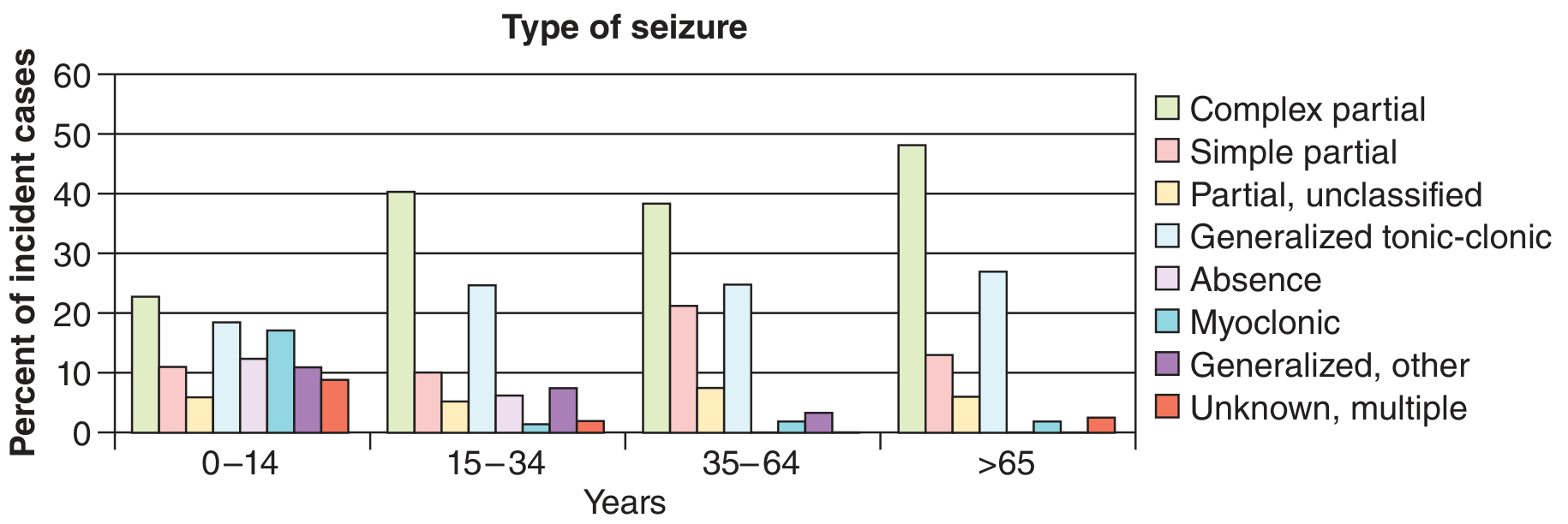
| Elderly | Focal seizures (from strokes, tumors, trauma) dominate |
Key Terminology Comparison: Old vs. New
| Old Term (pre-2017) | New ILAE 2017 Term |
|---|---|
| Simple partial seizure | Focal aware seizure (FAS) |
| Complex partial seizure | Focal impaired awareness seizure (FIAS) |
| Secondarily generalized | Focal-to-bilateral tonic-clonic (FBTC) |
| Grand mal | Generalized tonic-clonic |
| Petit mal | Absence seizure |
| Drop attack | Atonic seizure |
| Infantile spasms | Epileptic spasms / West syndrome |
Etiology-Based Classification (ILAE 2017 Parallel Framework)
| Category | Examples |
|---|---|
| Structural | Post-stroke, hippocampal sclerosis, cortical dysplasia, tumor, trauma |
| Genetic | SCN1A (Dravet), KCNQ2, SCN2A mutations; JME |
| Infectious | Neurocysticercosis, herpes encephalitis, meningitis |
| Metabolic | Hypoglycemia, hyponatremia, hypocalcemia, pyridoxine deficiency |
| Immune | Anti-NMDAR encephalitis, LGI1, CASPR2 antibodies |
| Unknown | No identifiable cause despite workup |
Provoked vs. Unprovoked Seizures
- Provoked (acute symptomatic): occur in the setting of an acute systemic or CNS insult (fever, hypoglycemia, alcohol withdrawal, drug toxicity). A single provoked seizure does NOT define epilepsy.
- Unprovoked: occur without an acute precipitant. Two or more unprovoked seizures separated by >24 hours = epilepsy.