
What is bassini repair and sholdice repair of hernia
- Bassini repair (1887): The classic technique - involves high ligation of the hernia sac and repair by suturing the conjoint tendon to the inguinal ligament in a single layer.
- Shouldice repair (1945): A refined, multi-layer technique using continuous non-absorbable suture to create 4 overlapping layers of the posterior wall - considered the gold standard open non-mesh repair with lower recurrence rates (~1-2%).
Bassini hernia repair inguinal canal surgical technique
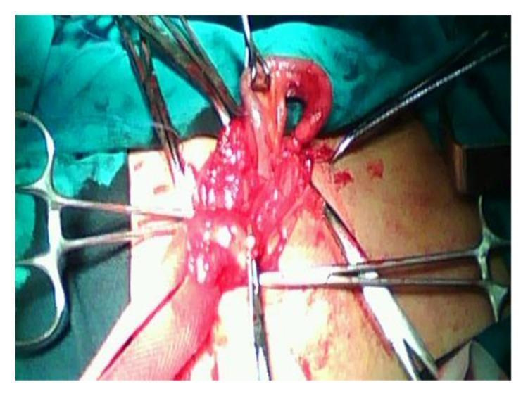
This clinical photograph shows an intraoperative view of an Amyand's hernia during a surgical repair. The image captures the right inguinal region where a hernia sac has been opened. Centrally, the vermiform appendix is clearly visualized as it is being mobilized from the inguinal canal. The appendix appears hyperemic, erythematous, and edematous, consistent with acute inflammation. Surrounding the appendix, part of the cecum and mesoappendix are visible. Multiple surgical instruments, including several hemostatic forceps (Kelly or Crile clamps) and retractors, are arranged radially to provide adequate exposure of the surgical field. The surgical site shows signs of fresh bleeding, typical of intraoperative dissection. This visual demonstrates a rare clinical entity where the appendix is found within an inguinal hernia sac, necessitating both appendectomy and hernia repair (in this case, via the Bassini technique). The image serves as a teaching tool for general surgery and emergency medicine regarding atypical hernia presentations.
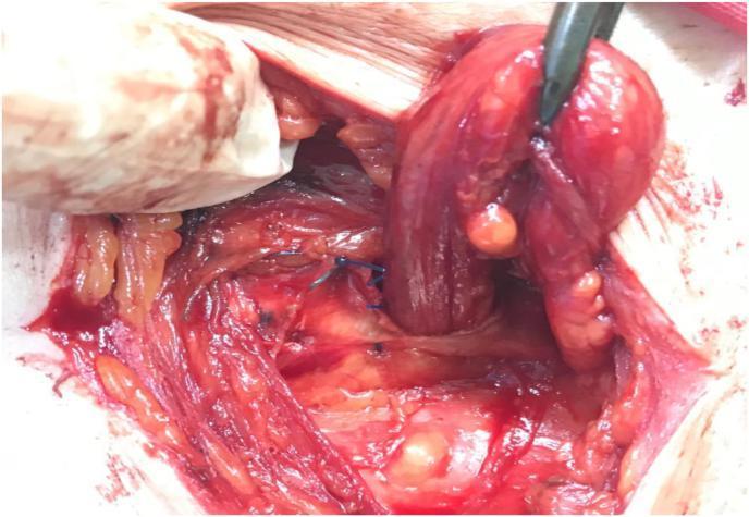
This clinical photograph captures an intraoperative view of an inguinal hernia repair, specifically demonstrating the 'double-breasting' technique of the External Oblique Aponeurosis (EOA). The surgical field shows exposed anatomical structures including the spermatic cord, which is being retracted superiorly and laterally by a surgical instrument (forceps) to reveal the underlying repair. The medial and lateral leaves of the EOA are visible; the lateral leaf is being sutured to overlap the medial leaf, effectively strengthening the posterior wall of the inguinal canal. Several blue, non-absorbable interrupted sutures (likely Prolene) are positioned in the center, approximating the fascial edges. The surrounding tissue includes striated muscle fibers, yellow adipose tissue (fat), and areas of surgical hemorrhage and serosanguinous fluid. This visual demonstrates the repositioning of the spermatic cord to a subcutaneous location as part of the canal's obliteration process to prevent hernia recurrence.
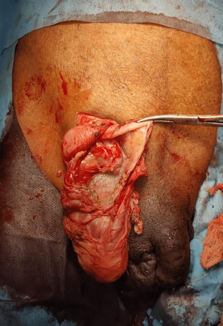
This clinical photograph captures an intraoperative view of an Amyand's hernia repair in a 47-year-old male. The surgical field focuses on the right inguinal region, where an incision has been made to expose the inguinal canal. A surgical hemostat or forceps is seen retracting a thin, membranous hernial sac. Protruding through the inguinal defect and within the sac is a clearly visible, elongated, and hyperemic (reddened) vermiform appendix, indicating inflammation (acute appendicitis). The surrounding tissue shows signs of surgical dissection, including minor hemorrhage and the presence of moist, glistening serosa. Anatomically, the base of the scrotum is visible below the surgical site, and the surrounding skin is prepped for a sterile procedure. This image is of high educational value for demonstrating a rare clinical entity where the appendix is found within an inguinal hernia sac, necessitating both appendectomy and hernia repair (modified Bassini's repair in this case).
Shouldice repair posterior wall inguinal canal four layer suture technique
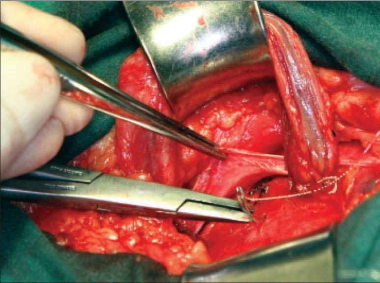
This clinical photograph shows an intraoperative view of an inguinal hernia repair, specifically demonstrating the third layer of a multi-layered anatomical reconstruction. The surgical field is exposed using a metal retractor, revealing several anatomical layers including the internal oblique muscle, the transversus abdominis, and the inguinal ligament. A pair of surgical forceps is seen elevating a thin, fibrous tissue layer—the external oblique aponeurosis—to facilitate suturing. A needle holder is actively positioning a curved needle with white non-absorbable suture to imbricate the tissues. The spermatic cord is visible and retracted to the side to ensure its protection during the posterior wall reinforcement. The procedure illustrates the technique of anchoring sutures from the internal oblique fascia to the shelving edge of the inguinal ligament, aiming to strengthen the inguinal canal's floor. The tissues appear healthy with normal vascularity, and the surgical focus is on re-establishing anatomical integrity to prevent hernia recurrence.
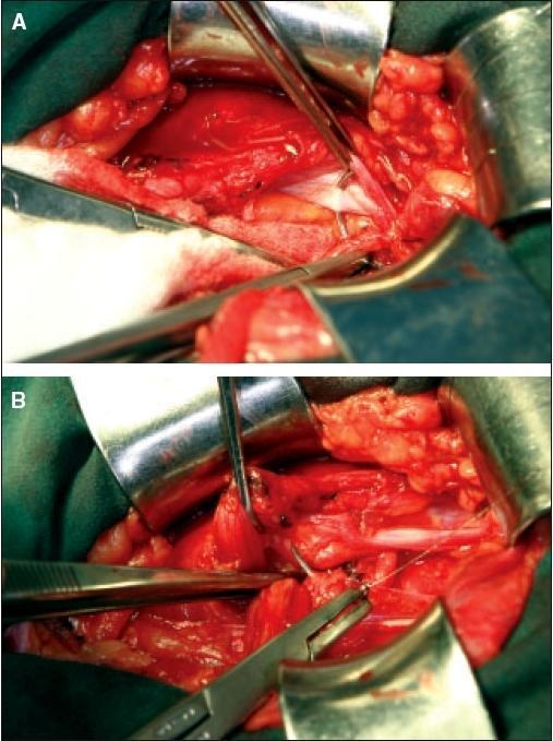
Clinical intraoperative photographs depicting the first layer of posterior wall reconstruction during an inguinal hernia repair. Panel A shows the initiation of the repair near the pubic tubercle, where the lateral flap of the transversalis fascia is being sutured to the lateral edge of the rectus sheath. A needle holder and surgical clamps are visible, demonstrating the technique of reaching underneath the superior-medial flap to anchor the tissues. Panel B illustrates the progression of the reconstruction toward the lateral aspect, redefining the internal inguinal ring. The image demonstrates the integration of the superior stump of the divided lateral cremasteric muscle flap into the suture line. This maneuver is used to buttress the medial edge of the new internal ring, with the suture eventually including a full-thickness bite of the internal oblique muscle and the transversus abdominis aponeurosis. These images highlight the anatomical layers involved in a multi-layered tissue-based hernia repair, specifically focusing on the imbrication of the transversalis fascia and stabilization of the internal ring.
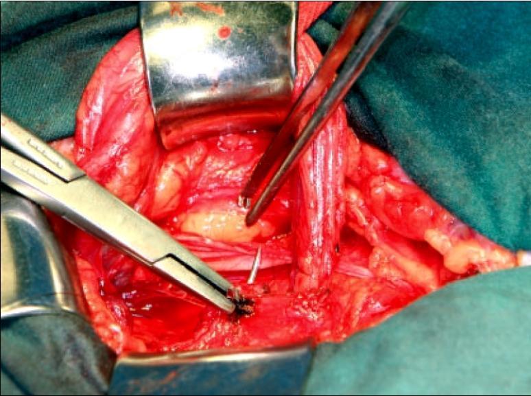
This clinical photograph captures an intraoperative view of an inguinal hernia repair, specifically demonstrating the second layer of a tissue-based reconstruction. The surgical field is exposed using metal retractors, revealing layered anatomical structures including the transversalis fascia and the inguinal ligament. A needle holder is seen in the foreground, manipulating a dark-colored suture to approximate the medial flap of the transversalis fascia to the shelving portion of the inguinal ligament. A second surgical instrument (tissue forceps) provides counter-traction to facilitate needle placement. The procedure illustrates an overlapping or imbrication technique, moving from the internal ring back toward the pubic tubercle to reinforce the posterior wall of the inguinal canal. The visual focus is on the meticulous placement of sutures to create a tension-free, multi-layered anatomical repair, emphasizing the relationship between the deep inguinal ring and the floor of the canal.
Bassini Repair and Shouldice Repair of Inguinal Hernia
1. Bassini Repair (1884/1890)
History
Steps of the Operation
- Opening the inguinal canal: The anterior wall (external oblique aponeurosis) is incised, entering the inguinal canal.
- Spermatic cord dissection: The cord is freed and retracted; lateral (indirect) vs. medial (direct) hernia is confirmed.
- Hernia sac management:
- Indirect sac: Separated from the cord, opened, contents reduced, sac suture-ligated at the neck, and excess sac excised.
- Direct sac: Inverted and transversalis fascia plicated.
- Posterior wall reconstruction (the key step): Interrupted sutures are placed between the conjoint tendon (internal oblique + transversus abdominis) superiorly and the inguinal (Poupart's) ligament inferiorly - running from the pubic tubercle laterally to the deep inguinal ring. This is a single-layer repair.
- The external oblique aponeurosis is closed over the cord.
Key Principle
Limitations
- Over 150 modifications have been described (showing its imperfections)
- Tension on suture line - a major cause of recurrence
- Recurrence rate: ~10% in general series
2. Shouldice Repair (1945)
History
Steps of the Operation
- The transversalis fascia is incised along the entire inguinal floor from the internal ring to the pubic tubercle, and cleaned posteriorly.
- The free lower flap is sutured in a continuous, imbricated (overlapping) fashion behind the upper flap - running from the pubic tubercle up to and through the internal ring, tightening the internal ring around the cord.
- The same suture is continued (not tied) in a running return pass from the internal ring back to the pubic tubercle - upper transversalis flap sutured to the base of the lower edge and the inguinal ligament. Tied at the pubic tubercle.
- A new suture begins at the tightened internal ring, bringing together the conjoint tendon (internal oblique + transversus abdominis aponeurosis) medially with the inguinal ligament laterally - run down to the pubic tubercle.
- The suture continues back to the internal ring, uniting the anterior rectus sheath medially with the posterior aspect of the external oblique aponeurosis laterally.
- The cord is replaced on the new inguinal floor.
- The external oblique aponeurosis is closed in 1-2 additional continuous layers.
The original Shouldice technique used continuous stainless steel wire for all four layers; modern surgeons use permanent synthetic suture (polypropylene/nylon).
Key Principle
Comparison Table
| Feature | Bassini Repair | Shouldice Repair |
|---|---|---|
| Year introduced | 1884/1890 | 1945 |
| Layers of repair | Single layer | 4 layers (continuous) |
| Suture type | Interrupted | Continuous (non-absorbable) |
| Transversalis fascia | Not formally opened | Opened and reconstructed |
| Tension distribution | Concentrated | Dispersed across canal |
| Recurrence rate (general) | ~10% | ~1-2% at expert centres |
| Recurrence vs mesh | Higher | Still higher (OR 3.8) |
| Technical difficulty | Moderate | High (steep learning curve) |
| Best setting | Resource-limited, mesh unavailable | High-volume specialist centres |
| Anesthesia | General/spinal | Can use local anesthesia |
Outcomes
- At Shouldice Hospital, recurrence rates are <1% in selected patients over 30 years - unmatched by any other pure tissue technique.
- A Cochrane meta-analysis (16 RCTs, 2566 hernias) showed Shouldice is significantly better than other non-mesh repairs (OR 0.62) but significantly worse than mesh repair (OR 3.8 recurrence).
- Importantly, recurrence with Shouldice depends heavily on surgical expertise: 9.4% (beginners) vs 2.5% (experienced) in one report.
- Shouldice is the repair of choice when mesh cannot be used (e.g., contaminated field, young patients, mesh allergy).
Intraoperative Views