
IHC markers
Asking for Preferences
IHC markers is a broad topic - what would you like to focus on?
Reading File
Finding Sources
Reading File
Immunohistochemistry (IHC) Markers — Overview
IHC uses antibodies against specific cellular antigens to stain tissue sections, helping classify tumors and other tissues that look ambiguous on routine H&E staining. Here's a breakdown by major category (Robbins Pathologic Basis of Disease, p. 349-353):
1. Lineage/Category Markers (for undifferentiated tumors)
When a tumor is too poorly differentiated to classify by morphology alone, panels distinguish carcinoma vs. lymphoma vs. melanoma vs. sarcoma:
| Marker | Lineage indicated |
|---|---|
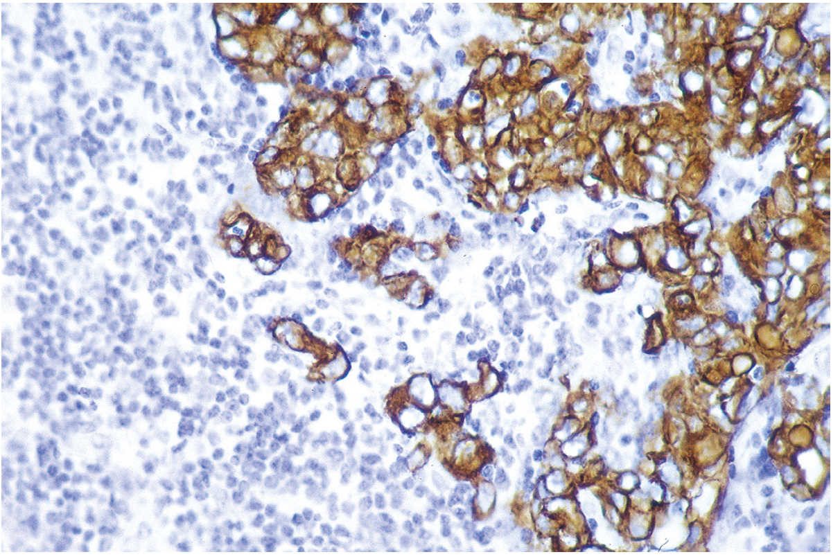
| Cytokeratins (pan-CK, CK7, CK20, AE1/AE3) | Epithelial (carcinoma) |
| Vimentin | Mesenchymal (sarcoma) |
| Desmin | Muscle origin |
| CD45 (LCA) | Hematopoietic/lymphoid neoplasms |
| SOX10, S100, Melan-A, HMB-45 | Melanoma |
2. Site-of-Origin Markers (for metastases of unknown primary)
Used when a metastasis is found but the primary tumor isn't obvious:
- PSA - prostate carcinoma
- Thyroglobulin, TTF-1 - thyroid (TTF-1 also positive in lung adenocarcinoma)
- CDX2 - colorectal
- GATA3, mammaglobin, GCDFP-15 - breast
- CK7/CK20 patterns - help narrow GI vs. lung vs. urothelial vs. ovarian origin
3. Lymphoid/Hematopoietic Panel
- CD45 - pan-leukocyte
- CD20, CD79a, PAX5 - B-cells
- CD3, CD5 - T-cells
- CD15, CD30 - Hodgkin (Reed-Sternberg cells)
- CD34, MPO - myeloid/blast lineage
4. Prognostic/Therapeutic (Predictive) Markers
These directly guide treatment decisions:
- ER/PR (estrogen/progesterone receptors) - breast cancer; positivity predicts response to antiestrogen therapy and confers better prognosis
- HER2 (ERBB2 product) - breast (and gastric) cancer; strong staining identifies candidates for anti-HER2 antibody therapy (often confirmed with FISH for gene amplification)
- ALK - identifies ALK-fusion-positive lung cancers and lymphomas, indicating eligibility for ALK inhibitors
- PD-L1 - predicts response to checkpoint inhibitor immunotherapy
- Ki-67 - proliferation index, prognostic across many tumor types
5. Neuroendocrine Markers
- Synaptophysin, chromogranin, CD56 - identify neuroendocrine tumors (carcinoids, small cell carcinoma, pancreatic NETs)
Example: Anti-cytokeratin stain confirming epithelial origin
This is a broad topic - IHC panels are typically tailored to a specific clinical question (e.g., "carcinoma of unknown primary," "lymphoma subtyping," "spindle cell tumor workup," "breast cancer receptor status"). If you have a specific tumor type or diagnostic scenario in mind, I can give you the exact panel and interpretation used for that workup.
This is a shared conversation. Sign in to Orris to start your own chat.