
Gene therapy for mbbs 1st year prof
gene therapy vectors viral delivery mechanism diagram
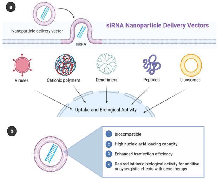
Educational diagram illustrating delivery systems for gene therapy and nucleic acid delivery. Panel (a) titled 'siRNA Nanoparticle Delivery Vectors' shows the mechanism of a nanoparticle vector entering a cell membrane to deliver siRNA. Below this, it categorizes five major types of delivery vectors: viruses, cationic polymers, branched dendrimers, cell-penetrating peptides, and cationic liposomes, all shown converging on a cell for 'Uptake and Biological Activity.' Panel (b) outlines the four essential characteristics of an engineered synthetic vector: 1) Biocompatibility, 2) High nucleic acid loading capacity, 3) Enhanced transfection efficiency, and 4) Desired intrinsic biological activity for additive or synergistic effects. This illustration serves as a pharmacological guide for understanding nanopharmaceutical drug delivery systems in molecular medicine and oncology.
![This medical schematic illustrates a non-viral transposon-mediated gene therapy system, detailing the mechanisms of delivery, excision, and chromosomal integration. The diagram identifies two primary vector configurations: 'trans' delivery, involving two separate plasmids (one for the therapeutic gene of interest [GOI] and another for the transposase enzyme), and 'cis' delivery, where both components are housed on a single plasmid. Key molecular components include the GOI (green arrow), an upstream promoter (orange box), and flanking Inverted Terminal Repeats (ITRs, red circles). The educational focus highlights the three-step 'cut and paste' mechanism: (1) Nonviral delivery of the DNA vector into the target cell nucleus; (2) Excision of the transposon cassette by the transposase enzyme (blue) at the ITR recognition sites; and (3) Site-specific chromosomal integration into the host's genomic DNA, specifically targeting TTAA-rich sites. This process demonstrates a method for stable transgene insertion in human regenerative medicine and oncology applications, emphasizing a bi-component DNA vector system as an alternative to viral-mediated delivery.](https://cdn.orris.care/cdss_images/pmc_clinical_VQA_4fe1b4ac5b86941a71c26ada6e54290a1cb05f89b524d5e00b55589513b11f27.jpg)
This medical schematic illustrates a non-viral transposon-mediated gene therapy system, detailing the mechanisms of delivery, excision, and chromosomal integration. The diagram identifies two primary vector configurations: 'trans' delivery, involving two separate plasmids (one for the therapeutic gene of interest [GOI] and another for the transposase enzyme), and 'cis' delivery, where both components are housed on a single plasmid. Key molecular components include the GOI (green arrow), an upstream promoter (orange box), and flanking Inverted Terminal Repeats (ITRs, red circles). The educational focus highlights the three-step 'cut and paste' mechanism: (1) Nonviral delivery of the DNA vector into the target cell nucleus; (2) Excision of the transposon cassette by the transposase enzyme (blue) at the ITR recognition sites; and (3) Site-specific chromosomal integration into the host's genomic DNA, specifically targeting TTAA-rich sites. This process demonstrates a method for stable transgene insertion in human regenerative medicine and oncology applications, emphasizing a bi-component DNA vector system as an alternative to viral-mediated delivery.
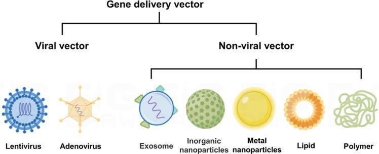
A medical infographic illustrating common gene delivery vectors for nucleic acid therapeutics. The classification is presented as a hierarchical flowchart, branching into two primary categories: Viral vectors and Non-viral vectors. The viral vector category includes two subtypes: Lentivirus, depicted as a blue spherical particle with external glycoprotein spikes and an internal helical genetic core; and Adenovirus, shown as a yellow icosahedral capsid with protruding fibers. The non-viral vector category displays five delivery systems: Exosomes (biological vesicles with membrane-bound proteins), Inorganic nanoparticles (represented as a green textured sphere), Metal nanoparticles (depicted as a smooth yellow gold-like sphere), Lipids (shown as an orange micellar ring), and Polymers (represented as a tangled light green molecular chain). This educational diagram summarizes the diversity of vehicle designs used to deliver pDNA, mRNA, siRNA, or miRNA to target cells, such as macrophages, highlighting the morphological differences between complex biological viruses and synthetic or cellular-derived delivery platforms.
![This medical infographic illustrates the mechanisms of ocular gene therapy delivery and intracellular processing. The top left features an anatomical diagram of the eye showing two primary administration routes: Intravitreal injection (into the vitreous humor) and Subretinal injection (between the retina and retinal pigment epithelium [RPE]). A cross-sectional inset details the diffusion of viral vectors through ocular barriers including the vitreous, retina, RPE, and choroid. The right and bottom panels depict a five-step cellular pathway for adenovirus-mediated delivery: 1) Diffusion through ocular barriers; 2) Evasion of immune responses, specifically non-specific phagocytosis by macrophages and degradation by nucleases; 3) Binding of the vector to target cell receptors; 4) Endosomal escape following internalization; and 5) Disassembly and cargo release within the cytoplasm. The diagram concludes by showing the fate of genetic material, where mRNA undergoes translation in the cytoplasm to produce desired proteins, while pDNA and RNP (ribonucleoprotein) complexes enter the nucleus for transcription and genome editing. This visual serves as a comprehensive educational resource for understanding ophthalmic gene therapy and molecular signaling pathways.](https://cdn.orris.care/cdss_images/pmc_clinical_VQA_7a396b23be942c1b8c81c0b00e86661a203159f14cfecd8a7c52bab410d1e8cf.jpg)
This medical infographic illustrates the mechanisms of ocular gene therapy delivery and intracellular processing. The top left features an anatomical diagram of the eye showing two primary administration routes: Intravitreal injection (into the vitreous humor) and Subretinal injection (between the retina and retinal pigment epithelium [RPE]). A cross-sectional inset details the diffusion of viral vectors through ocular barriers including the vitreous, retina, RPE, and choroid. The right and bottom panels depict a five-step cellular pathway for adenovirus-mediated delivery: 1) Diffusion through ocular barriers; 2) Evasion of immune responses, specifically non-specific phagocytosis by macrophages and degradation by nucleases; 3) Binding of the vector to target cell receptors; 4) Endosomal escape following internalization; and 5) Disassembly and cargo release within the cytoplasm. The diagram concludes by showing the fate of genetic material, where mRNA undergoes translation in the cytoplasm to produce desired proteins, while pDNA and RNP (ribonucleoprotein) complexes enter the nucleus for transcription and genome editing. This visual serves as a comprehensive educational resource for understanding ophthalmic gene therapy and molecular signaling pathways.
CRISPR Cas9 gene editing mechanism somatic cells
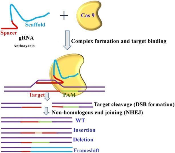
A pathophysiology diagram illustrating the CRISPR-Cas9 gene editing mechanism, specifically targeting anthocyanin pathways. The process begins with complex formation, where a guide RNA (gRNA)—comprised of a blue 'Scaffold' and a red 'Spacer' sequence—assembles with a yellow bean-shaped Cas9 endonuclease. The second stage demonstrates target binding, showing the Cas9-gRNA complex recognizing a double-stranded DNA target at the PAM (Protospacer Adjacent Motif) site. The next step illustrates 'Target cleavage' resulting in double-strand break (DSB) formation. The final section depicts the cellular repair mechanism 'Non-homologous end joining (NHEJ)' and its potential mutagenic outcomes: Wild Type (WT), Insertion (addition of nucleotides), Deletion (loss of DNA segments), and Frameshift mutations (disruption of the reading frame). The diagram serves as an educational summary of molecular biology techniques for precision genetic modification.
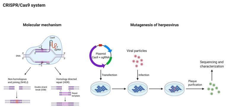
Educational diagram illustrating the CRISPR/Cas9 system and its application in herpesvirus mutagenesis. The left panel details the 'Molecular mechanism,' showing the Cas9 endonuclease guided by single-guide RNA (sgRNA) to a specific DNA target sequence upstream of a Protospacer Adjacent Motif (PAM). It depicts the creation of a double-strand break (DSB) and the two primary repair pathways: Non-homologous end joining (NHEJ), which results in random insertions/deletions, and Homology directed repair (HDR), which utilizes a repair template for precise genetic modification. The right panel outlines the workflow for 'Mutagenesis of herpesvirus.' The process begins with the transfection of a plasmid encoding Cas9 and sgRNA into host cells, followed by infection with viral particles. Within the cell, CRISPR-mediated viral DNA editing occurs. Subsequent steps include plaque purification to isolate recombinant viral clones and final sequencing and characterization to verify the mutation. This infographic is designed for intermediate to advanced molecular biology education, focusing on gene editing techniques in virology.
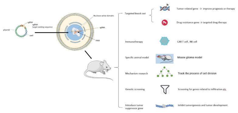
A medical infographic and pathophysiology diagram illustrating the application of the CRISPR/Cas9 gene-editing system in glioma research. The left side depicts the molecular mechanism: a plasmid containing sequences for sgRNA (composed of crRNA and tracrRNA) and Cas9 protein is introduced into a target cell. Within the nucleus, the sgRNA guides the Cas9 nuclease to a specific genomic target DNA site adjacent to a Protospacer Adjacent Motif (PAM), where the nuclease active domains create a double-strand break. The right side outlines six clinical and research applications of this technology in neuro-oncology: 1) Targeted knockout of tumor-related and drug-resistance genes to improve prognosis and therapy; 2) Immunotherapy, focusing on CAR T-cell and NK cell development; 3) Creation of specific animal models like mouse glioma models; 4) Mechanism research, such as tracking cell division; 5) Genetic screening for genes related to tumor infiltration; and 6) Introduction of tumor suppressor genes to inhibit tumorigenesis. The diagram effectively bridges basic molecular biology with translational oncology research for glioma.
Gene Therapy - MBBS 1st Year
1. Definition
- Emery's Elements of Medical Genetics and Genomics defines it as "the therapeutic delivery of nucleic acid polymers into a patient's cells as a drug to treat disease."
- Thompson & Thompson Genetics and Genomics in Medicine (9th ed.) - It is "the introduction of a biologically active gene into a cell to achieve a therapeutic benefit."
2. Types of Gene Therapy
A. Based on Target Cells
| Type | Target | Heritable? | Status |
|---|---|---|---|
| Somatic gene therapy | Non-reproductive (somatic) cells only | No - not passed to offspring | Permitted; in clinical use |
| Germline gene therapy | Germ cells (egg/sperm/zygote) | Yes - passed to all future generations | Worldwide moratorium in effect |
Somatic therapy changes only the targeted somatic cells, so the change is NOT passed to the next generation. Germline gene therapy modifies germ cells, so the change IS heritable - this raises profound ethical concerns and is currently banned.
B. Based on Route of Gene Delivery
| Type | Description | Example |
|---|---|---|
| Ex vivo | Cells removed from patient → genetically modified in lab → returned to patient | ADA-SCID: T lymphocytes removed, transduced with retroviral vector, reinfused |
| In vivo | Therapeutic gene delivered directly into patient's cells/tissues inside the body | CFTR gene therapy via nasal spray with liposomes or adenovirus for cystic fibrosis |
3. Vectors Used in Gene Therapy
Viral Vectors (most efficient)
| Vector | Key Features | Best For |
|---|---|---|
| Retrovirus | Integrates into host genome; only infects dividing cells | Hematopoietic stem cells (SCID, beta-thalassemia) |
| Lentivirus | Integrates into non-dividing cells too; derived from HIV | CNS (Parkinson), bone marrow |
| Adeno-associated virus (AAV) | Non-integrating (forms episome); low immunogenicity; long-lasting expression in post-mitotic cells | Muscle (lipoprotein lipase deficiency), retina (blindness), hemophilia |
| Adenovirus | High efficiency, large payload; does NOT integrate; causes immune response; expression is transient | Cancer gene therapy, respiratory (CF) |
Non-Viral Vectors (safer, less efficient)
- Liposomes - lipid-based nanoparticles; used in CFTR delivery (nasal spray for CF)
- Naked DNA / plasmids - direct injection into muscle; inefficient but safe
- Nanoparticles - polymers, dendrimers; used for siRNA delivery
4. Prerequisites for Gene Therapy (Technical Aspects)
- The genetic basis and pathophysiology of the disorder must be known
- The specific cells/tissue/organ affected must be accessible
- The method of gene introduction must be efficient AND safe
- Evidence from animal models that the inserted gene functions with proper regulatory/promoter/enhancer sequences
- The treated cell population must have a reasonable lifespan with continued gene expression
- The body must not react adversely to the gene product (no antibody production)
- Introduction of the gene must have no deleterious effects (no insertional mutagenesis or malignancy)
5. Mechanisms of Gene Therapy
a. Gene Addition / Transfer
b. Gene Editing (CRISPR-Cas9 and others)
- Allows a mutated gene to be repaired rather than just supplemented
- Combines DNA-binding molecules (guide RNA) + endonucleases (Cas9 protein) to identify and cleave mutated DNA sequences
- Cleavage activates homologous recombination repair, integrating the correct sequence
- CRISPR-Cas9 technology is currently in clinical trials for sickle cell anemia (modifying globin gene expression in hematopoietic stem cells)
c. Gene Silencing / Antisense Techniques
- RNA interference (RNAi), antisense oligonucleotides
- Used to suppress overactive/dominant genes
- Useful in cancer and neurodegenerative diseases
6. Diseases Amenable to Gene Therapy
- Single gene (monogenic) defect
- Gene and protein product well characterized
- Target tissue accessible
- No good conventional treatment
| Category | Specific Disorders |
|---|---|
| Immune deficiency | ADA deficiency (SCID), X-linked SCID, Chronic granulomatous disease |
| Hemophilia | Factor VIII deficiency (Hemophilia A), Factor IX deficiency (Hemophilia B) |
| Hemoglobinopathies | Sickle cell anemia, Beta-thalassemia |
| Lysosomal storage disorders | Gaucher disease, Mucopolysaccharidosis VII |
| Metabolic | Familial hypercholesterolemia (LDL receptor defect), Lipoprotein lipase deficiency |
| Respiratory | Cystic fibrosis (CFTR mutation) |
| Eye | Leber congenital amaurosis, Retinitis pigmentosa |
| Neurological | Parkinson disease (dopamine gene delivery), ALS |
| Cancer | Many solid tumors (p53, oncolytic viruses) |
| Emphysema | Alpha-1 antitrypsin deficiency |
7. Landmark Events in Gene Therapy
| Year | Event |
|---|---|
| 1990 | First human gene therapy trial - a 4-year-old girl with ADA deficiency (SCID). T lymphocytes corrected ex vivo with retroviral vector carrying ADA gene. |
| 2003 | China approved first gene therapy product for head and neck squamous cell carcinoma |
| 2012 | European Medicines Agency approved Alipogene tiparvovec (Glybera) for lipoprotein lipase deficiency - first gene therapy approval in Europe/US; AAV vector injected intramuscularly |
| 2017+ | Multiple approvals for spinal muscular atrophy, hemophilia, inherited retinal dystrophy (Luxturna), beta-thalassemia |
8. Target Organs and Approaches
| Organ | Approach | Example |
|---|---|---|
| Bone marrow | Ex vivo - stem cells corrected, reinfused | ADA-SCID (cured), Beta-thalassemia |
| Liver | Cells removed by partial hepatectomy → corrected in vitro → reinjected via portal vein | Familial hypercholesterolemia, Hemophilia |
| Muscle | Direct injection of DNA; AAV vectors | Lipoprotein lipase deficiency (Glybera) |
| CNS | Lentiviral/AAV vectors (can infect non-dividing neurons) | Parkinson disease (dopamine genes) |
| Lung | In vivo via liposomes or adenovirus (nasal spray) | Cystic fibrosis (CFTR) |
| Retina | Subretinal injection of AAV vector | Leber congenital amaurosis |
9. Modified mRNA (modRNA) - Emerging Approach
- Naturally occurring uridine in mRNA replaced by pseudouridine
- This limits recognition by RNase and innate immune system (Toll-like receptors 7/8), reducing cytokine toxicity
- Result: enhanced translation, non-immunogenic, does not integrate into host genome
- Being explored for heart failure (cardiac regeneration)
10. Challenges and Limitations
| Challenge | Details |
|---|---|
| Vector development | Achieving efficient delivery without immunogenicity |
| Immune response | Host may mount antibody response against viral vectors (especially adenovirus capsid) |
| Insertional mutagenesis | Retroviral vectors may insert near oncogenes - e.g., in XL-SCID trials, retrovirus inserted into LMO-2 oncogene (chromosome 11), causing leukemia in some children |
| Short-lived expression | Gene product expression may wane over time |
| Target accessibility | Some tissues (e.g., brain) difficult to reach |
| Cost | Extremely expensive (Zolgensma for SMA costs >$2 million/dose) |
| Ethical concerns | Germline editing, enhancement vs. therapy, equitable access |
11. Ethical Issues
- Somatic vs. germline therapy - major ethical divide; germline therapy is currently banned worldwide
- Enhancement vs. treatment - could be misused to enhance height, intelligence, athletic ability
- Informed consent - especially for pediatric patients
- Equity of access - treatments are very expensive
- Safety - 1999: death of Jesse Gelsinger in ornithine transcarbamylase deficiency trial highlighted risks
12. Regulatory Aspects
- UK Gene Therapy Advisory Committee (GTAC) oversees gene therapy trials
- All trials require ethics committee approval
- ~3,000 clinical trials have been approved globally to date
- Serious adverse events are rare but the 1999 patient death and leukemia cases in XL-SCID trials led to tighter international regulation
Quick Summary Table for Exams
| Feature | Details |
|---|---|
| Definition | Deliberate introduction of genetic material into somatic cells for therapeutic purposes |
| Types | Somatic (allowed) vs. Germline (banned) |
| Gene delivery | Ex vivo vs. In vivo |
| Best vectors | AAV (safest), Retrovirus (integrating), Lentivirus (non-dividing cells), Adenovirus (transient) |
| First trial | ADA deficiency (SCID), 1990 |
| First approved product | Alipogene tiparvovec (Glybera), 2012 |
| Gene editing tool | CRISPR-Cas9 |
| Key risk | Insertional mutagenesis → leukemia (XL-SCID trials) |
| Germline therapy | Worldwide moratorium - NOT permitted |
- Lippincott Illustrated Reviews: Biochemistry, 8th ed. - Chapter 34 (Biotechnology & Gene Therapy), p. 1368-1369
- Emery's Elements of Medical Genetics and Genomics - Chapter 15 (Gene Therapy), pp. 229-231
- Thompson & Thompson Genetics and Genomics in Medicine, 9th ed. - Chapter 14 (Gene Therapy), p. 328