
Miscarriage
"spontaneous abortion" OR "miscarriage" management
miscarriage spontaneous abortion types ultrasound
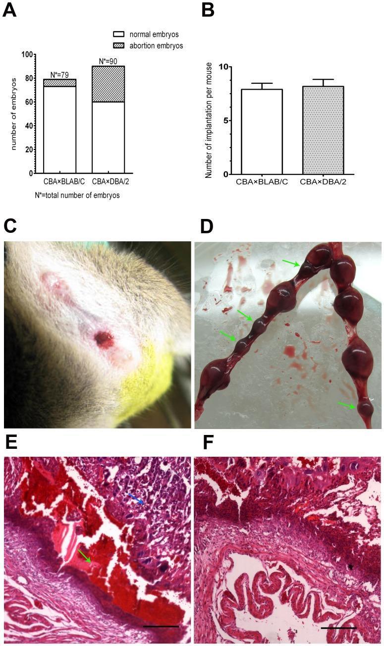
This composite image illustrates clinical and pathological findings in a murine model of spontaneous abortion, used as a surrogate for studying human miscarriage. Panel A displays a bar graph comparing embryo outcomes between two groups, showing a significantly higher abortion rate in the CBA!!!DBA/2 group. Panel B confirms comparable implantation rates between experimental and control groups. Panel C is a clinical photograph of a mouse exhibiting vaginal bleeding, a sign of threatened or inevitable abortion. Panel D shows a gross anatomical view of uterine horns; green arrows highlight abortion-prone embryos, which appear as dark, reddish-brown, dysplastic bulges with no clear distinction between fetus and placenta. Panels E and F provide light microscopy images (H&E stain) of uterine cross-sections. Panel E demonstrates significant pathology at an abortion-prone site, including extensive hemorrhage (green arrows) and necrotic tissue (blue arrow). In contrast, Panel F shows healthy uterine tissue with organized architecture and minimal vascular disruption. The educational focus is on the pathophysiology of pregnancy loss, specifically ischemia, hemorrhage, and necrosis at the implantation site.
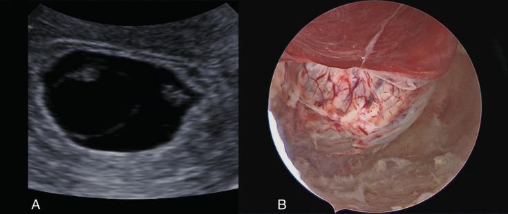
This composite clinical figure demonstrates a missed miscarriage through two distinct imaging modalities: transvaginal ultrasound and operative hysteroscopy. Panel A shows a transvaginal ultrasound image of a gestational sac (GS) appearing as a large, anechoic (black) fluid-filled structure with a prominent hyperechoic (white) decidual rim. Within the sac, irregular fetal poles or embryonic echoes are visible without cardiac activity, consistent with a missed abortion. Panel B displays a hysteroscopic view of the same gestational sac within the uterine cavity. The sac presents as a reddish, translucent, vascularized membrane with visible branching blood vessels. The surrounding endometrium is pinkish-tan, and thin, whitish fibrinous strands or tissue remnants are seen associated with the surface of the sac. This clinical photograph illustrates the preoperative findings of a monochorionic pregnancy failure, highlighting the correlation between radiological and direct visual anatomy in obstetric pathology.
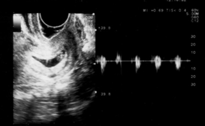
**Imaging Modality:** Transvaginal Ultrasound (TVUS) with Spectral Doppler. **Anatomical Region:** Female pelvis, specifically the cervix and endocervical canal. **Observed Pathology:** Cervical ectopic pregnancy. The image demonstrates a well-defined gestational sac containing a visible embryo implanted within the cervical stroma, below the level of the internal os. **Characteristic Visual Features:** * **Gestational Sac:** An anechoic, oval-shaped structure is situated within the cervical canal, separate from the uterine cavity. * **Embryonic Findings:** A fetal pole is visible within the sac, consistent with approximately seven weeks of gestation. * **Cardiac Activity:** The right panel displays a Spectral Doppler waveform (M-mode/Pulse Wave) confirming rhythmic embryonic cardiac activity, indicating a viable ectopic pregnancy. * **Uterine Landmark:** The empty uterine cavity (not fully visualized but implied by the low anatomical positioning) helps differentiate this from an intrauterine pregnancy. **Key Diagnostic Features:** The "hourglass" appearance of the uterus and the presence of a viable embryo within the cervix are pathognomonic for a cervical ectopic pregnancy, distinguishing it from an ongoing spontaneous abortion (miscarriage in progress) by the presence of peritrophoblastic flow and a fixed position within the cervical wall.
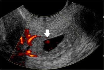
Transvaginal ultrasound (TVUS) with color Doppler demonstrating a cervical ectopic pregnancy. The image shows a well-defined, fluid-filled gestational sac located within the distended cervical canal, below the level of the internal os. Color Doppler imaging reveals prominent circumferential peripheral flow (the 'ring of fire' sign), indicating peritrophoblastic vascularization. A white arrow points to an internal focal Doppler signal within the gestational sac, representing blood flow associated with the developing embryo. This finding is critical for distinguishing a live cervical ectopic pregnancy from a spontaneous abortion in progress (miscarriage), where the gestational sac might be found in a similar cervical position but would lack active peritrophoblastic and internal embryonic vascularity.
Miscarriage (Spontaneous Abortion)
Definition
Epidemiology
- Occurs in 10-15% of clinically recognized pregnancies
- Using sensitive β-hCG assays, an additional 20% of early pregnancies terminate spontaneously before clinical recognition, meaning total loss rate approaches 20-40% of all conceptions
- 80% of miscarriages occur in the first trimester; 75% before 8 weeks of gestation
- 25% of pregnant patients experience some bleeding; up to 50% of those with first-trimester bleeding will miscarry
- Risk of miscarriage is fivefold higher in women >40 years vs. those aged 25-29
- Once fetal cardiac activity is confirmed on ultrasound, the risk of subsequent miscarriage drops to 3-6% - Rosen's Emergency Medicine, p. 3347
Types / Classification
| Type | Definition |
|---|---|
| Threatened abortion | Vaginal bleeding with closed internal cervical os; risk of miscarriage ~35-50% |
| Inevitable abortion | Vaginal bleeding with dilated (open) cervical os |
| Incomplete abortion | Products of conception at the cervical os or in vaginal canal; passage of only some tissue |
| Complete abortion | All fetal and placental material expelled; cervix closed, uterus contracted |
| Missed abortion | Fetal death <20 weeks without passage of fetal tissue for ≥4 weeks after death (term now largely replaced by "anembryonic gestation" or "delayed miscarriage") |
| Septic abortion | Any stage of abortion complicated by pelvic infection |
| Recurrent pregnancy loss | ≥3 consecutive spontaneous abortions |
Pathophysiology & Causes
Fetal/Embryonic Causes
- Chromosomal abnormalities are the single most common cause, present in approximately 50% of early abortions - aneuploidy, polyploidy, and translocations are the main types. More subtle genetic defects account for additional losses. - Robbins Pathologic Basis of Disease, p. 950
Maternal Causes
- Endocrine disorders: Luteal-phase defect, poorly controlled diabetes mellitus (T2DM increases risk by ~28%), thyroid disease, hyperprolactinemia
- Uterine structural anomalies: Submucosal leiomyomas, uterine polyps, congenital malformations, uterine scarring, cervical incompetence
- Immunologic factors: Antiphospholipid antibody syndrome, coagulopathies; fetal-maternal immune imbalance (the fetus as a semi-allograft requires active maternal immune tolerance)
- Systemic vascular disease: Hypertension, antiphospholipid syndrome
- Infections: Toxoplasma, Mycoplasma, Listeria, syphilis, HIV, certain viruses - ascending cervicovaginal infections are a particularly common cause of second-trimester losses
- Toxins & lifestyle: Tobacco, alcohol, cocaine, certain anesthetic agents, heavy metals, caffeine
- Advanced maternal and paternal age
- Prior miscarriage history, obesity, low pre-pregnancy BMI, maternal stress
Clinical Features
- Vaginal bleeding (with or without clots) - the most common presenting complaint
- Lower abdominal cramping or pelvic pain
- Loss of pregnancy symptoms (nausea, breast tenderness)
- Passage of tissue
- Abdominal exam: assess tenderness, peritoneal signs (to exclude ectopic pregnancy)
- Pelvic exam: cervical os open or closed, presence of tissue, degree of bleeding, uterine size/tenderness
- Note: In second/third-trimester bleeding, avoid cervical probing as the uterus is more vascular and the placenta may overlie the os.
Diagnosis
- Quantitative serum β-hCG (diagnostic and prognostic)
- CBC (assess blood loss)
- Blood type, Rh factor, and antibody screen
- Urinalysis (UTI associated with increased fetal wastage)
- Transvaginal ultrasound (TVUS) is the most sensitive modality
- Critical for locating pregnancy (intrauterine vs. ectopic), confirming viability, and identifying retained products of conception
| Finding | Gestational Age (from LMP) | β-hCG (mIU/mL) |
|---|---|---|
| Gestational sac (25 mm) | 5 weeks | ~1,000 |
| Discriminatory zone | 5-6 weeks | 1,000-2,000 |
| Yolk sac | 6 weeks | ~2,000 |
| Fetal pole | 7 weeks | ~5,700 |
| Fetal heart motion | 6-7 weeks | ~7,000 |
Differential Diagnosis
- Ectopic pregnancy - must always be excluded; can masquerade as threatened miscarriage
- Implantation bleeding
- Molar/gestational trophoblastic disease
- Cervical or vaginal lesions causing local bleeding
- Cervical ectopic pregnancy (sac in cervical canal with peritrophoblastic flow on Doppler, unlike a miscarriage in progress)
Management
Threatened Abortion
- Safe discharge if follow-up is ensured
- Bed rest has no proven benefit; cannot prevent a miscarriage
- Advise to avoid intercourse and tampons (reduce infection risk)
- Return precautions: heavy bleeding, severe pain, fever, tissue passage
- Serial β-hCG and repeat TVUS in 1-2 weeks
Inevitable / Incomplete Abortion
- Uterine evacuation required
- Medical management: Misoprostol (800 mcg intravaginal, single dose) - success rate 80-91%; oral misoprostol 600 mcg is an alternative
- Surgical management: Dilation and curettage (D&C) or suction curettage - reduces need for further visits/procedures, particularly if gestational sac or fetal pole visible on ultrasound
- Decision made in consultation with the patient and obstetrician
Complete Abortion
- Safe discharge if ectopic is excluded and endometrial stripe is thin on ultrasound
- 65% of first-trimester miscarriages complete without intervention
- Methylergonovine (0.2 mg orally twice daily) may be used to stimulate uterine involution
- Follow-up in 1-2 weeks with gynecologist
Missed Abortion
- Options: expectant management, medical (misoprostol), or surgical evacuation
- Decision based on patient preference, gestational age, and clinical stability
Septic Abortion
- IV fluid resuscitation
- Broad-spectrum antibiotics: Ampicillin/sulbactam 3 g IV, OR clindamycin 600 mg + gentamicin 1-2 mg/kg IV
- Early obstetric consultation for uterine evacuation
- Cover normal vaginal flora and STI pathogens
Rh-negative Patients
- All pregnant women with vaginal bleeding who are Rh-negative must receive RhoGAM (Rh₀[D] immunoglobulin) - ideally before discharge, or within 72 hours
Psychological Support
- Miscarriage causes significant psychological stress (grief, guilt, anxiety, depression)
- Follow-up counseling and support should be offered
- No proven benefit to routine antibiotics after D&C
Recurrent Pregnancy Loss (RPL)
- Karyotyping of both partners
- Uterine anatomy (hysteroscopy, saline sonogram, MRI)
- Antiphospholipid antibody syndrome workup (anticardiolipin antibodies, lupus anticoagulant)
- Thyroid function tests
- Evaluation for thrombophilias
- Hormonal assessment (progesterone, prolactin)
Key Pitfalls
- Always exclude ectopic pregnancy - it can present identically to a threatened miscarriage and is the third leading cause of maternal death
- A closed cervix after heavy bleeding does not confirm complete miscarriage - TVUS is required
- Rh status must be checked in every case of first-trimester bleeding
- Septic abortion is a life-threatening emergency requiring immediate antibiotics and uterine evacuation
- The severity of symptoms does not reliably correlate with the risk of miscarriage