
Neonatal respiratory distress syndrome md pathology exam give robbins based answer
Reading File
Finding Sources
Finding Sources
Searching PubMed
"Respiratory Distress Syndrome, Newborn"[MeSH Terms]
Searching the Web
Neonatal respiratory distress syndrome pathology hyaline membranes surfactant NIH
Reading File
Reading File
Searching Images
neonatal respiratory distress syndrome histology hyaline membranes surfactant deficient premature lung
Reading File
Neonatal Respiratory Distress Syndrome (NRDS)
Also called: Hyaline membrane disease
Definition
Neonatal respiratory distress syndrome is acute respiratory failure occurring predominantly in premature infants because the immature lung cannot synthesize enough pulmonary surfactant. The resulting diffuse alveolar collapse and epithelial injury cause formation of characteristic hyaline membranes.
Etiology and risk factors
The principal risk factor is prematurity, with incidence rising sharply as gestational age decreases.
Other associations:
- Male sex
- Maternal diabetes mellitus
- Cesarean delivery before onset of labor
- Congenital surfactant dysfunction, including mutations involving SFTPB or SFTPC
- Lack of intrauterine stress
Why maternal diabetes predisposes: fetal hyperinsulinemia suppresses surfactant synthesis.
Protective factors: antenatal corticosteroids and chronic intrauterine stress/fetal growth restriction can accelerate surfactant maturation through increased corticosteroid activity.
Pathogenesis
Surfactant is synthesized by type II pneumocytes. Its main phospholipid is dipalmitoyl phosphatidylcholine (DPPC, lecithin). Important surfactant proteins include:
- SP-B and SP-C: hydrophobic proteins that help reduce alveolar surface tension
- SP-A and SP-D: hydrophilic proteins involved in innate pulmonary host defense
Surfactant production increases substantially after about the 35th week of gestation.
Sequence of events
- Surfactant deficiency in immature lungs
- Increased alveolar surface tension
- Alveoli collapse at the end of expiration, causing widespread atelectasis
- Markedly reduced lung compliance: each inspiration requires high effort
- Hypoxemia, hypercapnia, and acidosis develop
- Hypoxia causes pulmonary vasoconstriction and endothelial/epithelial injury
- Leakage of plasma proteins and fibrinogen into air spaces, with necrotic epithelial cells
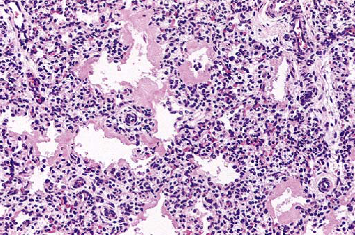
- Formation of eosinophilic hyaline membranes
- Hyaline membranes impair gas exchange further, worsening hypoxemia and suppressing surfactant synthesis: a vicious cycle
Morphology
Gross
- Lungs are normal-sized but heavy, firm, relatively airless, and mottled purple.
- They may appear solid because of diffuse atelectasis.
Microscopy
- Poorly expanded or collapsed alveoli, with alternating atelectasis and dilated alveoli.
- In infants who die very early, terminal bronchioles and alveolar ducts contain mainly necrotic cellular debris.
- Later, the characteristic lesion appears: thick, pink, eosinophilic hyaline membranes lining:
- Respiratory bronchioles
- Alveolar ducts
- Alveoli
Composition of hyaline membranes
They consist of:
- Necrotic type II pneumocytes
- Extravasated plasma proteins, particularly fibrinogen, which forms fibrin
There is generally little neutrophilic inflammation.
If the infant survives several days:
- Type II pneumocyte hyperplasia/proliferation
- Interstitial fibrosis
- Repair, or potentially chronic lung injury such as bronchopulmonary dysplasia
Clinical features
Typically presents soon after birth with:
- Tachypnea
- Dyspnea with retractions
- Nasal flaring
- Expiratory grunting
- Cyanosis
- Progressive hypoxemic respiratory failure
Untreated disease may be fatal. In uncomplicated cases treated appropriately, recovery often begins after about 3-4 days as endogenous surfactant production improves.
Prevention and treatment
- Antenatal glucocorticoids in threatened preterm delivery to accelerate fetal surfactant production
- Exogenous surfactant therapy after birth, especially in extremely preterm neonates
- CPAP or carefully controlled mechanical ventilation and oxygen support
Prolonged high-concentration oxygen and mechanical ventilation may cause:
- Bronchopulmonary dysplasia
- Retinopathy of prematurity
Exam answer in one line
NRDS is a disease of prematurity caused by deficient type II pneumocyte surfactant production, leading to diffuse atelectasis, hypoxemia, epithelial-endothelial injury, and fibrin-rich eosinophilic hyaline membranes lining alveoli.
Source: Robbins & Kumar Basic Pathology, Neonatal Respiratory Distress Syndrome, pp. 137-138; Robbins, Cotran & Kumar Pathologic Basis of Disease, neonatal RDS section, p. 416.
This is a shared conversation. Sign in to Orris to start your own chat.