
Carcinoma tongue
carcinoma tongue squamous cell oral cancer
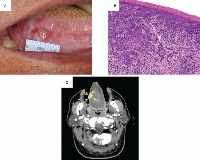
A multi-panel figure illustrating the clinical, histopathological, and radiological findings of oral squamous cell carcinoma. Panel A is a clinical photograph of the oral cavity showing a lesion on the right lateral border of the tongue. The lesion presents with an irregular, ulcerated surface and associated leukoplakia; a surgical ruler is positioned for scale, measuring approximately 7 x 10 mm. Panel B shows a histopathological section of the biopsy stained with hematoxylin and eosin (H&E) at 20x magnification. The tissue exhibits invasive nests of malignant epithelial cells with characteristic squamous differentiation and surrounding inflammatory cell infiltration, confirming squamous cell carcinoma. Panel C is an axial contrast-enhanced computed tomography (CT) scan of the head and neck. Yellow arrows highlight a hyperdense, contrast-enhancing mass on the right side of the tongue, measuring 18 mm in width and 11 mm in depth. This composite image is designed for educational use in oncology and oral surgery to demonstrate the diagnostic workflow for tongue cancer.
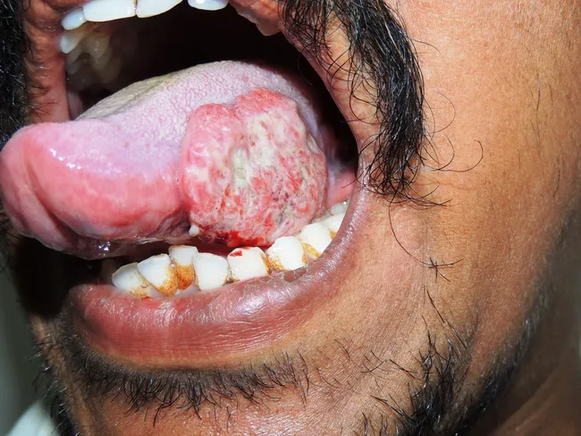
Intraoral clinical photograph of the left lateral tongue demonstrating an ulcerating, nodular mass approximately 3.0 cm in diameter. The lesion arises from the lingual mucosa along the left tongue margin and presents as a pink-erythematous, exophytic mass with focal surface ulceration. The patient is a 38-year-old male with over five years of chewing tobacco, a well-known risk factor for oral squamous cell carcinoma. The imaging modality is a standard white-light clinical photograph, acquired to document lesion morphology, localization, and size for baseline assessment and interdisciplinary discussion. The mass appears firm and indurated with surface irregularity and surrounding mucosal inflammation. Differential considerations include squamous cell carcinoma versus verrucous carcinoma or traumatic ulcer; however, the clinical history and lesion characteristics strongly support a malignant process. Definitive diagnosis requires histopathology from a biopsy, along with staging workup. This image is relevant for cancer screening, patient education, surgical and oncologic planning, and educational case discussions in otolaryngology/head-and-neck oncology. Keyword-rich descriptors: oral cancer, tongue cancer, oral SCC, left lateral tongue, intraoral lesion, ulcerating mass, tobacco use, risk factors, biopsy, histopathology, imaging documentation, staging, prognosis, therapy planning. Clinical images like this facilitate rapid recognition and timely management decisions. They also support patient counseling, multidisciplinary coordination, and education of trainees.
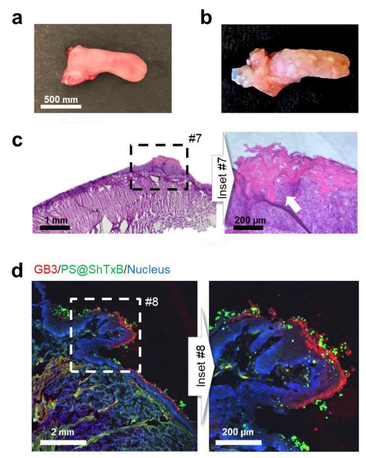
This figure illustrates the targeting of Shiga Toxin subunit B (ShTxB)-coated nanoparticles in a mouse model of oral carcinogenesis. (a) Macroscopic view of a healthy control tongue. (b) Macroscopic appearance of a tongue treated with the carcinogen 4NQO, displaying multifocal tumoral masses and leukoplakia characteristic of oral squamous cell carcinoma (OSCC). (c) Histological section (Hematoxylin and Eosin stain) of the 4NQO-treated tongue showing severe dysplasia and a focal area of invasive squamous cell carcinoma (indicated by the white arrow in inset #7). (d) Confocal immunofluorescence microscopy of a tissue cryosection demonstrating the specificity of targeted nanoparticles (PS@ShTxB). The image shows cell nuclei in blue (DAPI), the GB3 receptor—a known marker for certain HNC lesions—immunostained in red, and the green fluorescently labeled ShTxB-coated nanoparticles. The nanoparticles (green) are seen specifically clustering and coating the surface of GB3-positive (red) neoplastic lesions, highlighting the potential for targeted diagnostic imaging and therapeutic hyperthermia applications in head and neck cancer.
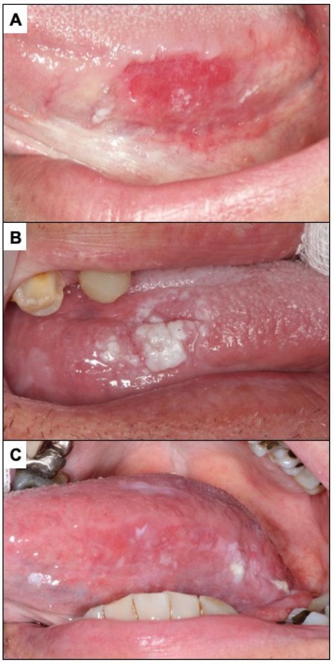
This composite of three clinical photographs (A, B, C) illustrates diverse clinical presentations of oral potentially malignant disorders and oral squamous cell carcinoma on the lateral border of the tongue. Panel (A) depicts erythroplasia, manifesting as a well-defined, intensely erythematous macule with an irregular, slightly granular surface texture and peripheral white striae. Panel (B) shows oral carcinoma presenting with a leukoplakia-like appearance, characterized by a thick, heterogeneous white plaque that is raised and irregular, contrasting sharply with the adjacent pink mucosa. Panel (C) demonstrates oral carcinoma with a mixed erythroleukoplakic appearance, featuring a combination of erythematous (red) and leukoplakic (white) components across the lateral tongue surface. These images serve as educational examples of incipient and established oral malignancies, highlighting the diagnostic importance of identifying persistent red, white, or mixed lesions in high-risk anatomical sites like the lateral tongue. The variations in color, texture, and border demarcation represent different clinical phenotypes within the spectrum of oral cancer development.
oral tongue squamous cell carcinoma management
Carcinoma of the Tongue
Anatomy Relevant to Tongue Cancer
- Oral tongue (anterior 2/3): The freely mobile portion, demarcated posteriorly by the circumvallate papillae (ICD-10 C02). Subdivided into tip, dorsum, lateral borders, and ventral surface.
- Base of tongue (posterior 1/3): Part of the oropharynx; generally presents at a later stage.
- Motor: Hypoglossal nerve (CN XII) - all muscles except palatoglossus (vagus, CN X)
- Sensation: Lingual nerve (branch of V3)
- Taste (anterior 2/3): Chorda tympani branch of CN VII
Epidemiology
- Oral tongue is the second most common site of oral cancer (30% of cases); in non-tobacco-chewing populations it accounts for 22-39% of oral cancers.
- Site distribution: Most tumors arise in the middle third, predominantly on the lateral border, followed by the ventral surface. Only 4-5% occur on the dorsum.
- Age: Usually presents in the 6th-8th decade; 90% of patients are over 40.
- Sex: Slightly more common in males; the male-to-female ratio has narrowed with increasing alcohol use in women.
- Risk factors: Tobacco (smoking and chewing), alcohol, immunosuppression, and possibly poor oral hygiene. Up to 70% of patients report significant tobacco and alcohol use.
- Emerging trend: Incidence in young adults (<40 years) has been increasing (from 4% in 1971 to 18% by the 1990s in the USA), often without identifiable risk factors - increased genetic susceptibility is postulated.
Clinical Presentation
Early Disease
- Erythroplakia (red inflammatory lesion) is the most common presentation of early SCC.
- Asymptomatic ulcer, lump, or persistent white patch on the lateral tongue.
- Most oral tongue cancers present at Stage I or II (unlike base of tongue tumors, which typically present at Stage III/IV).
Late/Advanced Disease
- Pain (may radiate to the ear via the lingual/auriculotemporal nerves)
- Ulceration with raised, rolled, indurated margins
- Tongue fixation (invasion of intrinsic/extrinsic muscles)
- Decreased tongue sensation
- Alteration in speech (dysarthria) and swallowing (dysphagia)
- Trismus (masseter/pterygoid involvement)
- Cervical lymphadenopathy
Cervical Metastases
- Clinically positive nodes at presentation: 21-34%
- Occult cervical metastasis: up to 53%
- Contralateral occult disease: up to 4.5%
- Tumors on the midline dorsum or ventral surface carry a greater risk of bilateral cervical node metastases
- Primary nodal drainage: upper jugular nodes (73%), submandibular (18%), middle jugular (18%), submental (9%)
Key prognostic factor: Depth of tumor invasion >2-4 mm significantly increases the risk of regional metastasis, recurrence, and mortality.
Clinical Images
Histopathology
- Vast majority are Squamous Cell Carcinoma (SCC).
- Most tongue tumors are well-to-moderately differentiated.
- Features that worsen prognosis: perineural invasion, lymphovascular invasion, positive/dysplastic margins, and advancing tumor thickness.
- Multifactorial histological malignancy grading of the invasive front helps assess cervical metastasis risk.
Staging (TNM - AJCC 8th Edition)
| T Stage | Description |
|---|---|
| T1 | Tumor ≤2 cm, depth of invasion (DOI) ≤5 mm |
| T2 | Tumor ≤2 cm with DOI >5 mm, or tumor 2-4 cm with DOI ≤10 mm |
| T3 | Tumor >4 cm, or any tumor with DOI >10 mm |
| T4a | Moderately advanced: invades cortical bone, inferior alveolar nerve, floor of mouth, or skin |
| T4b | Very advanced: invades masticator space, pterygoid plates, skull base; encases carotid |
- Depth of Invasion (DOI) was incorporated into AJCC 8th edition staging (not just tumor size), reflecting its major prognostic importance.
Workup / Investigations
- Biopsy: Most lesions are amenable to office biopsy; should include deep margin and peripheral mucosa. Deep biopsies provide tumor thickness information.
- Intraoral ultrasonography: Accurately assesses tumor thickness and aids in determining neck management.
- CT/MRI neck: To assess lymph node involvement (though sensitivity is moderate). MRI is preferred for soft tissue extent; CT for bone invasion.
- PET-CT: For advanced disease or suspected distant metastases.
- Chest imaging: To rule out pulmonary metastases and synchronous lung primaries.
- Panendoscopy: To assess for synchronous primary tumors.
Treatment
Primary Site Management
- Partial glossectomy with 1 cm margins in three dimensions is the treatment of choice.
- Reconstruction by primary closure, secondary intention, or skin graft.
- For patients unsuitable for surgery: external beam radiation ± brachytherapy.
- Ultrasonography can aid intraoperative margin assessment, particularly for the deep margin.
- Near-total or total glossectomy may be necessary.
- Glossectomy carries significant morbidity: dysphagia, aspiration; may require laryngectomy in select cases.
- For T4 with bone involvement: surgical resection ± composite resection (marginal or segmental mandibulectomy).
- Marginal mandibulectomy: For periosteal involvement.
- Segmental mandibulectomy: For medullary bone invasion (especially in edentulous patients with reduced bone stock).
- For T4: chemoradiation may be considered if surgery is not feasible.
Management of the Neck
| Depth of Invasion | Recommendation |
|---|---|
| <2 mm | Observation may be considered (though elective neck dissection gives better outcomes) |
| 2-4 mm | Elective neck dissection or radiation |
| >4 mm | Elective treatment mandatory (surgery or radiation) |
- Selective neck dissection (supraomohyoid): levels I-III minimum.
- Elective neck dissection (END) should include levels I-IV because of skip metastases.
- Bilateral neck dissections for midline tongue cancers.
- Sentinel lymph node biopsy (SLNB): Validated as feasible and accurate for T1-T2 N0 oral cavity cancers.
Adjuvant Therapy
- Post-operative radiotherapy (PORT): Indicated for positive margins, perineural invasion, lymphovascular invasion, multiple positive nodes, or extracapsular spread (ECS).
- Concurrent chemoradiotherapy (cisplatin-based): For ECS or positive margins.
Prognosis
| Stage | 5-Year Survival |
|---|---|
| Stage I-II | 60-75% |
| Stage III-IV | 25-40% |
| After salvage for regional metastasis | 35-40% |
- pN- (node negative): 5-year OS ~75%
- pN+/ECS- (node positive, no extracapsular spread): 5-year OS ~50%
- pN+/ECS+ (extracapsular spread): 5-year OS ~30%
- Tumor thickness >9 mm (5-year survival 66% vs. 100% for <3 mm)
- Perineural invasion
- Positive or dysplastic resection margins
- Cervical metastasis
- Young patients (<40 years) with tongue cancer paradoxically have a poorer long-term prognosis
Base of Tongue Carcinoma
- More commonly associated with HPV (especially p16-positive oropharyngeal SCC)
- Typically presents at Stage III/IV (later presentation)
- Often silent until advanced - first sign may be neck mass
- Management: primary chemoradiation or surgery (transoral robotic surgery, TORS)
- Better prognosis in HPV-positive cases
Key Points Summary
- Most common site: lateral border of the middle third of the oral tongue
- Erythroplakia is the earliest visible sign
- Depth of invasion is the single most important factor for neck management and prognosis
- Most oral tongue cancers present at Stage I/II - early detection is achievable
- Elective neck dissection (levels I-IV) is preferred even for clinically N0 necks when DOI >4 mm
- ECS (extracapsular spread) is a major adverse prognostic factor that mandates adjuvant chemoradiation
- Reconstruction after glossectomy must maximize residual tongue function
Umbilical hernia
umbilical hernia anatomy repair surgical
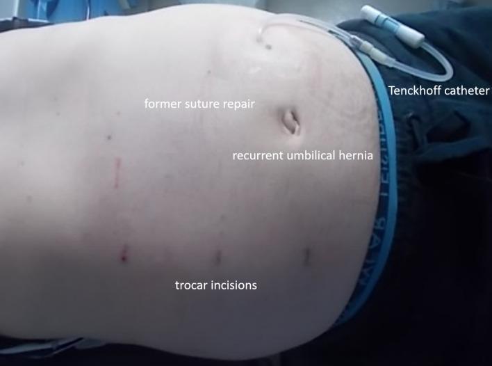
Clinical photograph of a patient's abdomen in a supine position, showing postoperative features following hernia repair and peritoneal dialysis catheter placement. In the upper right quadrant of the image, a Tenckhoff catheter is visible, exiting the abdominal wall with its external tubing and connector present. The central umbilical region displays a recurrent umbilical hernia, appearing as a slight protrusion and distortion of the umbilicus. Annotations on the skin identify a faint scar from a 'former suture repair' located superior and slightly lateral to the umbilicus. Several small, dark, punctate marks are visible across the lower abdominal wall, labeled as 'trocar incisions,' consistent with a minimally invasive or robotically-assisted surgical approach (eTEP). The image illustrates the complex abdominal wall anatomy in a patient with polycystic renal disease requiring both surgical hernia management and maintenance of peritoneal dialysis access.
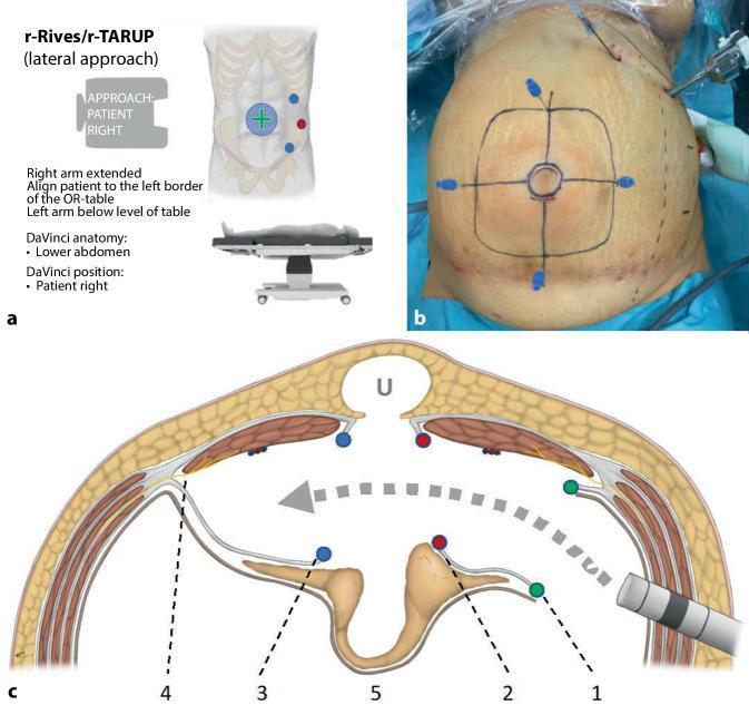
Educational graphic illustrating the surgical planning and anatomy for robotic transabdominal retromuscular umbilical prosthetic hernia repair (r-Rives or r-TARUP) via a left-lateral approach. Figure A shows an infographic detailing patient positioning on the operating table and port placement lateral to the left rectus muscle. Figure B is a clinical photograph of a patient's abdomen under pneumoperitoneum; ink markings delineate the umbilical hernia (central circle) and the anticipated mesh size (outer square). Transparietal needles are inserted at the square's perimeter to guide intra-abdominal preparation. Figure C is a cross-sectional anatomical diagram of the abdominal wall layers (skin, subcutaneous fat, rectus muscles, and posterior rectus sheaths). It maps the surgical dissection path (gray arrow) through the retrorectus space. Key steps are numbered: (1) green dots mark the lateral entry into the left posterior rectus sheath; (2) red dots signify the medial reopening of the sheath near the linea alba; (3) blue dots indicate the entry into the right posterior rectus sheath; (4) identifies lateral nerve preservation zones; (5) shows the mobilized hernia sac and peritoneal bridge. This content serves as a guide for surgical residents and fellows learning complex robotic ventral hernia repair techniques.
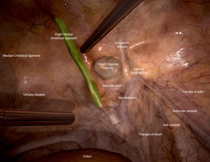
This didactic animation frame depicts the laparoscopic anatomy of the posterior abdominal wall and inguinal region during a Transabdominal Preperitoneal (TAPP) hernia repair simulation. The view shows a right-sided inguinal perspective with several labeled anatomical landmarks and surgical zones. Central to the image is a direct inguinal hernia defect, located medial to the epigastric vessels and internal inguinal ring. The median umbilical ligament is visible on the far left, with the urinary bladder adjacent. A surgical instrument is shown retracting the right medial umbilical ligament (highlighted in green). Further lateral, the iliopubic tract, vas deferens, and testicular vessels are identified. Crucially for surgical safety, the 'Triangle of Doom' (containing the external iliac vessels) is demarcated inferior to the vas deferens, while the 'Triangle of Pain' (containing sensory nerves) is identified superior to the testicular vessels. This image serves as an educational tool for identifying the 'Myopectineal Orifice of Fruchaud' and avoiding vascular or nerve injury during laparoscopic hernia surgery.
umbilical hernia repair adults mesh
Umbilical Hernia
Definition
Embryology and Anatomy
- The fascial margins of the umbilical defect form by the 3rd week of gestation.
- In the 6th week, the intestinal tract migrates outside the coelom through the umbilicus (physiological herniation); it returns at the 10th week as the midgut undergoes rotation.
- After this, the four folds of the somatopleure fuse inward, forming the tight umbilical ring that allows passage of only the umbilical vessels.
- At birth, ligation of the cord causes thrombosis of the umbilical arteries and vein, and the umbilical aperture closes.
- Any defect in this closure process results in an umbilical hernia.
Classification
1. Exomphalos (Omphalocele)
2. Congenital Umbilical Hernia (in infants)
- Comes out through the centre of a congenital weak umbilical scar.
- Appears within the first few months of birth.
- Higher incidence in premature babies and Black infants (up to 8x higher than White infants).
- Incidence: up to 10-30% of newborns in general; higher in premature infants.
- The neck is generally wide - strangulation is extremely uncommon below age 3.
- Swelling is easily reducible; increases in size on crying (classic conical shape); impulse on coughing.
- About 90-95% resolve spontaneously within the first 5 years as the umbilical scar thickens and contracts.
3. Acquired Umbilical Hernia (true umbilical hernia in adults)
- Protrudes directly through the umbilical scar in adult life.
- Relatively rare compared to paraumbilical hernia.
- Due to raised intra-abdominal pressure forcing through the umbilical scar.
- Causes: pregnancy, ascites, bowel distension, large ovarian cysts, fibroids.
4. Paraumbilical Hernia (most common acquired type)
- Occurs through a defect adjacent to (usually just above) the umbilicus, in the linea alba between the two recti.
- The lower half of the sac fundus is covered by the umbilicus.
- Affects middle-aged to older, obese women predominantly; female:male ratio ~3:1.
- Common symptoms: pain, swelling, discomfort.
- Many are irreducible when contents (omentum or bowel) become adherent to the sac.
- Due to the firm, fibrous, narrow neck - prone to incarceration and obstruction (though strangulation is less common than incarceration).
Clinical Features
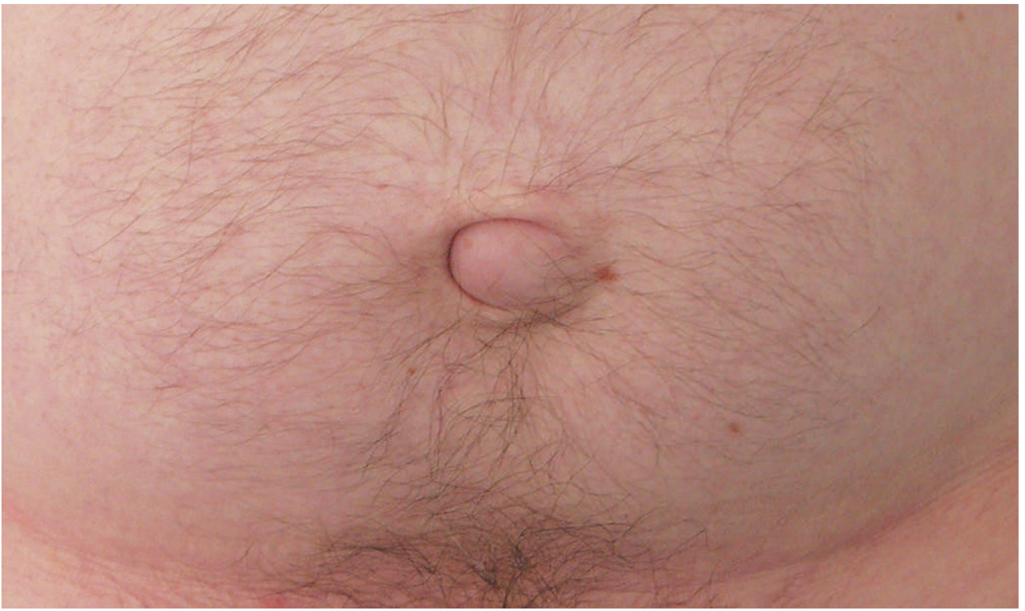
- Soft bulge at or adjacent to the umbilicus, often to one side, creating a crescent-shaped appearance.
- Usually reducible; the fascial defect can be palpated on reduction.
- Pain due to tissue tension or intermittent bowel obstruction.
- In large hernias, overlying skin becomes very thin; rarely, skin ulceration may occur; spontaneous rupture is extremely rare.
- Small hernias may contain only extraperitoneal fat or omentum (firm to palpation).
- Larger hernias may contain small or large bowel (soft, resonant on percussion).
- Abdominal wall varices (in portal hypertension/cirrhosis)
- Umbilical granuloma
- Sister Mary Joseph's nodule - metastatic tumor implant at the umbilicus (must be excluded in any atypical presentation)
- Urachal cyst or patent urachus (usually presents below the umbilicus)
Predisposing Conditions
| Factor | Mechanism |
|---|---|
| Obesity | Stretching and thinning of linea alba |
| Pregnancy | Repeated stretching of midline raphe |
| Liver cirrhosis with ascites | Raised intra-abdominal pressure + nutritional weakness |
| Peritoneal dialysis | Continuous pressure through catheter incision |
| Bowel obstruction | Repeated/persistent distension |
| Ventriculoperitoneal (VP) shunt | Raised intraperitoneal pressure |
Complications
- Incarceration (contents cannot be reduced) - common due to narrow fibrous neck
- Obstruction - intermittent or acute
- Strangulation - vascular compromise; may occur in one loculus of a multiloculated sac while other areas appear soft and non-tender
- Skin necrosis and ulceration - in very large hernias with thin overlying skin
- Rupture - extremely rare
The narrow neck relative to sac size makes adult umbilical hernias more prone to incarceration and strangulation than congenital ones in children.
Treatment
Children
| Age/Situation | Management |
|---|---|
| <2 years, asymptomatic | Conservative - parental reassurance; 95% resolve spontaneously |
| Defect <1 cm at birth | Spontaneous closure expected by age 4-5 years |
| Persists beyond age 2-3 years | Elective surgical repair |
| Large/symptomatic at any age | Earlier repair considered |
| Signs of incarceration (pain, bilious vomiting, hard tender mass) | Immediate surgical exploration |
- Small curvilinear "smile" incision just inferior to the umbilicus.
- Sac dissected free, contents reduced or inverted.
- Fascial defect closed transversely with interrupted slowly absorbable or permanent sutures.
- Skin closed subcuticularly; umbilical skin tacked to fascia.
- Day-surgery procedure under general anaesthesia; recurrence is rare.
Adults
- Hernia containing bowel (high strangulation risk - surgery advised)
- Symptomatic hernias (pain, obstruction)
- Enlarging hernias
- Small asymptomatic hernias may be observed, but tend to enlarge with time
- Repair is associated with significantly increased morbidity and mortality (haemodynamic instability, infection, electrolyte disturbance, high recurrence).
- Reserved for progressive symptoms or incarceration.
- Ascites should be controlled (medically or with transjugular intrahepatic portosystemic shunt) before elective repair.
Surgical Repair Techniques (Adults)
Open Repair
- Simple suture repair (0 polypropylene or nylon, interrupted) - as long as fascia is not closed under tension.
- "Darn" technique: non-absorbable monofilament suture criss-crossed across the defect.
- Transverse incision below the umbilicus.
- Sac dissected, opened, contents reduced, peritoneum closed.
- Defect extended transversely; fascial edges approximated with interrupted non-absorbable sutures.
- Mesh placement reduces recurrence (recommended by current guidelines for defects >1-2 cm).
- Onlay mesh: placed on the anterior surface of the closed defect.
- Sublay/retromuscular mesh (Rives-Stoppa principle): placed posterior to the rectus, superior to the posterior sheath - preferred for lower recurrence.
- Intraperitoneal onlay mesh (IPOM): used in laparoscopic repair.
- The classic overlapping fascial repair described in 1901.
- Transverse elliptical incision; upper flap sutured over lower flap in overlapping fashion.
- Now less commonly used as overlapping closures have been shown to weaken overall wound strength compared to mesh repair.
Laparoscopic Repair
- Indicated for larger defects and recurrent hernias.
- IPOM (intraperitoneal onlay mesh) is most common laparoscopic approach.
- The bulky falciform ligament must be taken down to create a smooth surface for mesh placement.
- Comparable or better outcomes vs. open for larger defects.
Emergency Repair
- Most performed by open surgery.
- In the presence of established strangulation: mesh should be avoided (high infection risk).
- Priority is to deal with the strangulated tissue (resect if necessary) and perform a suture repair.
- A more definitive mesh repair can be performed electively at a later date if needed.
Mesh vs. Suture - Current Evidence
Summary / Key Points
| Feature | Children | Adults |
|---|---|---|
| Common cause | Failure of umbilical closure | Raised intra-abdominal pressure |
| Sex | Equal | Female > Male (3:1) |
| Risk of strangulation | Very low (especially <3 yrs) | Higher - narrow fibrous neck |
| Spontaneous resolution | ~95% by age 5 | Does not close spontaneously |
| Primary treatment | Conservative (watchful waiting) | Surgical repair |
| Surgical approach | Suture closure | Suture (<2 cm) or mesh (>2 cm) |
| Preferred repair position | Transverse fascial closure | Sublay/retromuscular mesh preferred |
Proper relations of gangrene, necrosis, dead tissue, near dead tissue, ischemia and definitions of these
necrosis apoptosis cell death diagram pathology
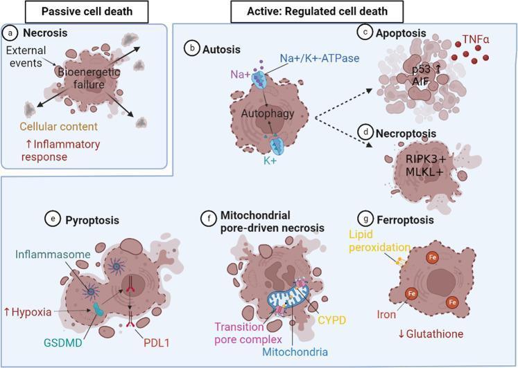
This pathophysiology diagram classifies types of cell death into two primary categories: Passive and Active (Regulated). Under Passive cell death, Necrosis (a) is shown as a result of bioenergetic failure and membrane rupture, leading to the release of cellular contents and an inflammatory response. The Active: Regulated cell death section includes several distinct pathways: Autosis (b), mediated by Na+/K+-ATPase and excess autophagy; Apoptosis (c), regulated by p53, TNFα, and AIF; and Necroptosis (d), involving RIPK3 and MLKL. Additionally, specialized forms of regulated death are depicted: Pyroptosis (e), characterized by hypoxia, inflammasome activation, GSDMD, and PDL1 translocation; Mitochondrial pore-driven necrosis (f), highlighting the transition pore complex and Cyclophilin D (CYPD); and Ferroptosis (g), driven by glutathione deficiency, iron accumulation, and lipid peroxidation. The diagram uses anatomical illustrations of cells to show specific morphological changes such as swelling, membrane blebbing, and organelle dysfunction, providing a comprehensive overview of cellular pathology and signaling pathways.
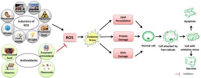
This pathophysiology diagram illustrates the link between Reactive Oxygen Species (ROS), oxidative stress, and cellular damage. The flow begins with 'Inductors of ROS,' categorized into external factors (smoking, pollution, radiation, UV light, heavy metals) and internal factors (metabolism, viruses, processed foods). These inductors drive ROS production, while an inhibitory pathway shows 'Antioxidants' (enzymatic, flavonoids, vitamins, and unprocessed foods) attempting to neutralize them. When production exceeds neutralization, it leads to 'Oxidative Stress,' resulting in a triad of cellular injuries: lipid peroxidation, protein damage, and DNA damage. The final sequence depicts cellular morphological progression from a 'Normal cell' to a 'Cell attacked by free radicals' (showing early membrane disruption), and finally a 'Cell with oxidative stress' (demonstrating severe fragmentation). This progression culminates in programmed cell death (Apoptosis) or premature cell death (Necrosis). The diagram is designed for intermediate medical education to explain the molecular mechanisms of oxidative pathology and the protective role of endogenous and exogenous antioxidants.
Gangrene, Necrosis, Dead Tissue, Near-Dead Tissue, and Ischemia: Proper Relations and Definitions
ISCHEMIA (reduced blood flow)
    ↓
HYPOXIA (reduced oxygen delivery to cells)
    ↓
REVERSIBLE CELL INJURY ("near-dead tissue")
    ↓  [point of no return]
IRREVERSIBLE CELL INJURY → NECROSIS (dead tissue at cell level)
    ↓  [when necrosis involves macroscopic tissue portions]
GANGRENE (a clinical/macroscopic form of necrosis, usually with specific context)
1. Ischemia
- It is the upstream cause - the initial insult - in the chain leading to necrosis and gangrene.
- Ischemia is distinct from simple hypoxia: in hypoxia, only oxygen is reduced (e.g., high altitude, anaemia), while metabolites are still washed away. In ischemia, both oxygen delivery and waste removal are impaired, making ischemia more damaging.
- Caused by: arterial thrombosis, embolism, atherosclerotic occlusion, external compression, vasospasm, prolonged hypotension.
- Consequences depend on duration and severity:
- Brief ischemia → reversible injury (stunning)
- Prolonged ischemia → irreversible injury → necrosis
- Restoration of blood flow after ischemia can paradoxically worsen injury: ischemia-reperfusion injury (mediated by reactive oxygen species, calcium overload, and inflammation).
- Oxygen falls → mitochondrial oxidative phosphorylation fails → ATP depletion
- Loss of ATP → failure of Na⁺/K⁺-ATPase pump → cell swelling (reversible stage)
- Anaerobic glycolysis → lactic acidosis, pH falls
- Ca²⁺ influx → activates destructive enzymes (phospholipases, proteases, endonucleases)
- Membrane phospholipid degradation → plasma membrane rupture → irreversible
2. Reversible Cell Injury ("Near-Dead Tissue")
- The cell is alive but impaired - this is "near-dead tissue."
- Morphology: cell swelling (oncosis), organelle swelling (mitochondria, ER), membrane blebbing, fatty change (in hepatocytes), reduced ATP, reduced pH.
- The cell membrane remains intact - this is the key difference from irreversible injury.
- Clinically this corresponds to a tissue that is viable but at risk: pale, cold, painful, not yet infarcted.
- Inability to reverse mitochondrial dysfunction - failure of oxidative phosphorylation and ATP generation persists even if the original injury is corrected.
- Profound disturbances in membrane function - irreversible damage to lysosomal membranes leads to enzymatic dissolution of the cell.
3. Necrosis (Dead Tissue at the Cellular Level)
- Necrosis is a passive, chaotic, unregulated process - "messy and destructive."
- It is the microscopic/cellular-level term for dead tissue.
- The inflammatory response it triggers is the key clinical feature - swelling, redness, pain around necrotic tissue.
Morphological Patterns of Necrosis
| Pattern | Appearance | Classic Context |
|---|---|---|
| Coagulative | Tissue architecture preserved for days; eosinophilic, anucleate "ghost cells"; firm texture | Ischemic infarcts of all solid organs EXCEPT brain |
| Liquefactive | Tissue completely digested into viscous liquid; architecture obliterated | Bacterial/fungal infections; hypoxic brain death; abscesses (pus) |
| Gangrenous | Coagulative necrosis of multiple tissue layers losing blood supply; superimposed infection → liquefactive component | Limb ischemia (see Gangrene below) |
| Caseous | Cheese-like, friable yellow-white; granular amorphous pink on H&E; granuloma surrounding | Tuberculosis (and other mycobacterial/fungal infections) |
| Fat necrosis | Focal fat destruction; chalky white saponification (Ca²⁺ + fatty acids); shadowy fat cell outlines on histology | Acute pancreatitis; abdominal trauma |
| Fibrinoid | Bright pink, amorphous material in vessel walls on H&E; only detectable microscopically | Immune vasculitis; malignant hypertension |
Nuclear Changes in Necrosis (histological hallmarks)
- Karyolysis - basophilia of chromatin fades (loss of DNA by enzymatic degradation)
- Pyknosis - nuclear shrinkage with increased basophilia (condensed, shrunken nucleus)
- Karyorrhexis - fragmentation of the pyknotic nucleus
- After 1-2 days, nucleus disappears completely.
Necrosis vs. Apoptosis
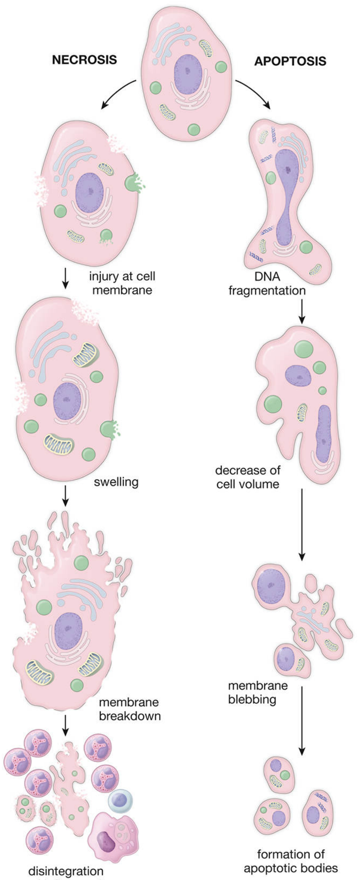
| Feature | Necrosis | Apoptosis |
|---|---|---|
| Trigger | Extreme injury, ischemia, toxins, trauma | Physiologic or programmed signal |
| Cell size | Swells | Shrinks |
| Membrane | Ruptures | Maintained until apoptotic bodies form |
| Nucleus | Karyolysis, pyknosis, karyorrhexis | DNA fragmentation (laddering) |
| Inflammation | Intense (leakage of lysosomal enzymes) | None (contents contained in apoptotic bodies, phagocytosed) |
| Nature | Passive, chaotic, "dies in a mess" | Regulated, "dies with dignity" |
4. Gangrene (Dead Tissue at the Macroscopic/Clinical Level)
- Gangrene is not a distinct pattern of cell death (Robbins) - it is a clinical/macroscopic term that describes what happens when coagulative necrosis (or coagulative + liquefactive in wet gangrene) involves a large volume of tissue, typically a limb.
- It represents the same process as necrosis but at a gross, visible scale.
- Usually affects the most distal part of a limb (poor collateral circulation): toes, feet, fingers.
- The black discoloration is not simply a colour of dead tissue - it is specifically from haemoglobin breakdown → iron sulphide formation.
Types of Gangrene
Dry Gangrene
- Mechanism: Gradual, slow reduction in arterial blood flow (e.g., atherosclerotic occlusion) without infection.
- Tissue state: Desiccated, mummified, shrunken, dry, leathery, black-brown.
- Infection: Absent or minimal.
- Line of demarcation: Clear, well-defined - separates dead tissue from living tissue; granulation tissue forms between the two zones; if proximal blood supply is good, demarcation appears within days and separation occurs neatly.
- Spread: Does not spread proximal; the process is self-limiting.
- Management: Can often be managed conservatively (wait for auto-amputation at demarcation); elective amputation when line is established.
Wet (Moist) Gangrene
- Mechanism: Arterial obstruction plus superimposed bacterial infection and putrefaction.
- Tissue state: Soft, moist, swollen, foul-smelling; partially perfused; retains some turgor.
- Infection: Present - bacteria (often mixed) cause liquefactive digestion superimposed on coagulative necrosis.
- Line of demarcation: Poorly defined or absent - infection spreads into adjacent living tissue, pushing the demarcation more proximally than in dry gangrene.
- Spread: Spreads rapidly proximally if untreated.
- Management: Surgical emergency - urgent debridement or amputation.
- Commonly seen in: diabetic foot, venous gangrene, intestinal ischemia.
Gas Gangrene
- Mechanism: Invasion of anaerobic bacteria (classically Clostridium species, e.g., C. perfringens) into devitalized muscle, releasing exotoxins (histotoxins, lecithinase) and gas (CO₂, H₂) locally and systemically.
- Clinical features: Crepitus on palpation (gas in tissues), rapidly spreading necrosis, systemic toxaemia, extreme toxicity.
- Management: Surgical emergency - immediate wide debridement, amputation, high-dose penicillin, hyperbaric oxygen.
Other Named Gangrenes
- Diabetic gangrene: Wet gangrene in diabetics - due to small vessel disease (microangiopathy), neuropathy (painless trauma), and impaired immunity combined.
- Fournier's gangrene: Rapidly spreading necrotizing fasciitis and gangrene of the perineum and genitalia; polymicrobial synergistic infection.
- Synergistic (Meleney's) gangrene: Progressive bacterial synergistic gangrene of the abdominal wall.
- Intestinal gangrene: Ischemic necrosis of bowel following mesenteric arterial occlusion or strangulated hernia.
Line of Demarcation
- A zone develops between truly viable and dead or dying tissue.
- Granulation tissue forms between the dead and living parts.
- In dry gangrene with adequate proximal blood supply: clear, neat, rapid demarcation.
- In moist gangrene: demarcation is proximal, irregular, and delayed due to infection extending into living tissue.
- If proximal blood supply is poor: line of demarcation is slow to form or absent; gangrene may spread or "skip" areas - skip lesions can appear higher up the limb.
- Bone demarcates more slowly than soft tissue (better blood supply); the stump tends to be conical.
5. Putting It All Together: The Hierarchy
ISCHEMIA
(reduced blood flow - cells alive but stressed)
        ↓ short duration / mild
REVERSIBLE CELL INJURY = "NEAR-DEAD TISSUE"
(cell swollen, ATP depleted, membrane blebbing - but membrane intact, can recover)
        ↓ prolonged / severe - point of no return crossed
IRREVERSIBLE CELL INJURY → NECROSIS
(membrane ruptures, lysosomal enzymes released, inflammation - cell is DEAD)
        ↓ when macroscopic - many cells / tissues involved
INFARCTION
(area of necrosis in an organ caused by ischemia; typically coagulative)
        ↓ when limb involved + blackening + macroscopic putrefaction
GANGRENE
(clinical/macroscopic term; coagulative necrosis ± infection in a limb or part)
Key Conceptual Distinctions
| Term | Level | Alive? | Reversible? | Key Feature |
|---|---|---|---|---|
| Ischemia | Tissue/organ (perfusion) | Yes | Yes (if corrected in time) | Reduced blood flow - cause, not consequence |
| Near-dead / reversible injury | Cell | Yes (barely) | Yes | Intact membrane; swollen; ATP depleted |
| Necrosis | Cell / microscopic | No | No | Membrane ruptured; inflammation; nuclear changes |
| Infarction | Tissue / organ | No | No | Macroscopic area of necrosis from ischemia (organ context) |
| Gangrene | Limb / macroscopic tissue | No | No | Macroscopic necrosis + blackening + putrefaction (limb context) |
| Dead tissue | Any level | No | No | Generic term for tissue beyond recovery |
Important Additional Points
- Ischemia does not equal necrosis - ischemia is the cause; necrosis is the outcome if ischemia is not corrected. Reversible ischemia resolves without necrosis.
- Necrosis does not equal gangrene - necrosis is the microscopic event. Gangrene is the gross/clinical manifestation when necrosis involves a large tissue volume in a specific pattern (limb, blackening, putrefaction).
- Gangrenous necrosis is not a separate cellular mechanism - it is coagulative necrosis ± liquefactive (when infected), just in a limb context at macroscopic scale.
- All gangrene involves necrosis, but not all necrosis is gangrene.
- All necrosis involves dead tissue, but dead tissue can arise from causes other than ischemia (infection, toxins, physical injury).
- Ischemia is the most common cause of gangrene and infarction, but gangrene can also follow trauma, cold injury, sepsis, or vasoconstriction.