
Ewing sarcoma pathology all info with diagrams
Reading File
Now let me run parallel searches across the library for comprehensive Ewing sarcoma coverage.
Finding Sources
Finding Sources
Searching PubMed
"Ewing sarcoma"[MeSH Terms] AND pathology
Excellent sources found. Now let me read the key pathology sections in parallel.
Reading File
Reading File
Reading File
Reading File
Excellent content. Now let me fetch the key images and also read the Quick Compendium pathology section and more Robbins content.
Loading Image
Loading Image
Reading File
Reading File
Now let me fetch the Robbins histology image and the Quick Compendium Ewing sarcoma section, and also get more on IHC/molecular from the pathology book.
Loading Image
Finding Sources
Reading File
Excellent. Let me get the Quick Compendium Ewing sarcoma family section and the Rheumatology / Miller's sections for IHC/staging detail.
Reading File
Reading File
Reading File
I have very comprehensive content. Let me fetch two more key images - the Miller's Review image with X-ray/MRI/histology composite and the Rheumatology X-ray image.
Loading Image
Loading Image
Reading File
I now have all the information needed for a comprehensive, well-illustrated answer. Let me compile it.
Ewing Sarcoma - Comprehensive Pathology
Overview
Ewing sarcoma is a malignant small round blue cell tumor (SRBCT) driven by EWSR1-ETS fusion proteins arising from balanced translocations. It is the second most common primary malignant bone tumor in children and adolescents (after osteosarcoma), and the most common in patients under 10 years old. It accounts for approximately 10-15% of primary malignant bone tumors.
- Age: Most cases occur in patients aged 5-25 years (85% aged 5-20 years); peak incidence in the second decade
- Sex: Slight male predominance
- Race: Exceedingly rare in individuals of African descent; predominantly affects Whites of European origin
- Incidence: ~200 new cases/year in the United States
- Location: 80% in bone, 20% extraosseous ("extraosseous Ewing sarcoma")
Molecular Pathogenesis
This is the defining characteristic of Ewing sarcoma:
| Translocation | Fusion Gene | Frequency |
|---|---|---|
| t(11;22)(q24;q12) | EWSR1-FLI1 | ~85-95% (most common) |
| t(21;22)(q22;q12) | EWSR1-ERG | ~5-10% |
| t(7;22)(p22;q12) | EWSR1-ETV1 | Rare |
| t(17;22)(q13;q12) | EWSR1-ETV4 | Rare |
| t(2;22)(q33;q12) | EWSR1-FEV | Rare |
- EWSR1 is located on 22q11 and encodes an RNA-binding protein
- The fusion protein binds chromatin and dysregulates transcription, leading to uncontrolled growth and abnormal differentiation
- All fusion partners belong to the ETS transcription factor family (FLI1, ERG, ETV1, ETV4, FEV)
- Cell of origin: Most likely mesenchymal stem cells or primitive neuroectodermal cells
- Additional somatic mutations: TP53, CDKN2A, and STAG2 mutations are detectable; most additional changes appear to be epigenetic
- An aggressive variant carries a CIC-DUX4 fusion (Ewing-like)
- Some cases harbor FUS rearrangements (related TET family member) instead of EWSR1
FISH Demonstration of EWSR1 Rearrangement
Dual-color break-apart FISH probes are used to detect EWSR1 rearrangement. A normal nucleus shows 2 red-green fusion signals (2F); an abnormal nucleus shows 1 red-green fusion + separate red and green signals (1R,1G,1F):
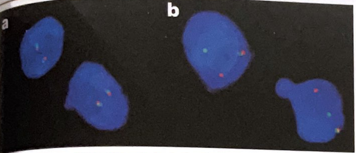
FISH dual-color break-apart probes: (a) normal nuclei with 2 fused signals; (b) abnormal nuclei showing separated red and green signals indicating EWSR1 rearrangement - Quick Compendium of Clinical Pathology, 5th ed.
Sites of Involvement
- Long bones: Metaphysis of the femur, tibia, fibula, and humerus (often with diaphyseal extension) - most common
- Flat bones: Pelvis and scapular girdle (femur + pelvis = one-third of all cases)
- Ribs: Classic site in adults
- Spine: Primary vertebral Ewing is rare (3.3-15%); sacral involvement in up to 50% of spinal cases
- Extraosseous: Soft tissue origin (20% of cases)
- Rarely involves small bones of hands/feet or skull
Gross Pathology / Macroscopy
- Arises in the medullary cavity and invades the cortex, periosteum, and surrounding soft tissue
- Soft, tan-white tumor with frequent areas of hemorrhage and necrosis
- Often associated with a large soft tissue mass adjacent to bone - this is characteristically much larger than the bony component visible on plain X-ray
- The periosteal reaction to advancing tumor produces layers of reactive bone
Histopathology (Microscopy)
Ewing sarcoma is the classic prototype of small round blue cell tumors (SRBCT) in children.
Key Histological Features:
- Sheets of uniform small round cells - slightly larger and more cohesive than lymphocytes
- Scant cytoplasm - may appear clear due to glycogen (PAS-positive, diastase-sensitive)
- Round nuclei with fine chromatin; nucleoli usually inconspicuous
- Minimal intercellular matrix - no bone or cartilage production (unlike osteosarcoma)
- Homer-Wright rosettes - circular groupings of cells with a central fibrillary core (when neuroectodermal differentiation is present, seen in peripheral PNET variant)
- Pseudorosettes (attempted formation of vascular channels)
- Areas of necrosis and hemorrhage are common
Classic H&E Photomicrograph - Robbins Pathology:
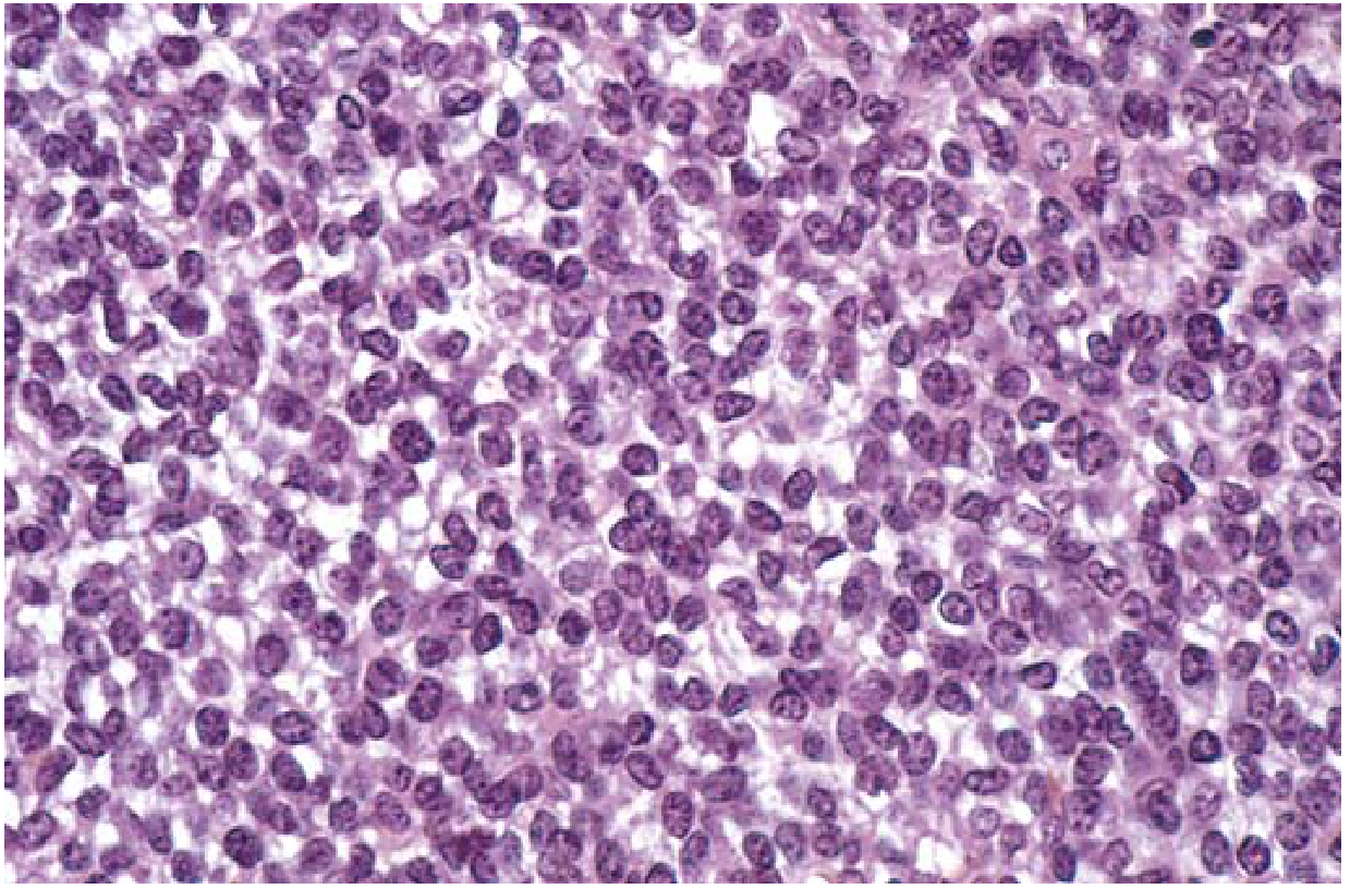
FIG. 19.23 Ewing sarcoma composed of sheets of small round cells with minimal clear cytoplasm - Robbins & Kumar Basic Pathology
Clinical + Imaging + Histology Composite - Miller's Review:
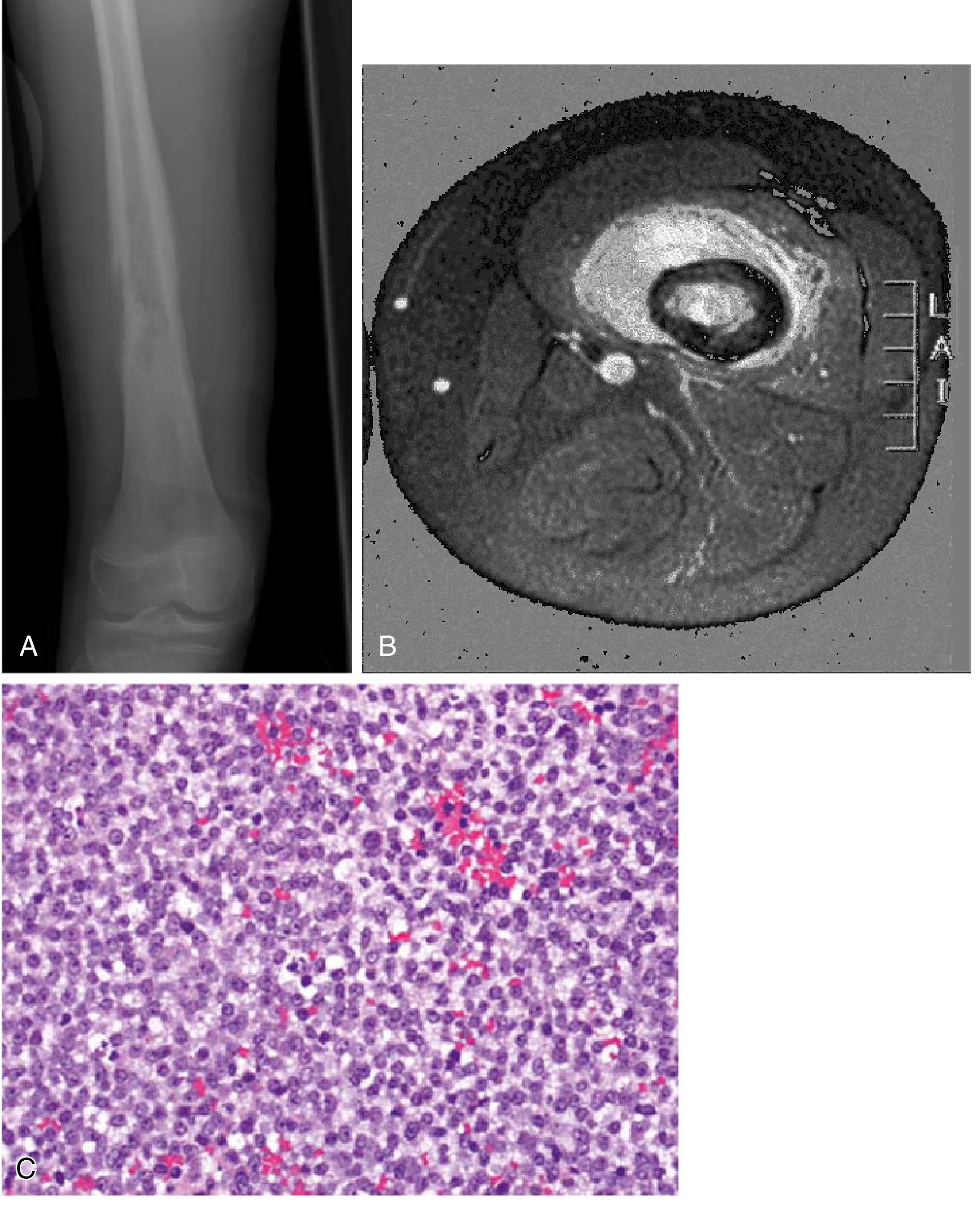
(A) Radiograph: destructive expansile lesion in distal femur with Codman triangle. (B) Axial MRI: large soft tissue mass with serial periosteal elevation ("onion skinning"). (C) Photomicrograph: sheets of monotonous small round blue cells - Miller's Review of Orthopaedics, 9th ed.
Immunohistochemistry (IHC)
IHC is required to differentiate Ewing sarcoma from other small round blue cell tumors:
| Marker | Ewing Sarcoma | Notes |
|---|---|---|
| CD99 (MIC2) | Strongly positive (diffuse membranous) | Most sensitive but not specific |
| FLI-1 | Nuclear positive | More specific than CD99 |
| Vimentin | Positive | Non-specific |
| NSE | Variable | Neuroectodermal cases |
| S100 | Negative | Helps exclude melanoma |
| Desmin | Negative | Helps exclude rhabdomyosarcoma |
| LCA (CD45) | Negative | Helps exclude lymphoma |
| CK (cytokeratin) | Negative | Helps exclude carcinoma |
CD99 and FLI-1 positivity together with the translocation studies are the cornerstone of diagnosis.
Radiological Features
Plain X-ray:
- Permeative / "moth-eaten" lytic destructive pattern
- "Onion-skin" periosteal reaction - multiple layers of reactive periosteal bone (most classic sign)
- "Sunburst" periosteal reaction - may also be seen
- Codman triangle - periosteal elevation at tumor margins
- Diaphyseal or metaphyseal location in long bones
- May appear near-normal on X-ray in highly permeative lesions (MRI then reveals marrow involvement)
MRI:
- Isointense on T1, iso- to hyperintense on T2
- Intense gadolinium enhancement
- Often reveals a large adjacent soft tissue mass (characteristic)
- Shows full extent of marrow involvement
- "Curtain sign" in spinal Ewing - invasion of spinal canal
Campbell's Operative Orthopaedics - Multimodal Imaging:
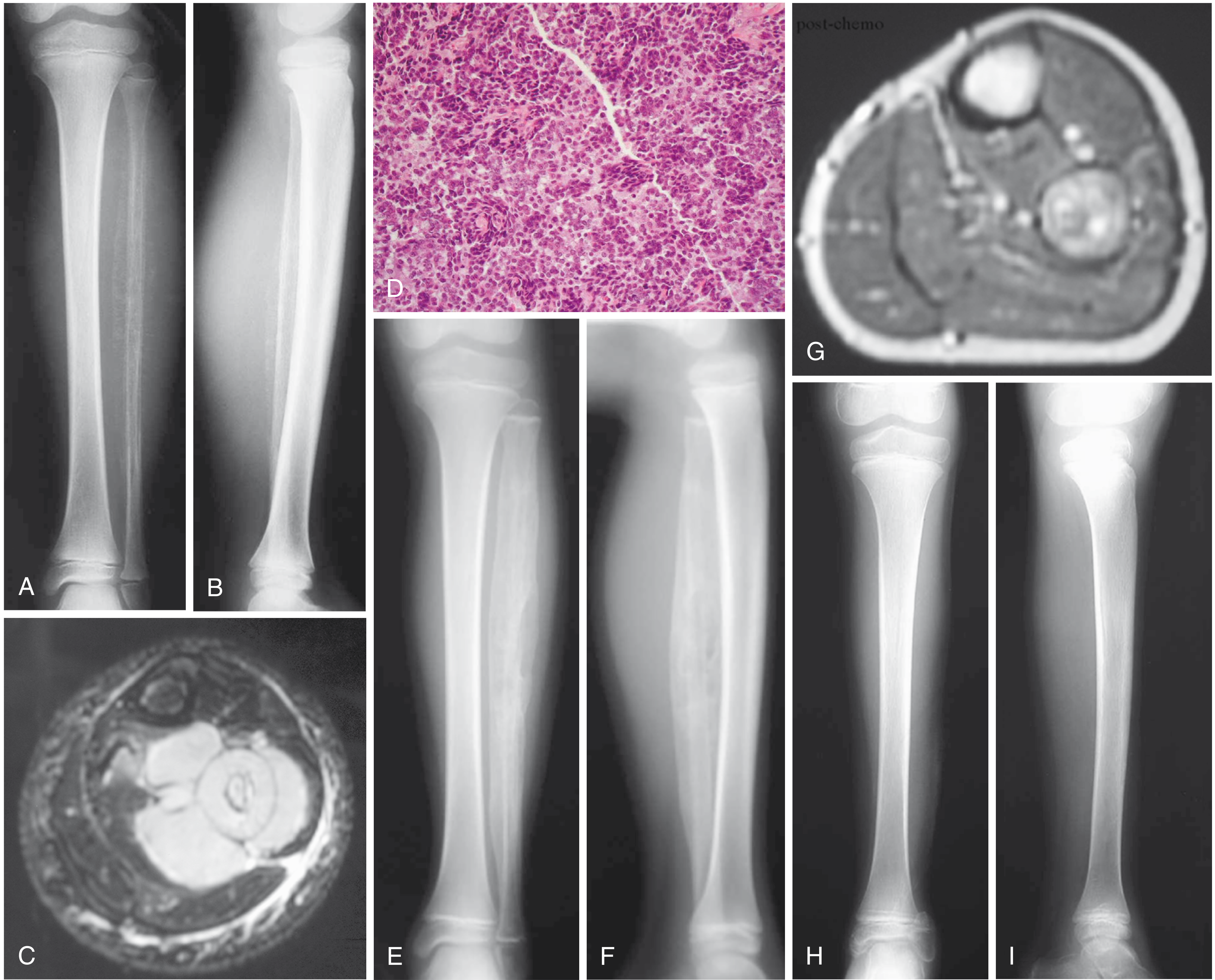
(A-B) AP/lateral radiographs of fibula in a 7-year-old: involvement of large bone portion typical of Ewing sarcoma. (C) Axial MRI: large soft-tissue mass. (D) Classic microscopic appearance. (E-F) Post-neoadjuvant chemotherapy: increased ossification. (G) Post-chemo MRI: marked reduction in soft-tissue mass. (H-I) Post-wide resection radiographs - Campbell's Operative Orthopaedics, 15th ed.
Spinal Ewing - MRI (L2):
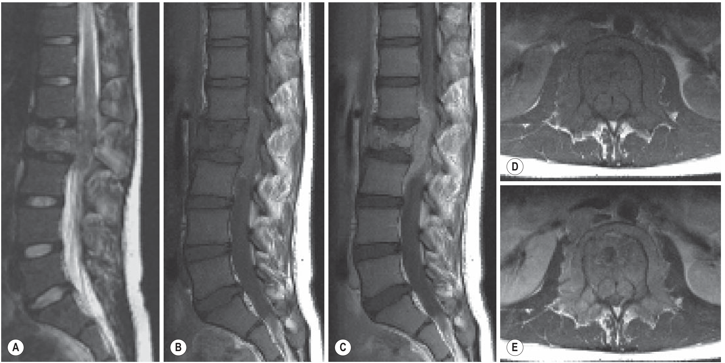
Fig. 49.39 Ewing Sarcoma of L2: (A) Sagittal T2 - slightly increased signal; (B,D) Sagittal and axial T1 - isointense; (C,E) Intense gadolinium enhancement; invasion of spinal canal ("curtain sign") and adjacent paraspinal soft tissues - Grainger & Allison's Diagnostic Radiology
Clinical Presentation
- Pain - nearly universal; often insidious onset; may be mild and intermittent initially
- Soft tissue mass - often warm and tender; can mimic infection
- Fever, leukocytosis, elevated ESR - mimics acute osteomyelitis (confounds diagnosis)
- Average delay from onset to diagnosis: ~34 weeks
- ~20% of patients present with metastatic disease at diagnosis
Staging / Workup
- Bone marrow biopsy is performed for staging
- CT chest for lung metastases
- Bone scan for skeletal metastases
- PET-CT: increasingly used for whole-body staging
- MRI of primary site: mandatory for local staging
Differential Diagnosis of Small Round Blue Cell Tumors
Ewing sarcoma must be distinguished from other SRBCTs, especially in children:
| Tumor | Key Distinguishing Features |
|---|---|
| Ewing sarcoma | CD99+, FLI-1+, EWSR1-FLI1 fusion, no matrix |
| Neuroblastoma | Most common in <5 yrs; adrenal/sympathetic chain; NSE+, MYCN amplification |
| Rhabdomyosarcoma | Desmin+, myogenin+, MyoD1+ |
| Lymphoma (primary bone) | LCA/CD45+, B/T cell markers |
| Small-cell osteosarcoma | Produces osteoid matrix |
| Mesenchymal chondrosarcoma | Cartilaginous islands present |
| DSRCT | EWS-WT1 fusion; t(11;22)(p13;q12); desmoplastic stroma; EMA/keratin/desmin+ |
| Neuroepithelioma/PNET | Homer-Wright rosettes; synaptophysin/chromogranin+ |
When a SRBCT is found in a child younger than 5 years, metastatic neuroblastoma and leukemia should be considered first.
Ewing Sarcoma Family (Unified Concept)
The "Ewing sarcoma family" unifies:
- Classic Ewing sarcoma - non-matrix-forming bone tumor
- Peripheral PNET (pPNET) - more prone to neuroepithelial rosettes; arises from viscera or soft tissue
- Extraosseous Ewing sarcoma - same genetics, soft tissue origin
- Askin tumor - chest wall PNET in children
All share: proliferatively active small round blue cells + EWSR1 gene anomalies
Ewing-like Tumors with BCOR Alterations (Newer Entity):
- Previously grouped with Ewing sarcoma family based on similar morphology
- Characterized by BCOR-CCNB3 fusions (paracentric inversion on X chromosome short arm)
- Composed of small round blue cells (like Ewing) or small spindle blue cells in whorled/hemangiopericytomatous pattern
- Distinct from classic Ewing sarcoma despite overlap
Prognosis and Prognostic Factors
- 5-year event-free survival: ~70% with localized disease treated with multimodal therapy
- Long-term cure rate: ~50-75% with neoadjuvant chemo + surgery ± radiation
- Patients with clinically detectable metastatic disease at presentation have poor outcomes
Poor Prognostic Factors:
- Spine and pelvic tumor location
- Tumor size >100 cm² (or >200 mL volume)
- Poor histological response to chemotherapy (<90% tumor necrosis)
- TP53 mutation
- Fusion genes other than EWS-FLI1
- Metastatic disease at presentation
Metastasis Pattern:
- Lungs - 50%
- Bone - 25%
- Bone marrow - 20%
Treatment Summary
- Neoadjuvant chemotherapy (induction) - VAC/IE: vincristine + doxorubicin + cyclophosphamide alternating with ifosfamide + etoposide
- Local control: Wide surgical resection (R0) is preferred; radiation for pelvic/spinal lesions where resection is morbid
- Adjuvant chemotherapy (consolidation)
- High-dose therapy + peripheral blood stem cell transplant (PBSCT): For large tumors (>200 mL) with poor chemo response
- Salvage regimens: Cyclophosphamide/topotecan, irinotecan/temozolomide, gemcitabine/docetaxel
Sources: Robbins & Kumar Basic Pathology; Campbell's Operative Orthopaedics 15th ed.; Miller's Review of Orthopaedics 9th ed.; Quick Compendium of Clinical Pathology 5th ed.; Grainger & Allison's Diagnostic Radiology; Goldman-Cecil Medicine; Rheumatology 2022 (Elsevier)
This is a shared conversation. Sign in to Orris to start your own chat.