
can you teach me everything about cell division in class 11 biology
Reading File
Now let me search the medical library for cell division content, running multiple parallel searches to cover all aspects.
Finding Sources
Finding Sources
Searching the Web
cell division class 11 biology NCERT mitosis meiosis complete notes
Good. Let me now read the most relevant textbook sections in parallel - Emery's Genetics (dedicated chapter on chromosomes and cell division) and the Ganong's physiology section on mitosis/meiosis.
Reading File
Reading File
Finding Sources
Let me now read the main cell division chapter from Emery's Genetics and the Ganong's physiology section in parallel.
Reading File
Reading File
Finding Sources
Excellent! I have a great cell cycle diagram image URL. Let me read more of the Emery's chapter on mitosis and meiosis phases, and also fetch that diagram image.
Reading File
Loading Image
There's an image URL for the mitosis stages diagram. Let me get it and also read the meiosis section.
Reading File
I have everything I need. Let me also fetch the cell cycle diagram image to include it.
Reading File
Loading Image
I also have the meiosis diagram URL. Let me fetch it quickly.
Loading Image
Reading File
I now have everything needed to write a complete, thorough Class 11 biology lesson on cell division. Let me compile this into a detailed, well-structured response.
Cell Cycle and Cell Division - Complete Class 11 Notes
This is the full topic as per the NCERT Class 11 Biology syllabus (Chapter 10), drawn from authoritative medical and biology textbooks.
1. Why Do Cells Divide?
Every multicellular organism starts as a single fertilized cell (zygote). Rapid and controlled cell division builds the entire body - a human adult contains approximately 10^14 (100 trillion) cells. Cell division also:
- Replaces worn-out cells (skin, blood cells)
- Repairs damaged tissues
- Allows growth during childhood and adolescence
- Transmits genetic information to the next generation
2. The Cell Cycle
The cell cycle is the sequence of events a cell goes through from the time it is formed until it completes division and produces two daughter cells.
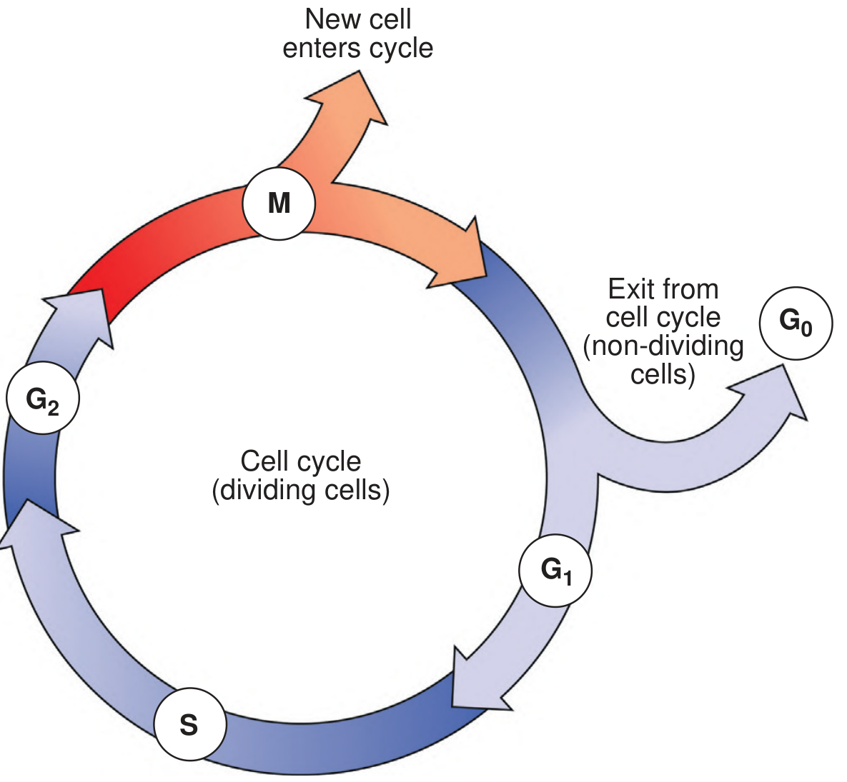
Fig. 3.12 - Stages of the cell cycle: G1 and G2 are gap/resting phases of interphase. S is DNA synthesis. M is mitosis. G0 is the non-dividing arrest phase.
The cell cycle has two major phases:
A. Interphase (the "resting" phase - but actually very busy!)
Interphase is the period between two successive mitoses. In rapidly dividing cells it lasts 16-24 hours. It has three sub-phases:
| Sub-phase | Full Name | What Happens |
|---|---|---|
| G1 | Gap 1 (First Gap) | Cell grows in size. Proteins, organelles, and RNA are synthesized. Chromosomes are thin and extended. |
| S | Synthesis | DNA replication occurs. Each chromosome duplicates - each chromosome now consists of two identical sister chromatids joined at the centromere. |
| G2 | Gap 2 (Second Gap) | More cell growth. Chromosomes begin to condense in preparation for division. |
G0 Phase (non-dividing state): Cells that have stopped dividing (like neurons) exit the cycle at the end of G1 and enter a resting state called G0. They remain permanently non-dividing.
Key fact: The G1 phase is the most variable in length - this is what controls how fast different cell types divide. Cancer cells often have a very short G1 phase.
DNA content: At the start of S phase, the cell is 2n (diploid). After S phase, DNA content doubles to 4n (tetraploid), even though the chromosome number remains 2n=46 - because each chromosome now has two chromatids.
3. Chromosome Structure (Essential Background)
Before studying division, you need to understand chromosomes:
- Chromosome = tightly coiled DNA + histone proteins. The word comes from Greek: chroma (color) + soma (body) - they take up stain.
- Humans have 46 chromosomes (44 autosomes + 2 sex chromosomes) in every somatic cell.
- Each chromosome has:
- Centromere - the primary constriction; separates chromosome into short arm (p) and long arm (q)
- Chromatids - after S phase, each chromosome consists of two identical sister chromatids joined at the centromere
- Telomeres - protective caps at the tips of chromosome arms; maintain structural integrity; they shorten with each division, limiting cells to ~50-60 divisions (cellular aging)
Types by centromere position:
| Type | Centromere Position |
|---|---|
| Metacentric | Central - equal arms |
| Submetacentric | Off-center - unequal arms |
| Acrocentric | Near one end - has satellite bodies |
4. MITOSIS (Equational Division)
Definition: Mitosis is the process of somatic (body) cell division in which one parent cell divides to produce two genetically identical daughter cells, each with the same chromosome number as the parent (46 in humans - diploid, 2n).
Duration: Usually 1-2 hours.
Mitosis has 5 stages (continuous, but described separately):
Stage 1 - Prophase
- Chromosomes condense and become visible under a microscope
- Each chromosome can be seen as two sister chromatids joined at the centromere (X-shape)
- Spindle formation begins - two centrioles move to opposite poles, and microtubules radiate outward
Stage 2 - Prometaphase
- The nuclear membrane breaks down (disintegrates)
- Chromosomes spread throughout the cell
- Each chromosome attaches at its centromere to a microtubule of the forming spindle
Stage 3 - Metaphase (most important for chromosome counting!)
- Chromosomes line up along the equatorial plate (middle of the cell)
- Chromosomes are at maximum contraction - most visible here
- Each chromosome resembles the letter X
- The full spindle (mature) is formed - chromosomes attached to both poles by microtubules
Exam tip: Karyotyping (chromosome analysis) is done at metaphase because chromosomes are most contracted and visible.
Stage 4 - Anaphase
- Centromeres split longitudinally
- Sister chromatids separate and move to opposite poles of the cell, pulled by spindle fibers
- Each pole now gets one copy of each chromosome
- The cell starts to elongate
Stage 5 - Telophase
- Chromatids (now independent chromosomes) have completely separated to the two poles
- Nuclear membranes reform around each set of chromosomes
- Chromosomes decondense (become thin again)
- Cytokinesis begins - the cytoplasm divides, producing two daughter cells
Cytokinesis
- In animal cells: a cleavage furrow forms (cell membrane pinches inward)
- In plant cells: a cell plate forms in the middle (from vesicles of the Golgi apparatus), which becomes the new cell wall
Result of Mitosis: 2 daughter cells, each diploid (2n = 46), genetically identical to the parent cell.
5. MEIOSIS (Reductive Division)
Definition: Meiosis is the specialized cell division that occurs during gamete formation (eggs and sperm). It produces four genetically unique haploid cells (n = 23 chromosomes each) from one diploid parent cell.
Meiosis has TWO successive divisions: Meiosis I (reduction division) and Meiosis II (equational division).
Key difference from mitosis at the start:
Before meiosis I begins, each chromosome has already been duplicated (just like before mitosis), so each chromosome consists of two sister chromatids. However, in Meiosis I, homologous chromosome pairs come together - this is unique to meiosis.
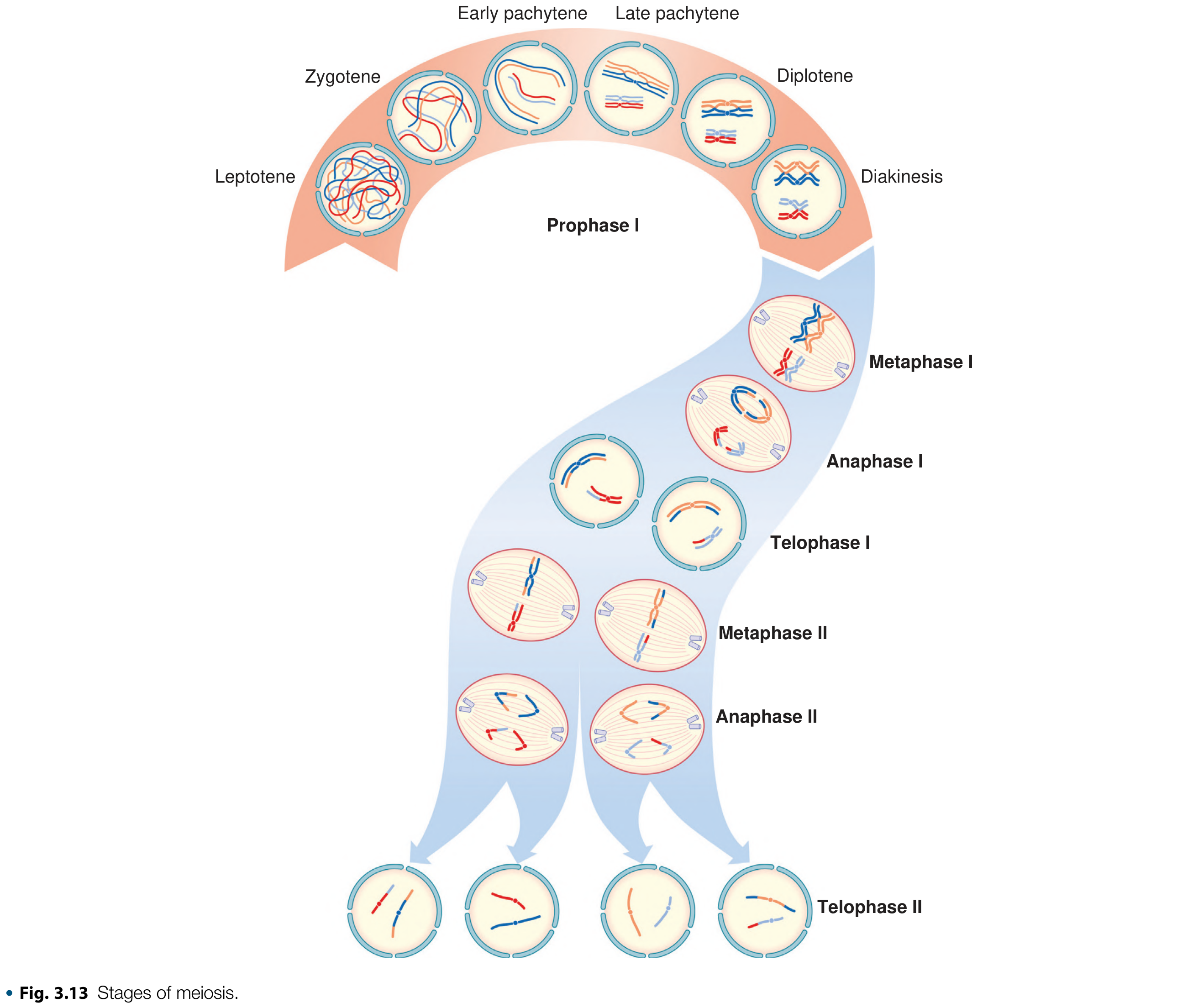
Fig. 3.13 - Complete stages of meiosis, showing Prophase I sub-stages, through to four haploid daughter cells at Telophase II.
MEIOSIS I (Reduction Division)
Prophase I - The Most Complex Stage (5 sub-stages)
This is the most important and longest stage in all of meiosis. Homologous chromosomes pair up (a process called synapsis).
| Sub-stage | What Happens | Memory Trick |
|---|---|---|
| Leptotene | Chromosomes become visible as they start to condense | Lepto = thin |
| Zygotene | Homologous chromosomes align and pair up (synapsis); held together by synaptonemal complexes | Zygo = joined |
| Pachytene | Paired chromosomes (bivalents) become tightly coiled. Crossing over (recombination) occurs - homologous segments of DNA exchange between non-sister chromatids | Pachy = thick |
| Diplotene | Homologous chromosomes begin to separate but remain attached at chiasmata (points of crossing over) | Diplo = double |
| Diakinesis | Maximum condensation. Chromosomes prepare to separate | Dia = across |
Bivalent (Tetrad): During Prophase I, each pair of homologous chromosomes (with their sister chromatids) forms a structure of 4 chromatids = a bivalent or tetrad.
Crossing Over: Exchange of genetic material between non-sister chromatids of homologous chromosomes at Pachytene. This is the single most important source of genetic variation. The sites of crossing over are called chiasmata (singular: chiasma).
Metaphase I
- Nuclear membrane disappears
- Bivalents (pairs of homologous chromosomes) align at the equatorial plate
- Each homolog is attached to spindle fibers from opposite poles
Anaphase I
- Homologous chromosomes separate to opposite poles (centromeres do NOT split here - this is different from mitosis!)
- Each chromosome still consists of two chromatids
Telophase I
- Each pole now has a haploid set of chromosomes (23 in humans), but each chromosome still has 2 chromatids
- Cell divides - two secondary spermatocytes or oocytes are formed
- Chromosome number is halved - this is why Meiosis I = Reduction Division
MEIOSIS II (Equational Division)
This is essentially the same as mitosis. No new DNA replication occurs between Meiosis I and II.
Prophase II
Chromosomes condense again, spindle forms.
Metaphase II
Chromosomes (each still with 2 chromatids) align at equatorial plate.
Anaphase II
Centromeres split - sister chromatids separate to opposite poles.
Telophase II
Nuclear membranes reform. Cytokinesis.
Result: From each secondary gametocyte, 2 more cells are produced.
Final Products of Meiosis
| Males (Spermatogenesis) | Females (Oogenesis) | |
|---|---|---|
| Starts | Puberty | Early embryonic life |
| Duration | 60-65 days | 10-50 years |
| Products per meiosis | 4 spermatids (each becomes a sperm) | 1 ovum + 3 polar bodies |
| Gametes produced | 100-200 million/ejaculate | 1 ovum per menstrual cycle |
Why only 1 ovum? During Meiosis I, the oocyte divides unequally - one cell gets most of the cytoplasm (secondary oocyte) while the other becomes a polar body (degenerates). The same happens in Meiosis II. This ensures the ovum has enough resources to support embryo development.
6. Mitosis vs. Meiosis - Comparison Table
| Feature | Mitosis | Meiosis |
|---|---|---|
| Where it occurs | Somatic (body) cells | Gonads (ovaries/testes) |
| Purpose | Growth, repair, asexual reproduction | Sexual reproduction - gamete formation |
| Number of divisions | 1 | 2 (I + II) |
| Daughter cells produced | 2 | 4 |
| Chromosome number in daughter cells | Same as parent (2n = 46, diploid) | Half of parent (n = 23, haploid) |
| Genetic identity | Daughter cells identical to parent | Daughter cells genetically unique |
| Synapsis / Crossing over | Does NOT occur | Occurs in Prophase I |
| Prophase length | Relatively short | Very long (especially Prophase I) |
| Centromere splits | At Anaphase | At Anaphase II (not Anaphase I) |
7. Significance of Cell Division
Significance of Mitosis:
- Enables growth of the organism
- Repairs and regenerates tissues
- Maintains the same chromosome number in all somatic cells
- Basis of asexual reproduction in lower organisms
Significance of Meiosis:
- Maintains the chromosome number of a species across generations (halving + fusion at fertilization = constant 2n)
- Generates genetic diversity in two ways:
- Independent assortment - Homologs separate independently during Meiosis I; probability of two identical gametes = 1 in 2^23 (≈ 1 in 8 million)
- Crossing over - Exchanges segments of DNA between homologous chromosomes at Pachytene; makes the probability of two genetically identical gametes virtually zero
- These two mechanisms together are the reason no two people (except identical twins) are genetically identical
8. Quick Memory Aids
Phases of Mitosis: Prophase → Prometaphase → Metaphase → Anaphase → Telophase
- Mnemonic: "Please Pass Me A Trophy"
Prophase I sub-stages (Meiosis): Leptotene → Zygotene → Pachytene → Diplotene → Diakinesis
- Mnemonic: "Lazy Zebras Play During Daylight"
Key numbers to remember:
- Human somatic cells: 2n = 46 (diploid)
- Human gametes: n = 23 (haploid)
- Telomeres allow about 50-60 cell divisions maximum
- Crossing over: ~40 recombination events per meiosis per gamete
9. Common Exam Questions (NCERT / NEET)
Q: What is the significance of G0 phase?
Cells that have permanently stopped dividing (neurons, heart muscle cells) exit the cycle at G1 and enter G0. They remain metabolically active but do not divide.
Q: Why is meiosis I called the reduction division?
Because the chromosome number is halved during Meiosis I - homologous chromosomes separate, reducing from 2n (46) to n (23).
Q: What is a synaptonemal complex?
A protein filament structure that holds homologous chromosomes together during Zygotene of Prophase I, facilitating synapsis and crossing over.
Q: Why is crossing over important?
It shuffles genetic material between homologous chromosomes, creating new combinations of alleles in gametes - the primary source of genetic variation in sexually reproducing organisms.
Q: What happens to polar bodies?
They degenerate (and are not fertilized). Their purpose is to get rid of the extra chromosomes after meiosis while keeping the cytoplasm concentrated in the one functional egg.
Q: At which stage of mitosis do chromosomes appear most distinct?
Metaphase - chromosomes are maximally contracted and aligned at the equatorial plate. This is why karyotyping is done at this stage.
Sources: Emery's Elements of Medical Genetics and Genomics (9780702079665), Ganong's Review of Medical Physiology 26e (9781260122404)
This is a shared conversation. Sign in to Orris to start your own chat.