

What is this
herpes zoster shingles dermatomal grouped vesicles on erythematous base shoulder
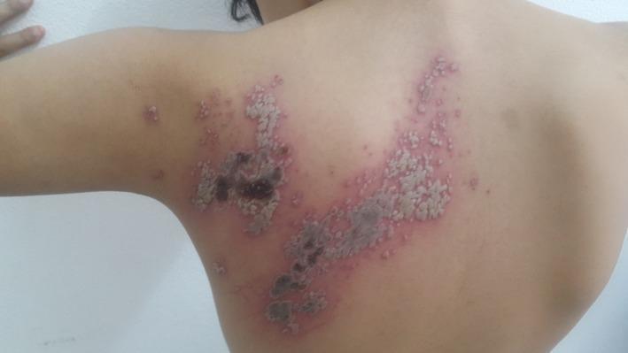
This clinical photograph displays a dermatomal distribution of skin lesions across the left upper back and posterior shoulder of a child. The lesions are characteristic of Herpes Zoster (shingles), appearing as multiple grouped vesicles in a 'bouquet' arrangement along a specific thoracic metamere (likely T4). The morphology varies, showing a progression of disease stages: clusters of clear or whitish vesicles on an erythematous base, alongside areas of dark, necrotic crusting and scabbing. The rash exhibits a distinct band-like, unilateral pattern that does not cross the spinal midline. This visual serves as a primary example of Herpes Zoster in a pediatric patient, illustrating typical cutaneous manifestations including vesiculation, local necrosis, and the characteristic neuro-cutaneous distribution following a dermatome.
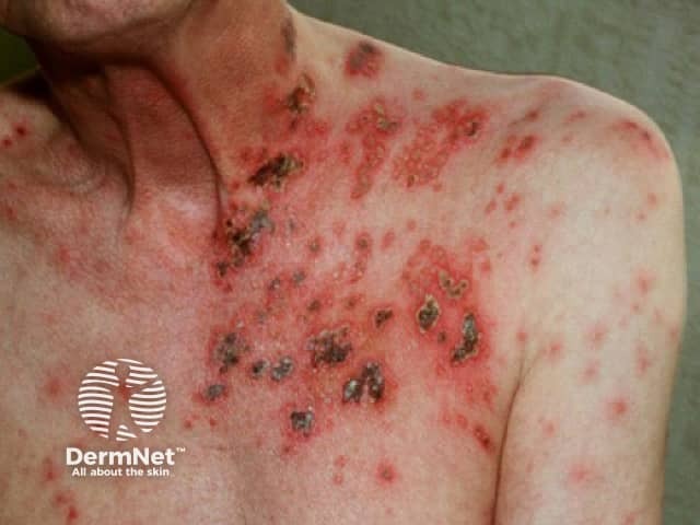
Imaging modality: Clinical photography of the cutaneous surface. Perspective: Anterior view of the left shoulder and anterior chest. The skin demonstrates a vesiculopapular eruption along a unilateral dermatomal distribution, with clusters of small fluid-filled vesicles on an erythematous base and variably crusted lesions. Lesions appear in different stages, including active vesicles, rupture-related erosions, and crusts, consistent with herpes zoster (varicella-zoster virus reactivation). The distribution follows a thoracic dermatome, with sharp demarcation from unaffected skin; surrounding areas show mild edema and erythema. This pattern—acute onset of painful, grouped vesicles in a girdle-like belt—supports a clinical diagnosis of shingles. In such images, differential considerations include herpes simplex, chickenpox, contact dermatitis, and other vesiculobullous conditions; however, dermatomal confinement and age-related risk favor VZV reactivation. Clinical significance centers on prompt antiviral therapy (acyclovir, valacyclovir, famciclovir) within 72 hours of rash onset to reduce duration and risk of postherpetic neuralgia; vaccination history and immune status are relevant for prognosis. This image has educational value for dermatology, infectious disease, and primary care training; it demonstrates typical morphology of shingles and the dermatomal distribution essential for accurate diagnosis and triage in suspected varicella-zoster infection. Clinical relevance emphasized.
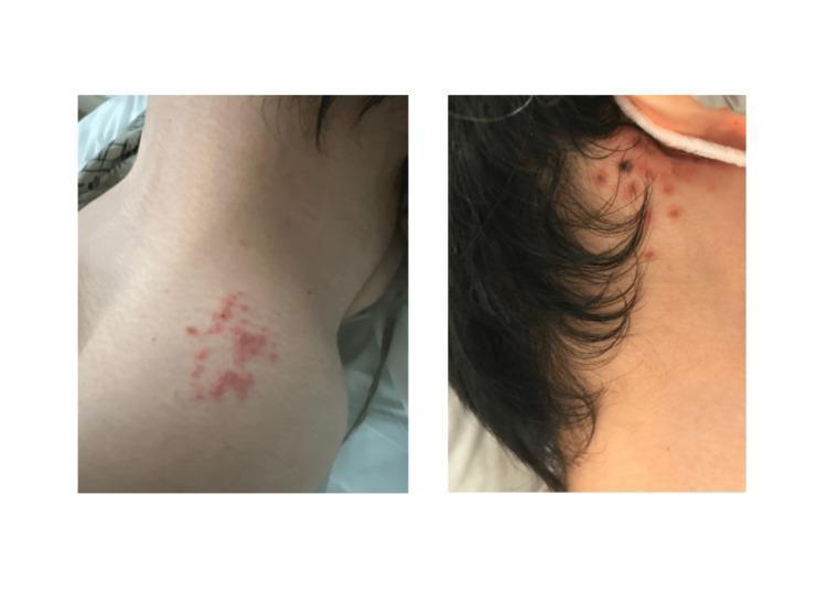
Two-panel clinical photograph demonstrating a localized vesicular rash consistent with Varicella-Zoster Virus (VZV) infection. The left panel shows a cluster of erythematous, fluid-filled vesicles on the left shoulder. These lesions are grouped in an irregular, somewhat branching morphology on an erythematous base, characteristic of 'dewdrops on a rose petal.' The right panel displays the posterior auricular region (behind the right ear), featuring multiple discrete vesicles. Some of these post-auricular lesions exhibit darker pigmentation and crusting, suggesting an older stage of progression compared to the shoulder lesions. Mild surrounding inflammation is visible, and the distribution is suggestive of a dermatomal pattern. This visual serves as a classic educational example of Herpes Zoster (shingles) presentation, illustrating different stages of lesion evolution—from active vesicles to crusting—and typical localized distribution.
"herpes zoster"[MeSH Terms] AND "antiviral agents"[MeSH Terms]
- Red, inflamed skin on the shoulder and upper back, in a band-like patch on one side.
- Small clustered blisters and pustules on the red base. Some have crusted or eroded, so the lesions are at different stages.
- The pattern follows a dermatome, which is the strip of skin supplied by one nerve root.
- Herpes simplex (zosteriform).
- Contact dermatitis.
- Impetigo.
- Insect bites.
- Scabies.
- See a doctor within 72 hours of the rash starting. Antivirals (valacyclovir, famciclovir or acyclovir) work best if started in that window. They shorten the illness and lower the risk of lasting nerve pain (postherpetic neuralgia). Treatment later than that is still often given.
- Seek care urgently if the rash is near the eye or on the face or ear, if you are immunocompromised, pregnant or older, if the pain is severe, or if the rash spreads widely.
- Until the blisters crust over, keep the area covered. Fluid from the blisters can give chickenpox to anyone who hasn't had it or been vaccinated, so avoid pregnant women, newborns and immunosuppressed people.
- Cool compresses and simple painkillers can ease discomfort. Don't pop the blisters.
- The recombinant zoster vaccine (Shingrix) helps prevent recurrence, once the acute episode has settled.
It seems painful
- See a doctor today or tomorrow if you can. Antivirals work best when started within 72 hours of the rash appearing. A same-day urgent care or GP visit is reasonable, because this is a Saturday and the window may be closing.
- Treatment a doctor will likely give:
- An antiviral such as valacyclovir, famciclovir or acyclovir, usually for 7 days. The dose depends on your kidney function and age, so don't pick one yourself.
- Pain relief, starting with paracetamol/acetaminophen or an NSAID if you can safely take them.
- Stronger options such as gabapentin, pregabalin or a short opioid course for severe pain.
- Lidocaine patches or cream once the skin is intact. Don't put them on open sores.
- At home:
- Cool, damp compresses for 15 to 20 minutes.
- Loose, soft clothing. The strap or fabric rubbing on that shoulder may hurt.
- Keep the rash clean and covered. Don't pop or scratch the blisters.
- Avoid pregnant women, newborns and immunosuppressed people, and anyone who hasn't had chickenpox or the vaccine, until all the blisters have crusted over.
- Rash or pain near the eye, on the tip of the nose, or around the ear, or any new vision change, facial droop or hearing loss.
- Fever, confusion, a severe headache or a stiff neck.
- Weakened immunity, for example from steroids, chemotherapy, HIV or a transplant.
- Spreading redness, warmth, pus or swelling, which suggests a bacterial infection.
- Pain so severe that you can't control it.