
Development female internal genitalia with with complete anatomy lymphatics blood circulation applies aspects with simpke diagrams for exam
female internal genitalia anatomy diagram uterus ovary fallopian tube
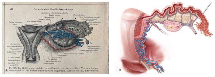
A side-by-side comparison of female pelvic anatomy illustrations. Figure A is a historical anatomical diagram from a 1903 atlas (Dr. Carl Toldt), providing a posterior view of the uterus, adnexa, and parametrium with extensive German and Latin labeling. Key structures include the uterine wall layers (Tunica muscularis and serosa), the cervical canal (Canalis cervicis), and the fallopian tube (Pars uterina, Isthmus, and Ampulla tubae). It details the epoophoron, mesosalpinx, and the fimbriated end near the ostium abdominale. Figure B is a modern medical illustration by Markus Voll, showing a coronal sectional view of the uterus and adnexa. This modern rendering emphasizes the intricate vascular supply, depicting the dense network of uterine arteries and veins within the myometrium. It also highlights the internal mucosal folds of the fallopian tube and the anatomical relationship with the ovary. Both images serve to demonstrate the consistency of anatomical knowledge and the evolution of medical visualization techniques in gynecology and pelvic surgery.
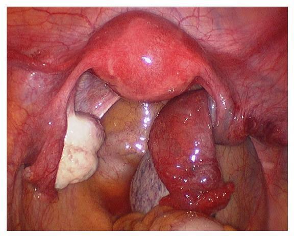
This diagnostic laparoscopic photograph illustrates the female internal pelvic reproductive organs, providing a clear comparison between normal anatomy and adnexal pathology. Centrally located is the uterus, appearing smooth, pear-shaped, and pinkish-red. To the viewer's left, the left ovary is visible as a pale, white, nodular structure with a healthy appearance, accompanied by a normal-looking fallopian tube. In stark contrast, the right adnexa (viewer's right) exhibits signs of acute pathology. The right ovary and fallopian tube are significantly enlarged, edematous, and demonstrate a dusky, deep-red to purple discoloration, consistent with venous congestion and hemorrhagic infarction. The right fallopian tube appears thickened and twisted upon its pedicle. This visual presentation is characteristic of ovarian torsion, a surgical emergency where the twisting of the adnexa compromises blood supply. The image is highly relevant for medical education in gynecology and emergency medicine, demonstrating the clinical signs of ischemic adnexal torsion compared to contralateral healthy structures.
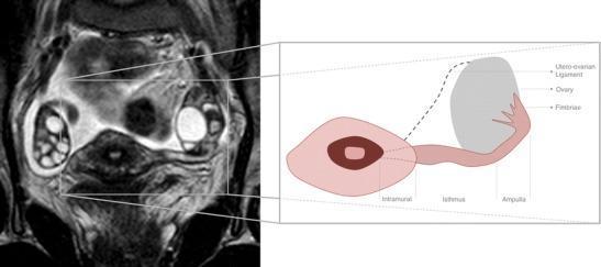
This composite educational graphic features a diagnostic MR image alongside an anatomical diagram, focusing on the female reproductive system. The left panel shows a T2-weighted coronal oblique MRI section of the pelvis from a 32-year-old female. Visible structures include a centrally positioned uterus, bilateral ovaries containing multiple high-signal intensity (bright) follicles, and the full longitudinal course of the left fallopian tube. The fallopian tube appears as a thin, convoluted, slightly hyperintense structure extending laterally toward the ovary. The right panel is a stylized anatomical illustration that maps and labels the segments of the fallopian tube based on the adjacent MRI. Labeled segments include the intramural (interstitial) portion within the uterine wall, the isthmus, the ampulla, and the fimbriae at the lateral infundibulum near the ovary. The diagram also identifies the utero-ovarian ligament. This visual is designed to teach normal pelvic anatomy and segmental division of the fallopian tubes, which are typically difficult to visualize on MRI unless surrounded by small amounts of peritoneal fluid.
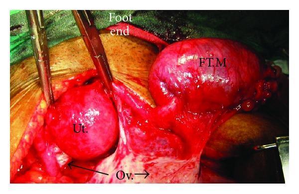
This clinical surgical photograph captures an intraoperative view of the female pelvic anatomy during a surgical procedure, specifically highlighting primary fallopian tube carcinoma. The image shows three major labeled structures: the uterus (Ut.), which appears as a reddish, spherical organ held by surgical retractors; the right ovary (Ov.), visible as a smaller, whitish, oval-shaped structure situated inferiorly; and a significant right tubal mass (FT.M). The tubal mass is markedly enlarged, elongated, and exhibits a hyperemic, irregular surface texture compared to the smoother uterus. Surgical instruments and retractors are positioned to expose the adnexa and uterine corpus. Annotations including 'Foot end' provide anatomical orientation. The image illustrates the gross morphological appearance of an adnexal mass that has originated from the fallopian tube, demonstrating its size relative to normal pelvic organs and its high vascularity, which are key diagnostic indicators for gynecologic oncology. This visual serves as an educational reference for distinguishing tubal pathology from primary ovarian or uterine lesions during laparotomy.
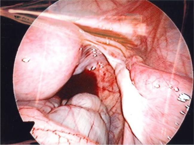
This endoscopic clinical photograph displays an intraoperative view of the female pelvic cavity during a laparoscopic ovarian cortex biopsy. Centrally, the uterus is visible as a smooth, rounded, pinkish organ. Extending from the right uterine horn is a healthy right Fallopian tube, characterized by its elongated, tubular structure and fine superficial vascularity. Adjacent to the Fallopian tube is the right ovary, which appears relatively small but shows typical pinkish coloration and a slightly nodular surface texture. Supporting ligaments, including the broad ligament, are seen as thin, translucent membranous tissues. The anatomical landmarks demonstrate a normal pelvic orientation with no evidence of major adhesions or large cysts. This image serves as a clinical example of pelvic anatomy in the context of fertility preservation procedures, particularly for patients with mosaic Turner syndrome where ovarian reserve may be diminished despite seemingly normal gross anatomy.
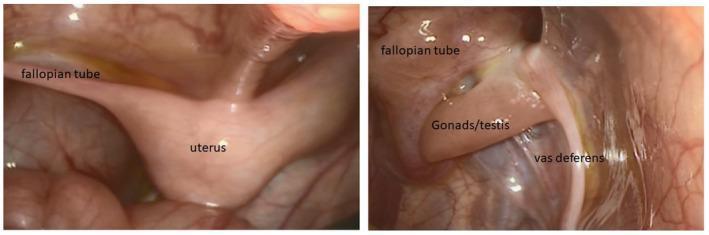
This diagnostic image consists of two labeled laparoscopy frames showcasing internal pelvic anatomy in a patient with ambiguous genitalia (46,XY DSD). The left panel illustrates a central, pear-shaped uterus-like structure with a fallopian tube extending laterally. The right panel displays complex internal reproductive anatomy, including a gonad identified as a testis, positioned adjacent to both a fallopian tube and a cord-like vas deferens. The presence of both Müllerian structures (uterus, fallopian tubes) and Wolffian structures (vas deferens) in an individual with testes is characteristic of persistent Müllerian duct syndrome or specific genetic variants affecting sex development, such as MAP3K1 mutations. The endoscopic view demonstrates the morphological appearance and spatial relationships of these ductal structures within the pelvic cavity, providing critical clinical evidence for surgical planning and diagnostic classification in pediatric endocrinology and urology.
Mullerian duct paramesonephric duct development uterus embryology
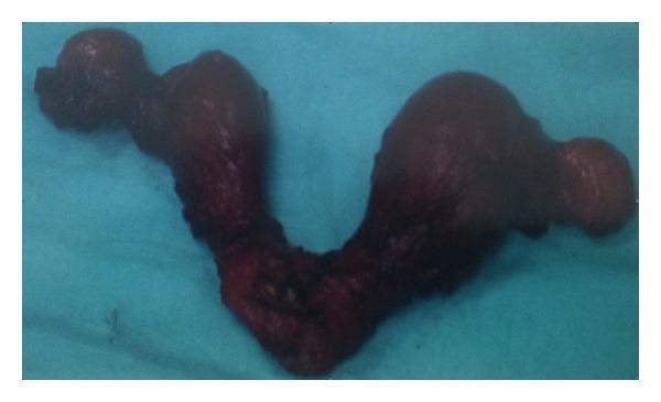
This clinical photograph shows a posterior view of a gross pathological specimen of a didelphys uterus. The image displays a complete Müllerian duct anomaly characterized by two entirely separate uterine bodies (horns), each with its own fundus, resulting in a distinct 'V' or bilobed shape. The specimen appears as dark, reddish-brown tissue against a blue surgical drape. The morphology demonstrates total non-fusion of the paramesonephric ducts, leading to the formation of two distinct uterine cavities and two cervices, which converge at a common lower point. The surface of the tissue is irregular and nodular, particularly in the fundal regions, which is consistent with the clinical diagnosis of uterine carcinosarcoma in one of the horns. This visual serves as a clear educational example of a Class III Müllerian duct anomaly (uterine didelphys) and its associated pathological findings.
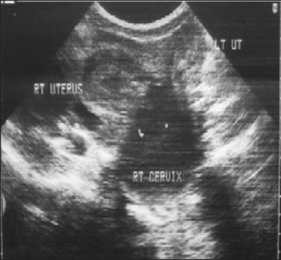
Diagnostic Image: Transabdominal ultrasound of the female pelvis in a transverse plane. The imaging reveals a Müllerian duct anomaly, specifically a didelphys uterus characterized by two distinct, widely divergent uterine horns labeled 'RT UTERUS' and 'LT UT'. There is no visible communication between the two endometrial cavities. Below the right uterine horn, the 'RT CERVIX' is shown to be significantly distended and enlarged, containing low-level internal echoes consistent with hematocervix or fluid accumulation. The anatomy demonstrates a failure of fusion of the paramesonephric ducts. This visual is clinically significant for diagnosing obstructive reproductive tract anomalies and teaching the sonographic presentation of uterine malformations associated with possible cervical stenosis or vaginal septa.
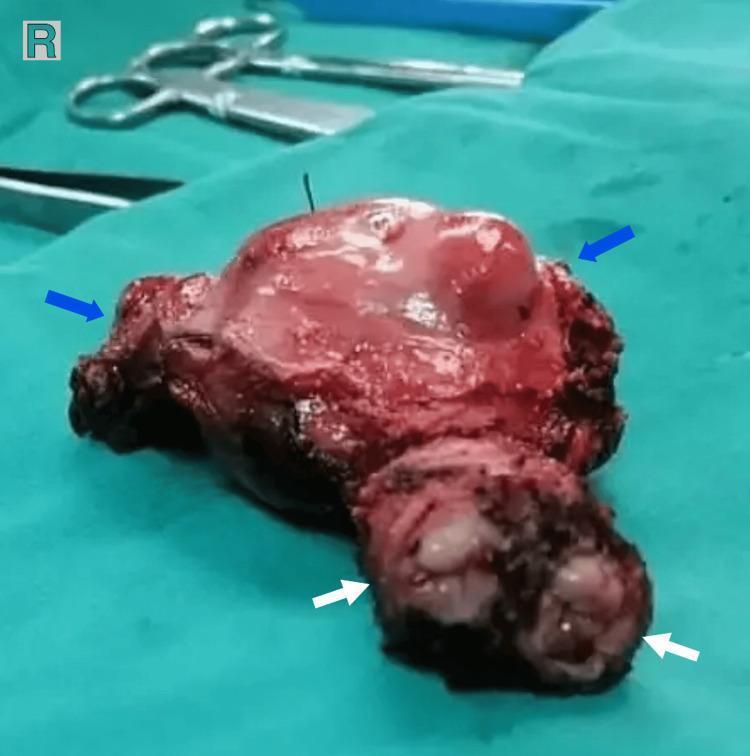
This clinical photograph shows a gross pathological specimen of a total abdominal hysterectomy featuring a congenital Mullerian duct anomaly, specifically a didelphys uterus. The specimen, measuring approximately 9 x 5 x 4 cm, is displayed on a green surgical drape. Blue arrows indicate two distinct uterine horns: the left horn (40 mm) and the right horn (35 mm). The upper portion of the specimen shows a smooth, reddish-pink serosal surface with visible hemorrhagic areas. White arrows at the inferior aspect of the specimen point to two separate cervixes, which share a common cervical canal. This anatomical variation is characteristic of uterine didelphys, a class III Mullerian anomaly resulting from the failure of the paramesonephric ducts to fuse during embryogenesis. The image provides significant educational value for gynecology and pathology, illustrating both the gross external morphology of a double uterus and the associated cervical duplication, which are key diagnostic features in reproductive tract malformations.
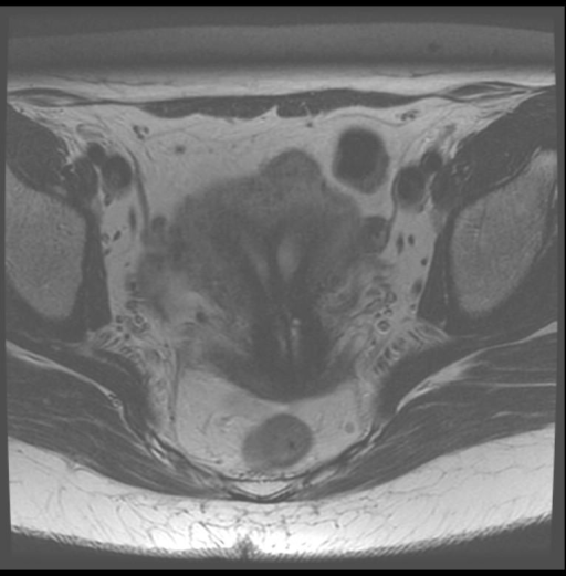
**Imaging Modality:** Magnetic Resonance Imaging (MRI) **Anatomical Region:** Female pelvis, axial cross-section **Observed Pathology:** Müllerian duct anomaly, specifically consistent with a **bicornuate uterus**. **Characteristic Visual Features:** The image displays a deep indentation of the external fundal contour, splitting the superior aspect of the uterus into two distinct cornua (horns). Centrally, a prominent muscular septum is visible, which separates the two diverging endometrial cavities. The pelvic musculature, iliac bones, and subcutaneous fat are visible in the periphery. **Key Diagnostic Features:** - **External Contour:** Deep fundal cleft (>1 cm), a primary feature used to differentiate a bicornuate uterus from a septate uterus. - **Endometrial Cavities:** Two separate, widely divergent hyperintense endometrial signals. - **Uterine Morphology:** "Heart-shaped" appearance of the uterine fundus on this axial slice. **Clinical Significance:** This radiological finding represents a fusion anomaly of the paramesonephric (Müllerian) ducts. It is essential for obstetric and gynecological indexing, as it is associated with increased risks of malpresentation, recurrent pregnancy loss, and preterm labor.
female reproductive tract blood supply lymphatic drainage anatomy
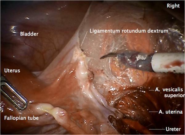
This clinical photograph captures a laparoscopic view of the female pelvic anatomy during a surgical procedure, specifically highlighting the lateral peritoneal incision and preparation of the vascular mesometrium. The field shows the uterus, fallopian tube, and bladder as primary landmarks. Labeled structures include the right round ligament (ligamentum rotundum dextrum), which is being divided by a surgical dissecting instrument. Key vascular and ductal structures are identified in the retroperitoneal space: the superior vesical artery (A. vesicalis superior), the uterine artery (A. uterina), and the ureter. The image demonstrates the spatial relationships between the Müllerian compartment and the underlying pelvic vasculature and urinary tract. This educational visual is intended for gynecologic oncologists and residents to understand the surgical steps of radical pelvic mesometrial resection (rPMMR) and the importance of identifying anatomical landmarks to ensure safe dissection and complete resection of lymphatic drainage pathways.
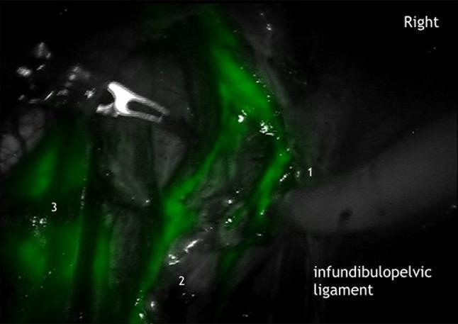
This intraoperative clinical photograph demonstrates Indocyanine Green (ICG) fluorescence imaging used for real-time lymphography during a surgical procedure. The image depicts the mesonephric lymphatic drainage pathways within the female pelvic anatomy. Bright green linear fluorescence highlights active lymphatic trunks tracking along the right ovarian vein (labeled 1) and the right ovarian artery (labeled 2). These vessels are seen within the infundibulopelvic ligament, indicating the transport of lymphatic fluid from the uterine corporal sub-compartment toward higher nodes. A more diffuse and concentrated area of fluorescence is visible in the common iliac lymph basin (labeled 3), representing a primary drainage station. This visualization assists in identifying the mesonephric pathway to para-aortic nodes, distinguishing it from the pelvic pathways. The use of ICG here facilitates precise anatomical mapping of sentinel lymph nodes and lymphatic networks in oncological or gynecological surgery, allowing for better identification of the Müllerian and mesonephric compartments.
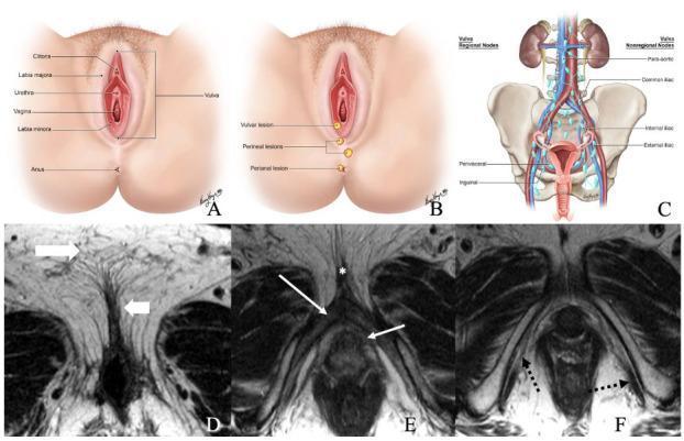
A multi-panel educational figure illustrating the normal anatomy of the female vulvar region and its lymphatic drainage. Panels A and B are anatomical illustrations showing external structures including the clitoris, labia majora and minora, urethra, vagina, and anus, with markers for typical lesion locations. Panel C is a schematic diagram of the lymphatic system, detailing regional (inguinal, perivisceral) and non-regional (external iliac, internal iliac, common iliac, and para-aortic) lymph nodes relative to the pelvis and kidneys. Panels D–F present axial non-fat saturated T2-weighted MRI scans of a 36-year-old female. Image D identifies the labia majora and labia minora through their signal intensities. Image E highlights the deep structures including the bulb of the vestibule, glans clitoris, and crus of the clitoris. Image F demonstrates the ischiocavernosus muscle. This content provides foundational anatomical context for the staging and clinical evaluation of vulvar cancer.
This clinical photograph shows a detailed anatomical dissection of the human stomach's lymphatic system in a cadaveric specimen. The image highlights the complex network of gastric lymph nodes and their associated vessels without the use of standard injections like cedar oil, though some blue-green vital dye staining is visible on specific lymphatic structures to aid visualization. The central anatomy includes the lesser curvature and retroperitoneal space, showing fleshy, tan-to-pinkish-brown visceral organs interspersed with numerous lymph nodes of varying sizes. Significant branching of mesenteric and gastric blood vessels is observed, with lymphatic channels following the vascular supply. The spatial organization demonstrates the high density of the lymphatic network near the left gastric, hepatic, and splenic drainage areas, which is clinically significant for understanding the pathways of gastric cancer metastasis. The textures are glistening and moist, typical of a surgical or high-fidelity cadaveric dissection, illustrating the intimate relationship between the lymphatic vessels and the surrounding adipose and connective tissues.
Development of Female Internal Genitalia - Complete Anatomy, Blood Supply & Lymphatics
PART 1 - EMBRYOLOGICAL DEVELOPMENT
A. Indifferent Stage (up to week 6)
- Mesonephric (Wolffian) duct - runs lateral to the mesonephros
- Paramesonephric (Mullerian) duct - arises as a longitudinal invagination of the urogenital ridge epithelium, opens cranially into the abdomen with a funnel-shaped end
B. Female Genital Duct Development (Week 6 onwards)
| Structure | Embryological Origin |
|---|---|
| Uterine tubes | Cranial 2 parts of paramesonephric duct |
| Uterus (body + cervix) | Caudal fused paramesonephric ducts |
| Upper vagina (~upper 1/3) | Caudal fused paramesonephric ducts |
| Lower vagina (~lower 2/3) | Sinovaginal bulbs from urogenital sinus |
| Broad ligament | Transverse pelvic fold formed when fused ducts pull urogenital ridges to lie transversely |
| Ovary | Gonadal ridge (genital ridge) mesoderm + primordial germ cells from yolk sac |
- Epoophoron (in broad ligament near ovary)
- Paroophoron (in broad ligament, medial to epoophoron)
- Gartner's cyst (along lateral vaginal walls) - clinically important!
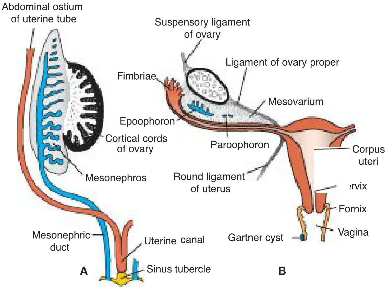
C. Vaginal Development (Sinovaginal Bulbs)
INDIFFERENT STAGE (wk 4-6)
Both ducts present
       ↓
  NO testosterone / NO AMH
       ↓
Wolffian duct → DEGENERATES (leaves Gartner's cyst, epoophoron, paroophoron)
Mullerian duct → DEVELOPS
       ↓
Cranial 2/3 → Uterine tubes (each side separate)
Caudal 1/3 → Fuses in midline
             ↓
    Uterovaginal canal
    (becomes uterus + upper vagina)
             ↓
    Sinus tubercle projects into urogenital sinus
             ↓
    Sinovaginal bulbs → Vaginal plate → Canalizes → Vagina (lower 2/3)
    Hymen = junction of the two
PART 2 - COMPLETE ANATOMY OF FEMALE INTERNAL GENITALIA
A. Uterus
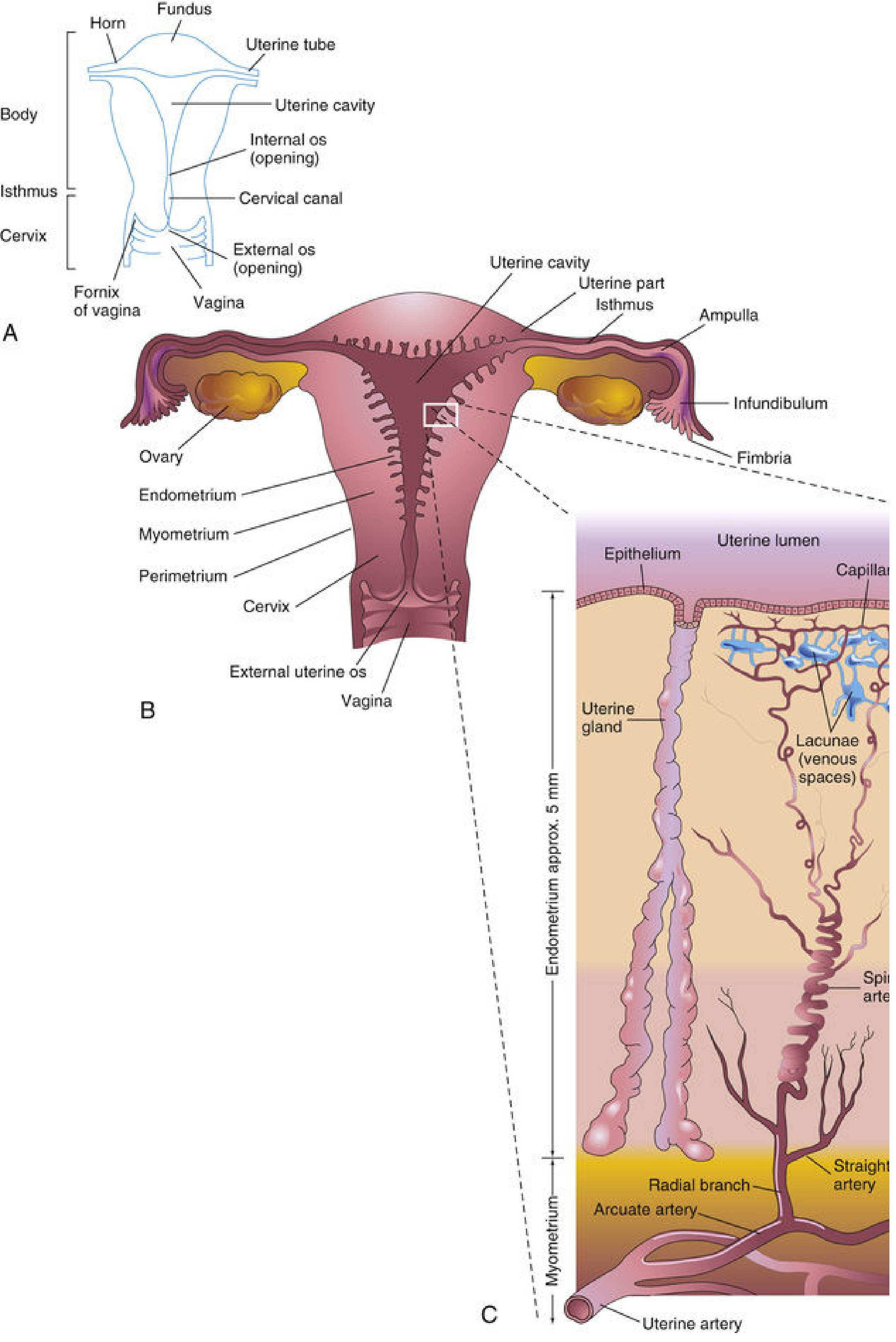
| Part | Description |
|---|---|
| Fundus | Dome above the uterine tube openings |
| Body (Corpus) | Main flattened part; cavity is an inverted triangle when viewed anteriorly |
| Isthmus | Narrow junction between body and cervix (clinically = lower uterine segment in pregnancy) |
| Cervix | Cylindrical lower part projecting into vagina; has internal os (above) and external os (below) |
- Endometrium - mucosa; functional layer shed in menstruation, basal layer regenerates
- Myometrium - thick smooth muscle
- Perimetrium - peritoneal covering
- Anterior: Vesicouterine pouch, bladder base
- Posterior: Rectouterine pouch (Pouch of Douglas), rectum
- Lateral: Broad ligament, ureter, uterine artery (ureter passes 1.5 cm lateral to cervix - "water under the bridge")
B. Fallopian Tubes (Uterine Tubes)
UTERUS ---[Intramural]---[Isthmus]---[Ampulla]---[Infundibulum/Fimbriae]--- OVARY
            (narrowest)   (narrow)  (widest,     (funnel-shaped; fimbriae
                                    fertiliz-    sweep egg into tube;
                                    ation site)  one large ovarian fimbria
                                                 attaches to ovary)
C. Ovaries
- Mesovarium - peritoneal fold attaching ovary to posterior broad ligament (contains hilum)
- Suspensory ligament (infundibulopelvic ligament) - connects upper pole to lateral pelvic wall; carries ovarian vessels and nerves
- Ligament of the ovary (ovarian ligament proper) - connects lower pole to uterine horn
- Germinal epithelium → Tunica albuginea → Cortex (follicles, corpus luteum) → Medulla (vessels, hilar/Leydig-like cells)
D. Vagina
- Fibromuscular tube ~8 cm (posterior wall slightly longer than anterior)
- Lined by stratified non-keratinized squamous epithelium (no glands; lubrication is transudate + cervical secretion)
- Vaginal vault/fornix = recess around cervix: posterior fornix (deepest - related to Pouch of Douglas), anterior fornix, lateral fornices
- pH 4.0-4.5 (lactic acid from Lactobacillus/Doderlein bacilli acting on glycogen)
PART 3 - LIGAMENTS & SUPPORTS
| Ligament | Attachment | Clinical Significance |
|---|---|---|
| Broad ligament | Uterus to lateral pelvic wall - contains uterine tube (mesosalpinx), ovary (mesovarium), round ligament (mesometrium) | Contains uterine artery, ureter |
| Round ligament | Uterine horn → inguinal canal → labia majora | Derived from gubernaculum; analogous to gubernaculum testis in male |
| Cardinal (Mackenrodt's) ligament | Cervix/upper vagina to lateral pelvic wall | Primary support against prolapse |
| Uterosacral ligament | Cervix to sacrum (S2-S4) | Keeps cervix posteriorly directed |
| Pubocervical ligament | Cervix to pubis | Anterior support |
PART 4 - BLOOD SUPPLY
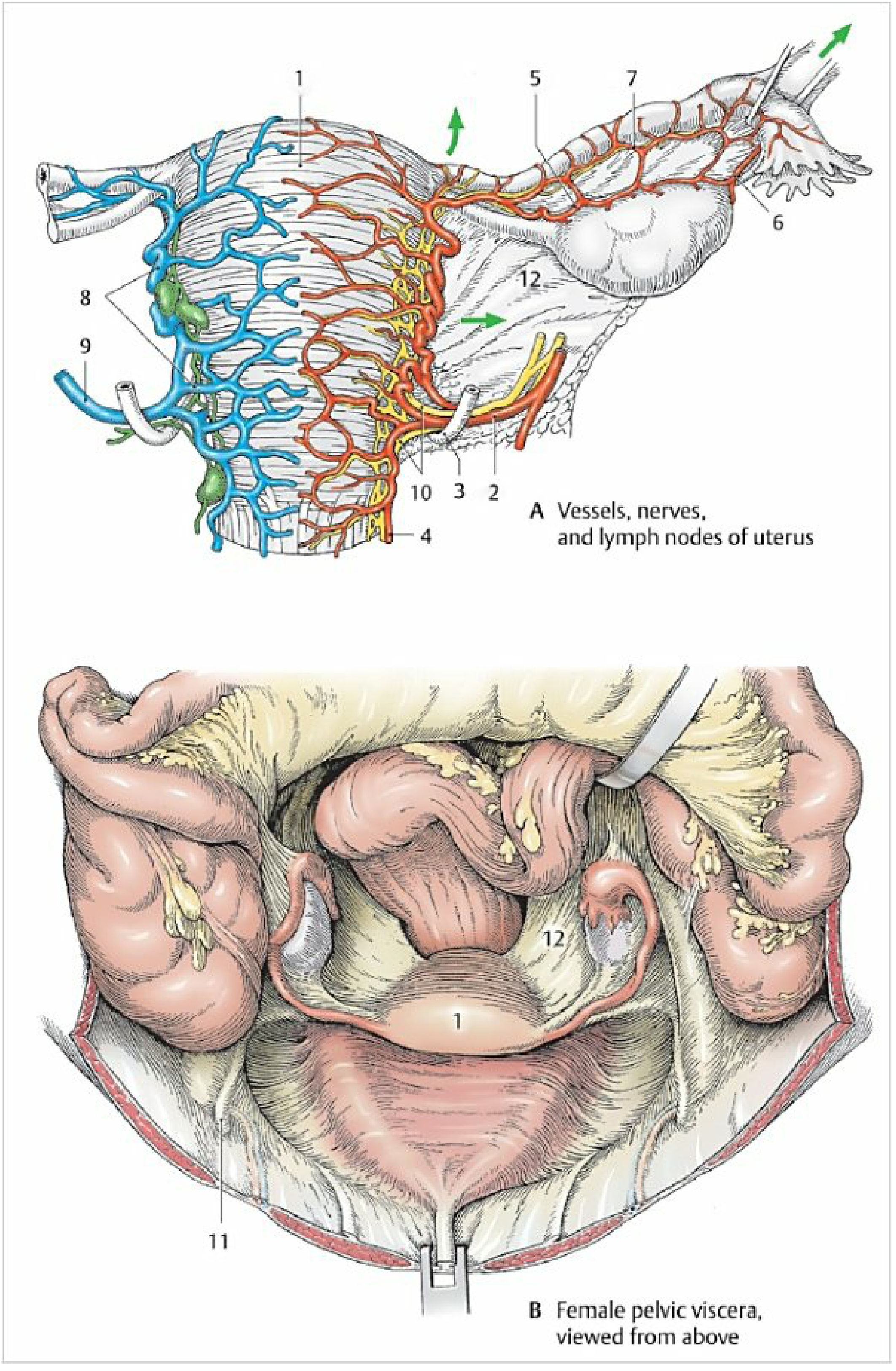
Uterus
- Branch of internal iliac artery (anterior division)
- Courses in subperitoneal connective tissue, crosses over the ureter at the base of broad ligament (~1.5 cm lateral to cervix)
- At uterine wall near cervix → divides into:
- Ascending branch - tortuous, runs along lateral uterine wall → fundus → gives ovarian branch (anastomoses with ovarian artery) and tubal branch
- Descending branch (vaginal artery) - supplies cervix and upper vagina
- Valveless uterine venous plexus (in parametrium) → uterine veins → internal iliac veins
Ovary
- Direct branch of abdominal aorta at L1 level (equivalent to testicular artery in male)
- Descends in suspensory ligament (infundibulopelvic ligament) → anastomoses with uterine artery ovarian branch
- Pampiniform venous plexus → ovarian vein
- Right ovarian vein → IVC (directly)
- Left ovarian vein → left renal vein (then to IVC) - explains why left ovarian vein thrombosis is more common
Fallopian Tube
- Medial 1/3 - uterine artery tubal branch
- Lateral 2/3 - ovarian artery
Vagina
- Upper part - vaginal branches from uterine artery
- Middle part - inferior vesical artery branches
- Lower part - internal pudendal artery
AORTA (L1)
├── Ovarian artery (direct) ────→ Ovary + lateral tube
│
INTERNAL ILIAC ARTERY
└── Uterine artery ─────────────→ Uterus (main)
    ├── Ascending branch
    │    ├── Ovarian branch ──── anastomoses with ovarian a.
    │    └── Tubal branch ────── medial tube
    └── Descending (vaginal) ──→ Cervix + upper vagina

INFERIOR VESICAL A. ──────────→ Middle vagina
INTERNAL PUDENDAL A. ─────────→ Lower vagina
PART 5 - LYMPHATIC DRAINAGE
Uterus (Most important for exam!)
FUNDUS & BODY OF UTERUS
├── 1. Along suspensory ligament of ovary (infundibulopelvic)
│        → Para-aortic (lumbar) nodes (at L1)
├── 2. Along round ligament
│        → Superficial inguinal nodes
└── 3. Via broad ligament
         → Common iliac nodes (at iliac bifurcation)

CERVIX
├── 1. External iliac nodes
├── 2. Internal iliac nodes
├── 3. Obturator nodes
└── 4. Sacral nodes (posterior)
Ovary
- Para-aortic (lumbar) nodes at L1 level (following ovarian vessels)
- Note: Same nodes as testis in male (both drain to para-aortic, NOT pelvic nodes)
Fallopian Tube
- Medial portion → uterine plexus → internal iliac nodes
- Lateral 2/3 → para-aortic nodes (via ovarian vessels)
Vagina
| Part | Drainage |
|---|---|
| Upper | Internal iliac nodes |
| Middle | Internal iliac nodes |
| Lower (below hymen) | Superficial inguinal nodes |
PART 6 - NERVE SUPPLY
- Parasympathetic: Pelvic splanchnic nerves (S2-S4)
- Sympathetic: T10-L1 via inferior hypogastric (pelvic) plexus
- Uterovaginal plexus (Frankenhäuser ganglion) - formed lateral to cervix; transmits uterine pain
- T10 (aortic plexus via ovarian vessels) - hence ovarian pain is referred to the umbilicus (T10 dermatome)
PART 7 - CONGENITAL ANOMALIES (Quick Exam Summary)
| Anomaly | Mechanism | Result |
|---|---|---|
| Uterus didelphys | Complete non-fusion | Double uterus + double cervix + often double vagina |
| Uterus bicornis | Partial non-fusion | Two horns entering one cervix and vagina |
| Uterus arcuatus | Slight midline indentation only | Mildest form; often asymptomatic |
| Uterus septus | Failed resorption of septum | Normal external shape, internal septum |
| Cervical/vaginal atresia | Both sides fail to fuse or sinovaginal bulbs fail to develop | Hematocolpos/hematometra |
| Gartner's cyst | Remnant of Wolffian duct | Cyst along anterior-lateral vaginal wall |
| Mayer-Rokitansky-Kuster-Hauser (MRKH) syndrome | Aplasia of Mullerian ducts | Absent uterus and upper vagina; normal ovaries and female karyotype |
- "Water under the bridge" - Ureter passes UNDER uterine artery (surgical danger in hysterectomy)
- Ovary/testis → Para-aortic nodes (not pelvic) - because they descended from lumbar region
- Cervix → Obturator nodes (most commonly affected in cervical cancer spread)
- Left ovarian vein → Left renal vein (not IVC directly) - explains left-sided varicocele/thrombosis more common
- Fertilization occurs in the ampulla of the fallopian tube
- Mullerian = female default; Wolffian = needs testosterone