
Extensor tendons of hand
I'll look up the extensor tendons of the hand from the medical library for a thorough answer.
Searching sources...
Reading File
Finding Sources
Searching Images
extensor tendons of hand anatomy diagram
Finding Sources
Reading File
Finding Sources
Finding Sources
Loading Image
Loading Image
Now let me get the key anatomical text on extensor compartments, zones, and the dorsal digital expansion:
Reading File
Reading File
Reading File
Reading File
Now let me get the extensor compartments text and zone descriptions from Miller's and Sabiston:
Finding Sources
Reading File
Now let me get the zones of injury from Sabiston and the Miller's extensor anatomy section:
Reading File
Reading File
I now have comprehensive information from multiple authoritative textbooks. Let me also fetch the zones-of-injury image from Sabiston:
Loading Image
Loading Image
I now have excellent material from multiple authoritative sources. Here is the comprehensive answer:
Extensor Tendons of the Hand
Overview
The extensor tendons of the hand form a complex system on the dorsum that extends the wrist, hand, and digits. They are all innervated by the radial nerve (posterior interosseous branch) and pass through six synovial-lined compartments beneath the extensor retinaculum at the wrist.
The Six Dorsal Compartments (Radial → Ulnar)
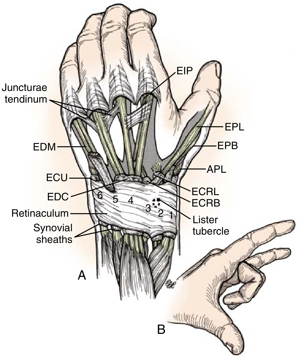
These are arranged from radial to ulnar, separated by septa of the extensor retinaculum. The Lister tubercle on the radius separates compartments 2 and 3.
| Compartment | Tendons | Notes |
|---|---|---|
| 1st | Abductor pollicis longus (APL), Extensor pollicis brevis (EPB) | Radial border of the anatomic snuffbox; site of de Quervain's tenosynovitis |
| 2nd | ECRL, ECRB | Insert on bases of 2nd and 3rd metacarpals; radial wrist deviators and extensors |
| 3rd | Extensor pollicis longus (EPL) | Ulnar border of the anatomic snuffbox; hooks around Lister's tubercle; inserts on distal phalanx of thumb |
| 4th | EDC (II-V), Extensor indicis proprius (EIP) | The major finger extensor; EIP always lies ulnar to the EDC tendon of the index finger |
| 5th | Extensor digiti minimi (EDM) | Lies over the distal radioulnar joint; always ulnar to the EDC of the little finger |
| 6th | Extensor carpi ulnaris (ECU) | Ulnar wrist deviation and extension |
- Miller's Review of Orthopaedics, 9th Ed.; Roberts and Hedges' Clinical Procedures in Emergency Medicine; Sabiston Textbook of Surgery
Key Tendons and Their Insertions
Extensor Digitorum Communis (EDC)
- Extends fingers II-V via a single muscle belly splitting into four tendons
- Inserts loosely at the base of the proximal phalanx (via sagittal bands), then trifurcates over the proximal phalanx into:
- A central slip → inserts at the base of the middle phalanx
- Two lateral branches → join the interosseous/lumbrical lateral bands → fuse to form the terminal extensor mechanism (TEM) → inserts at the base of the distal phalanx
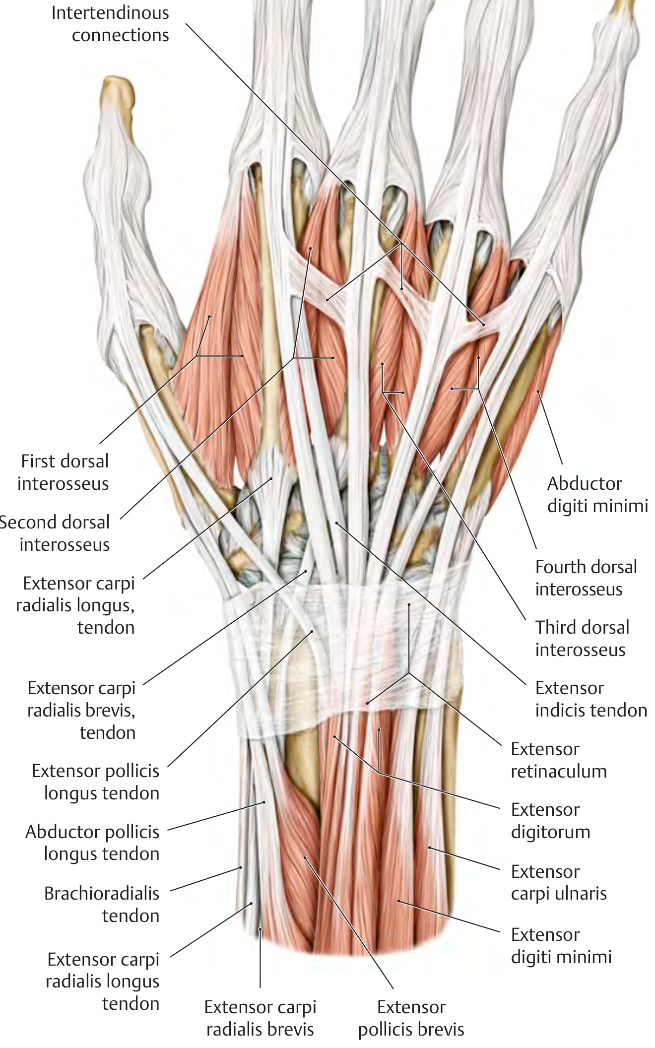
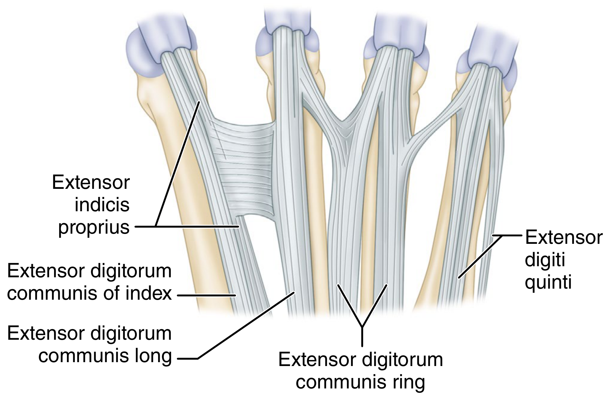
- Tendons are interlinked on the dorsum by oblique bands called juncturae tendinum (intertendinous connections) - this is why a complete EDC laceration may not abolish extension entirely
Extensor Indicis Proprius (EIP) and Extensor Digiti Minimi (EDM)
- The index and little fingers have their own dedicated extensor muscles, allowing independent extension
- Their tendons always lie on the ulnar side of the corresponding EDC tendon
- No junctura tendinum is present on the EIP tendon
Thumb Extensors
- EPL: extends IP joint; forms ulnar border of anatomic snuffbox
- EPB: extends MCP joint; forms radial border of anatomic snuffbox
- APL: abducts thumb; inserts on base of 1st metacarpal
The Dorsal Digital Expansion (Extensor Hood)
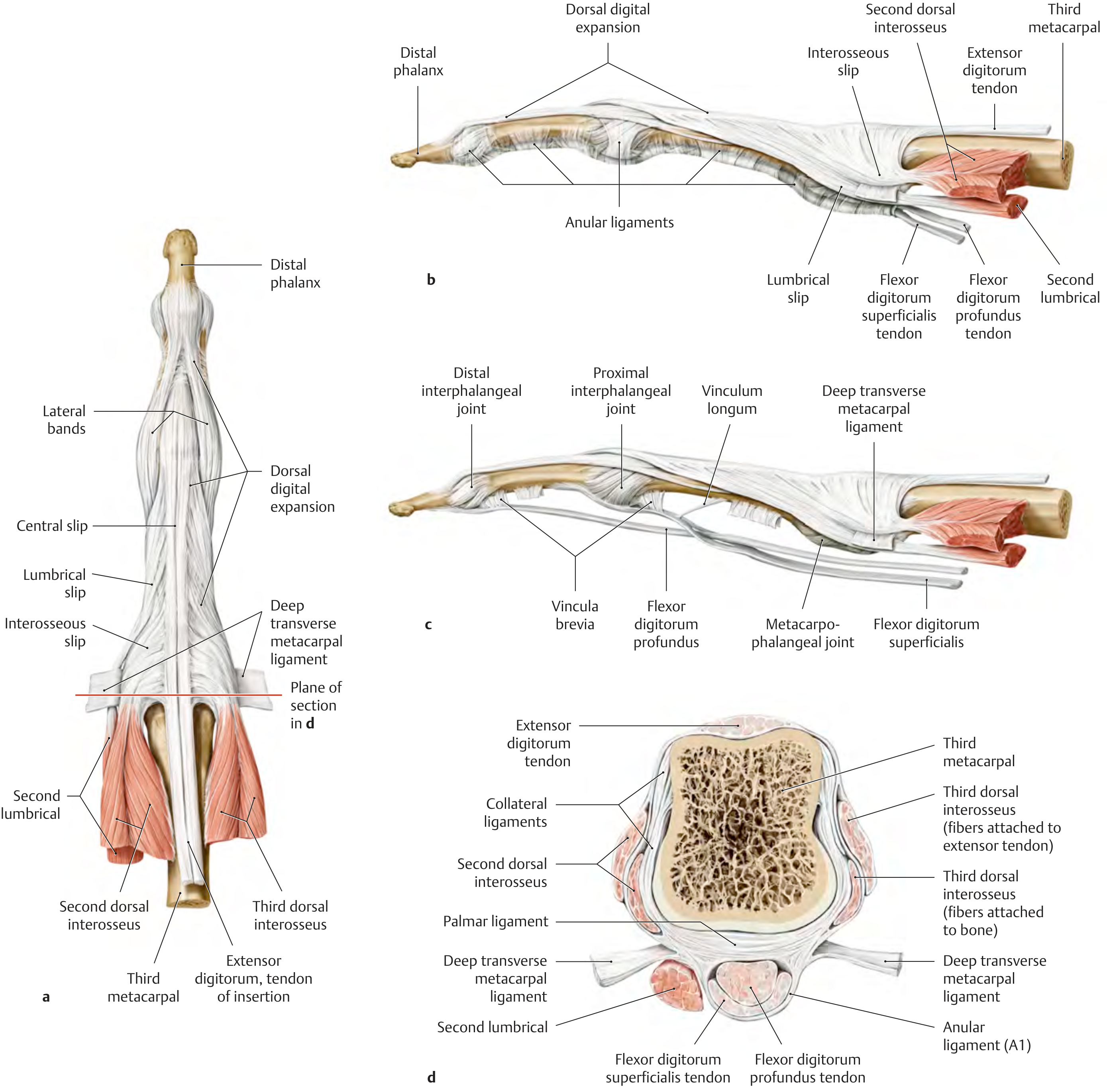
This is not merely an aponeurosis but a complex interwoven system of fiber bands that allows coordinated action of extrinsic and intrinsic muscles over all three finger joints.
Key components:
-
Sagittal bands (proximal hood at MCP level): loop around the joint and blend into the volar plate; keep the EDC tendon centered over the metacarpal head; extend the MCP joint
-
Central slip: EDC central continuation to the base of the middle phalanx; extends the PIP joint
-
Lateral bands: contributions from lumbricals and interossei; lie dorsal to the axis of the PIP joint so they act as PIP extensors and MCP flexors
-
Triangular ligament: connects the two lateral bands over the middle phalanx, preventing volar subluxation of the lateral bands (injury → boutonniere deformity)
-
Terminal tendon (terminal slip): fusion of the conjoined lateral bands; inserts at the base of the distal phalanx; extends the DIP joint
-
Oblique retinacular ligament (Landsmeer's ligament): links PIP and DIP extension; relaxed in PIP flexion (allows DIP flexion), taut in PIP extension (assists DIP extension)
-
General Anatomy and Musculoskeletal System, THIEME Atlas of Anatomy; Sabiston Textbook of Surgery
Zones of Extensor Tendon Injury
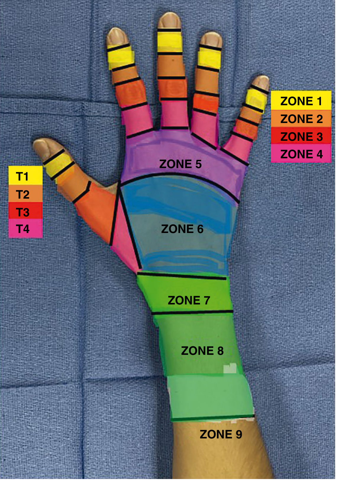
The extensor surface is divided into 9 zones (I-IX) for clinical and surgical communication. Odd-numbered zones overlie joints; even-numbered zones overlie bones. Zone IX is the forearm (muscle belly level).
| Zone | Location | Clinical Significance |
|---|---|---|
| I | DIP joint | Mallet finger (terminal tendon avulsion); treat with 6 weeks DIP extension splinting |
| II | Middle phalanx | May injure lateral bands or triangular ligament |
| III | PIP joint | Central slip injury → untreated → Boutonniere deformity |
| IV | Proximal phalanx | Partial trifurcation of EDC |
| V | MCP joint | Sagittal band injury → EDC subluxation; human bite lacerations common here |
| VI | Dorsum of hand (metacarpals) | Juncturae tendinum may mask complete EDC laceration - must test strength, not just movement |
| VII | Wrist (extensor retinaculum) | Retinaculum usually repaired to prevent bowstringing |
| VIII | Distal forearm | Muscle-tendon junction injuries |
| IX | Forearm | Muscle belly injuries |
- Campbell's Operative Orthopaedics, 15th Ed.; Sabiston Textbook of Surgery
Dorsal Hand Overview
Key Clinical Points
| Concept | Detail |
|---|---|
| Juncturae tendinum | Allow apparent extension despite complete EDC laceration; always test grip strength, not just active motion |
| Mallet finger (Zone I) | Treat closed injuries with continuous DIP extension splinting x 6-8 weeks; surgical fixation if >50% joint surface involved or volar subluxation |
| Boutonniere deformity (Zone III) | Central slip rupture → lateral bands migrate volar → PIP flexion + DIP hyperextension; untreated chronic mallet can lead to swan-neck deformity |
| Zone V (fight bite) | High infection risk; MCP joint often penetrated |
| Zone VI | Patient may still extend due to juncturae; test for weakness |
| Zone VII | Repair retinaculum to prevent bowstringing, but do not make too tight |
| EPL rupture | Can occur with undisplaced distal radius fracture (attrition over Lister's tubercle) |
| De Quervain's | Tenosynovitis of 1st compartment (APL + EPB) |
Sources: General Anatomy and Musculoskeletal System - THIEME Atlas; Miller's Review of Orthopaedics, 9th Ed.; Campbell's Operative Orthopaedics, 15th Ed.; Sabiston Textbook of Surgery; Roberts and Hedges' Clinical Procedures in Emergency Medicine
Extensor zones and compartments of hand
This was covered in detail in my previous answer. Let me pull the key focused content with the best images.
Diving deeper...
Finding Sources
Searching Images
extensor tendon zones of hand injury diagram
Finding Sources
Finding Sources
Reading File
Reading File
Reading File
Loading Image
Loading Image
Reading File
Loading Image
I now have everything needed for a comprehensive, image-rich focused answer on zones and compartments.
Extensor Zones and Compartments of the Hand
Part 1 - The Six Dorsal Compartments (at the Wrist)
The extensor tendons pass beneath the extensor retinaculum through 6 compartments from radial to ulnar. The Lister's tubercle of the radius separates compartments 2 and 3. A useful mnemonic for the tendon count: 2 - 2 - 1 - 5 - 1 - 1 (radial to ulnar).
| Compartment | Tendons (count) | Key Details |
|---|---|---|
| 1st | APL + EPB (2) | Forms radial border of anatomic snuffbox; site of de Quervain's tenosynovitis; APL has multiple slips in 56-98% of dissections; 1st compartment may be septated in 20-60% |
| 2nd | ECRL + ECRB (2) | Insert on bases of 2nd and 3rd metacarpals; radial wrist deviation + extension; "mobile wad" |
| 3rd | EPL (1) | Hooks around Lister's tubercle; forms ulnar border of snuffbox; inserts on distal phalanx of thumb; vulnerable to attrition rupture after distal radius fracture |
| 4th | EDC (II-V) + EIP (5) | Largest compartment; EIP always ulnar to EDC of index finger |
| 5th | EDM / Extensor digiti quinti (1) | Overlies distal radioulnar joint; always ulnar to EDC of little finger |
| 6th | ECU (1) | Ulnar wrist deviation + extension |
- Campbell's Operative Orthopaedics, 15th Ed.; Sabiston Textbook of Surgery
Anatomic variations are common: double EIP, double or triple EDC to the long/ring finger, absent EDC to the small finger (with double EDM), etc.
Part 2 - Extensor Zones of Injury
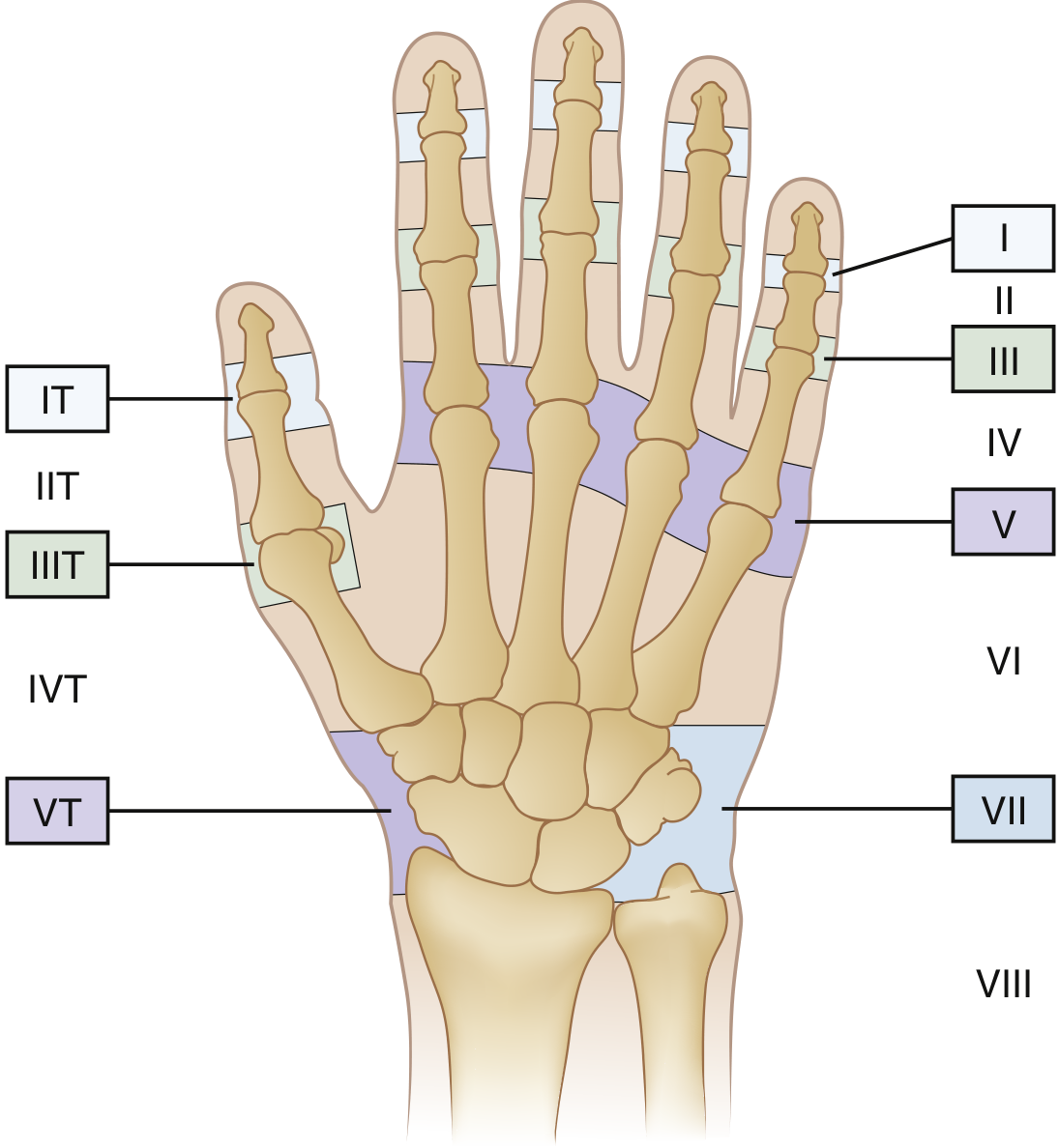
The extensor surface is divided into 9 zones (I-IX) for fingers, plus 5 thumb zones (TI-TV). The system is based on the principle:
- Odd-numbered zones = over joints
- Even-numbered zones = over bones
- Zone VIII and IX = proximal to the wrist
Zone-by-Zone: Anatomy, Injury, and Management
Zone I - DIP Joint
- Structure at risk: Terminal extensor tendon (insertion at base of distal phalanx)
- Injury: Mallet finger - forced DIP flexion avulses the tendon (sometimes with a bony fragment); common in ball sports ("jammed finger")
- Signs: DIP extensor lag, pain/tenderness over DIP; check X-ray for avulsion fracture
- Treatment:
- Closed: DIP extension splinting x 6-8 weeks continuously, then night-only splint for 2-4 more weeks
- Surgical: K-wire DIP fixation; suture anchor if complete tendon avulsion; operative fixation if >50% joint surface in fragment or volar subluxation present
- Splinting within 2 weeks is as effective as delayed splinting; can work up to ~12 weeks post injury
Zone II - Middle Phalanx
- Structure at risk: Tendon over middle phalanx, triangular ligament, lateral bands
- Injury: Usually sharp lacerations; rarely closed
- Treatment: Repair with figure-of-eight or running sutures + static splinting
Zone III - PIP Joint ⚠️ Most Clinically Important
- Structure at risk: Central slip (± lateral bands, triangular ligament)
- Injury: Boutonniere deformity - central slip rupture → lateral bands migrate volar to PIP axis → PIP flexion + DIP hyperextension
- Mechanisms: Forced PIP flexion, blunt dorsal PIP trauma, volar PIP dislocation
- Key test: Elson's test - PIP held in 90° passive flexion; ask patient to extend against resistance. If central slip intact: DIP cannot extend (lateral bands go lax). If central slip ruptured: DIP extends/hyperextends + PIP extends (positive test)
- Note: Deformity may not be apparent acutely if the triangular ligament is intact
- Treatment: 6 weeks PIP extension splinting (DIP left free); surgical repair if open injury
Zone IV - Proximal Phalanx
- Structure at risk: Flat, broad portion of EDC where it begins to trifurcate
- Injury: Usually partial lacerations (tendon is wide here)
- Treatment: Partial injuries may be managed conservatively; complete lacerations require repair
Zone V - MCP Joint
- Structure at risk: Sagittal bands (hold EDC tendon centered over metacarpal head)
- Injury: Sagittal band rupture → EDC subluxation/dislocation (usually ulnar); "snapping" with extension
- Boxer's knuckle: Closed sagittal band rupture from punching
- Most common at the radial sagittal band of the middle finger
- Human fight bite: MCP puncture from opponent's tooth; high infection risk, may penetrate joint
- Classification: Type I (no instability), Type II (subluxation), Type III (complete dislocation)
- Treatment: Type I/II: 4-6 weeks MCP extension splinting; Type III/open: surgical repair
Zone VI - Dorsum of Hand (Metacarpals)
- Structure at risk: EDC tendons + juncturae tendinum (intertendinous connections)
- Injury: Usually lacerations
- Critical pitfall: Juncturae tendinum transmit extension forces from adjacent EDC tendons → patient can STILL extend the digit despite complete tendon laceration
- Rule: Always test strength of extension, not just presence of active motion
Zone VII - Wrist (Extensor Retinaculum)
- Structure at risk: Tendons within their synovial compartments beneath the retinaculum
- Injury: Lacerations over the carpus; closed injuries with distal radius fractures
- Treatment: Surgical repair; extensor retinaculum usually repaired (to prevent bowstringing) but not too tightly (avoid compartment syndrome)
Zone VIII - Distal Forearm
- Structure at risk: Muscle-tendon junction
- Injury: Lacerations, crush; junctions are less amenable to primary repair
Zone IX - Forearm
- Structure at risk: Extensor muscle bellies
- Injury: Lacerations and crush of muscle body
Thumb Zones (TI - TV)
| Zone | Location |
|---|---|
| TI (IT) | Thumb IP joint |
| TII (IIT) | Proximal phalanx of thumb |
| TIII (IIIT) | Thumb MCP joint |
| TIV (IVT) | Thumb metacarpal |
| TV (VT) | Carpus / wrist level |
Part 3 - Classic Deformities at a Glance
| Deformity | Zone | Mechanism | Appearance |
|---|---|---|---|
| Mallet finger | I | Terminal tendon rupture | DIP drop (flexion), cannot actively extend DIP |
| Swan-neck | Sequela of zone I | Chronic untreated mallet → lateral bands sublux dorsally at PIP | PIP hyperextension + DIP flexion |
| Boutonniere | III | Central slip rupture → lateral bands migrate volar | PIP flexion + DIP hyperextension |
| EDC subluxation | V | Sagittal band rupture | Snapping/clicking at MCP with extension |
- Rosen's Emergency Medicine; Sabiston Textbook of Surgery; Campbell's Operative Orthopaedics, 15th Ed.
This is a shared conversation. Sign in to Orris to start your own chat.