
Teach me and exam efficient notes in med school elaborate and well explained manner Microbiology INTRODUCTION INTO MEDICAL MICROBIOLOGY Definition Medical microbiology is the branch of microbiology that deals with the study of microorganisms (bacteria, viruses, fungi, and parasites) that cause diseases in humans, as well as their diagnosis, treatment, and prevention. Scope of Medical Microbiology Study of Pathogens Bacteria Viruses Fungi Parasites Host–Pathogen Interaction How microbes cause disease (pathogenesis). How the human body responds (immunity). Diagnosis Laboratory methods to identify microbes. Microscopy, culture, serology, molecular tests. Treatment Antibiotics, antivirals, antifungals, antiparasitic drugs. Prevention Vaccines, sterilization, disinfection, infection control. Importance of Medical Microbiology Bacteriology – Study of disease-causing bacteria. Virology – Study of viruses Mycology – Study of pathogenic fungi. Parasitology – Study of parasites Immunology – Study of body defenses against infection. History (Ancient Period) Diseases were often believed to be caused by spirits, miasma (“bad air”), or curses. No clear understanding of microorganisms. 17th Century Antonie van Leeuwenhoek (1674): First to observe living microorganisms with a simple microscope. Laid the foundation of microbiology. 19th Century – The Birth of Medical Microbiology Louis Pasteur (1822–1895): Disproved “spontaneous generation.” Showed microbes cause fermentation and spoilage. Developed vaccines (rabies, anthrax). Robert Koch (1843–1910): Proved that specific microbes cause specific diseases (Koch’s postulates). Discovered causative agents of tuberculosis, anthrax, and cholera. Introduced pure culture techniques. -Late 19th – Early 20th Century Development of Gram stain (Hans Christian Gram, 1884). Discovery of many pathogens (e.g., Neisseria, Mycobacterium). Paul Ehrlich (1909): Introduced the first antimicrobial drug (Salvarsan for syphilis). 20th Century -Alexander Fleming (1928): Discovered penicillin, the first true antibiotic. -Development of many antibiotics and vaccines. -Growth of immunology and virology (discovery of viruses, electron microscope). 21st Century -Advances in molecular microbiology, genomics, and rapid diagnostics. -Development of new vaccines (e.g., mRNA vaccines for COVID-19). -Global focus on antimicrobial resistance and emerging infections. Characteristics of Bacteria Size and Shape -Very small: usually 0.2 – 2 µm in diameter, 1 – 10 µm in length. Shapes: Cocci – spherical Bacilli – rod-shaped Spirilla / Spirochetes – spiral-shaped Vibrio – comma-shaped Structure Prokaryotic cells → no true nucleus, DNA is in nucleoid. Cell wall (most have peptidoglycan). Some have capsule/slime layer (protection, virulence). Flagella, pili, fimbriae for movement and attachment. Ribosomes (70S) for protein synthesis. Plasmids → extra DNA carrying resistance/virulence genes. Reproduction Mainly by binary fission (asexual reproduction). Some exchange DNA via: Conjugation (plasmid transfer) Transformation (uptake of DNA from environment) Transduction (bacteriophage-mediated). Metabolism Can use different energy sources: Autotrophs – make their own food. Heterotrophs – depend on organic material. Based on oxygen use: Obligate aerobes – need oxygen. Obligate anaerobes – oxygen is toxic.(Can not survive with the presence of oxygen) Facultative anaerobes – can grow with or without oxygen. Growth Requirements Need nutrients, water, suitable pH, and temperature. Most human pathogens grow best at 37°C (body temperature) Pathogenicity Some bacteria are harmless/beneficial (normal flora, gut microbiota). Others are pathogenic → cause disease by: Producing toxins (endotoxins, exotoxins). Damaging host tissues. Evading immunity (capsule, antigenic variation). staining Characteristics Gram-positive bacteria → thick peptidoglycan, stain purple. Gram-negative bacteria → thin peptidoglycan, outer membrane, stain pink. Acid-fast bacteria (e.g., Mycobacterium) → waxy cell wall. Viruses – Characteristics Nature Acellular (not made of cells). Considered obligate intracellular parasites – can only reproduce inside living host cells. Contain either DNA or RNA (never both) Size Much smaller than bacteria: 20 – 300 nanometers. Cannot be seen with a light microscope, only with electron microscope. Replication Do not divide like cells. Use host cell machinery to replicate: Attachment Entry (penetration) Uncoating of genome Replication & synthesis of viral proteins Assembly of new viral particles Release (lysis or budding) Classification Based on: Type of nucleic acid (DNA vs RNA) Shape (helical, icosahedral, complex) Presence/absence of envelope Pathogenicity Cause a wide range of diseases: Respiratory: Influenza, COVID-19 Neurological: Rabies, Polio Hepatic: Hepatitis viruses Oncogenic: HPV (cervical cancer), EBV (Burkitt lymphoma) Characteristics of Fungi Cell Type Eukaryotic → have nucleus and membrane-bound organelles. Structure Cell wall made of chitin (not peptidoglycan like bacteria). Cell membrane contains ergosterol (target of antifungal drugs). Exist as: Yeasts – unicellular (e.g., Candida). Molds – multicellular, filamentous (hyphae, mycelium). Dimorphic fungi – can switch between yeast and mold forms. Characteristics of Parasites Definition Organisms that live on or in a host and derive nutrients at the host’s expense. Types Protozoa (unicellular eukaryotes): e.g., Plasmodium (malaria), Giardia. Helminths (worms): multicellular, e.g., roundworms, tapeworms, flukes. Ectoparasites: live on body surface (lice, ticks, fleas). Cell Type Eukaryotic organisms (like fungi). Reproduction Protozoa → usually asexual (binary fission), some sexual stages. Helminths → complex life cycles, often with sexual reproduction. Transmission Through contaminated food, water, soil, insect vectors, or direct contact. Medical Importance • Cause parasitic diseases (malaria, amoebiasis, helminth infections). • Many have complex life cycles with intermediate and definitive hosts. Sterilization Definition Sterilization is the process of completely removing or destroying all forms of microorganisms, including bacteria, viruses, fungi, spores, and parasites, from an object oar surface. Types of Sterilization Physical Methods Heat Sterilization Moist Heat (steam): Autoclaving (121°C, 15 psi, 15–20 min) → most reliable, kills spores. Boiling (100°C) → kills most microbes, not spores. Pasteurization (milder heat for food, not full sterilization). Dry Heat: Hot air oven (160–170°C for 2 hours). Incineration (burning contaminated material). Radiation Ionizing radiation (X-rays, gamma rays): sterilizes disposable medical supplies (syringes, catheters). Non-ionizing (UV light): disinfects surfaces, air, water (limited penetration). Filtration Removes microbes using membrane filters (0.22 µm). Used for heat-sensitive liquids (antibiotics, vaccines). Chemical Methods Gaseous Sterilization Ethylene oxide gas → for heat-sensitive items (plastic, medical instruments). Formaldehyde gas. Liquid Chemicals Glutaraldehyde, formalin, peracetic acid → high-level disinfectants that can sterilize with enough exposure time. Used for surgical instruments, endoscopes.
bacteria shapes cocci bacilli spirochetes morphology diagram
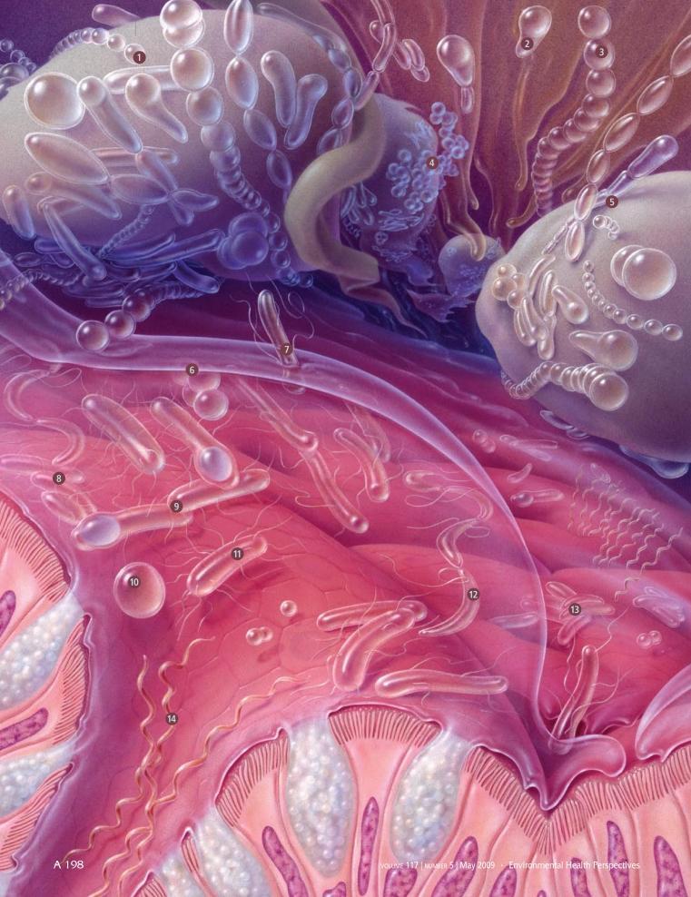
This medical illustration provides a microscopic visualization of the human gut microbiota and the intestinal mucosal environment. The diagram highlights the morphological diversity of bacteria found within the gastrointestinal tract, categorized by numbered labels 1 through 14. Visible bacterial morphologies include cocci in chains (Bifidobacterium, Lactobacillus), staphylococcal clusters (Staphylococcus), bacilli or rod-shaped bacteria (Escherichia, Eubacterium, Clostridium), and spirochetes with spiral symmetry (Brachyspira). The background depicts the physiological structure of the intestinal wall, featuring pink-hued tissue layers and finger-like projections representing intestinal villi and microvilli. These anatomical structures are shown interacting with a translucent mucosal layer where the microbes reside. The illustration serves as an educational tool for microbiology and gastroenterology, demonstrating the complex relationship between commensal microorganisms and the host's epithelial surface. Key genera represented include Bacteroides, Fusobacterium, and Ruminococcus, illustrating the variety of the human microbiome's anaerobic and facultative aerobic constituents.
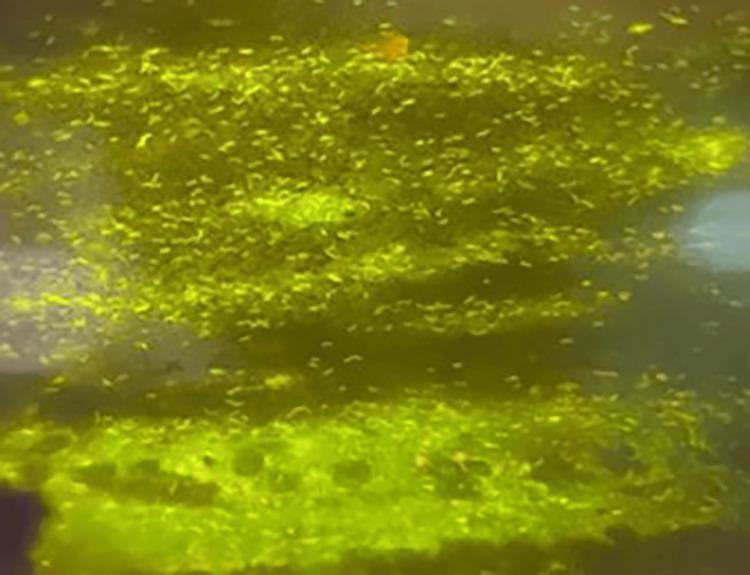
This diagnostic micrograph displays a fluorescent staining pattern used in microbiology for the detection of Mycobacteria. The image shows numerous acid-fast bacilli (AFB) identified as tubercle bacilli, which exhibit a brilliant, bright greenish-yellow fluorescence against a contrasting dark background. The morphology of the microorganisms is characterized by small, slightly curved, beaded bacillus shapes. These bacteria are distributed across the field of view, appearing both as individual rods and in dense clusters. This visual represents an auramine-rhodamine fluorochrome stain viewed under a fluorescent microscope, a highly sensitive method for diagnosing pulmonary and extrapulmonary tuberculosis. The educational focus is on the distinctive visual characteristics of Mycobacterium tuberculosis when using fluorescent microscopy, emphasizing the importance of color, shape, and contrast in the rapid identification of pathogenic mycobacteria in clinical specimens.
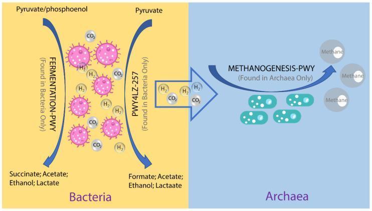
A pathophysiology diagram illustrating the symbiotic metabolic relationship between gut bacteria and archaea, specifically focusing on hydrogen (H2) and carbon dioxide (CO2) exchange. The diagram is divided into two sections. The left side (yellow background) depicts bacterial fermentation processes: 'FERMENTATION-PWY' and 'PWY4LZ-257'. These pathways utilize Pyruvate and phosphoenolpyruvate as inputs to produce Succinate, Acetate, Ethanol, Lactate, and Formate, while releasing H2 and CO2 as metabolic byproducts. The bacteria are represented as pink cocci with surface projections. A large arrow indicates the transfer of these gases to the right side (blue background), which depicts Archaea. The archaeal section shows teal-colored bacilli performing 'METHANOGENESIS-PWY', a pathway that consumes the H2 and CO2 produced by bacteria to generate Methane. This illustration demonstrates the concept of syntrophy and interspecies hydrogen transfer within the microbiome, highlighting the specialized metabolic niches of bacteria (hydrogen producers) and archaea (methanogens).
Gram stain positive negative bacteria peptidoglycan cell wall diagram
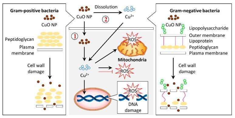
A pathophysiology diagram illustrating the antibacterial mechanisms of copper oxide nanoparticles (CuO NPs) on Gram-positive and Gram-negative bacteria. The left panel shows Gram-positive bacteria with a thick peptidoglycan layer above a plasma membrane. The right panel shows Gram-negative bacteria with a complex cell wall including lipopolysaccharides, an outer membrane, lipoproteins, and a thin peptidoglycan layer. Both panels demonstrate CuO NPs causing cell wall damage and lysis. The central panel details the intracellular bactericidal pathway labeled as mechanism 1 and 2. Mechanism 1 involves the internalization of CuO NPs into the cytosol, while mechanism 2 illustrates the dissolution of CuO NPs into copper ions (Cu2+) which then permeate the cell. Inside the cell, both Cu2+ ions and CuO NPs induce the production of reactive oxygen species (ROS). The resulting oxidative stress leads to mitochondrial disruption and DNA damage, ultimately resulting in cell death. This infographic highlights the role of nanoparticle morphology and ion release in targeting bacterial structural and metabolic integrity.
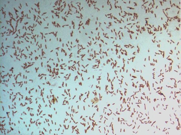
This diagnostic image shows a light microscopy view of a bacterial culture following Gram staining. The specimen displays numerous small, rod-shaped (bacilli) bacteria distributed across the field. The microorganisms exhibit a consistent pink-to-red hue, identifying them as Gram-negative. This staining characteristic indicates a cell wall structure with a thin peptidoglycan layer and an outer membrane that does not retain the crystal violet stain. The arrangement of the bacilli is predominantly individual or scattered, with occasional small clusters and pairs. Specifically identified as D. fastidiosa strain JC13T, this image serves as a primary microbiological reference for the morphological and staining properties of this novel genus within the family Erysipelotrichaceae. The visual demonstrates key diagnostic features including cell morphology, arrangement, and Gram reaction, which are essential for clinical microbiology classification and initial pathogen identification.
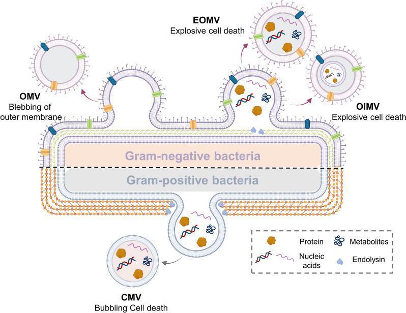
A medical microbiology diagram illustrating the biogenesis and classification of bacterial extracellular vesicles (BEVs) from Gram-negative and Gram-positive bacteria. The top half depicts a Gram-negative bacterium with its triple-layered cell envelope (outer membrane, periplasmic space, and cytoplasmic membrane). Three vesicle formation mechanisms are shown: blebbing of the outer membrane creating Outer Membrane Vesicles (OMV), and explosive cell death leading to Explosive Outer Membrane Vesicles (EOMV) and Outer-Inner Membrane Vesicles (OIMV). The bottom half represents a Gram-positive bacterium characterized by a thick peptidoglycan cell wall and a single cytoplasmic membrane. This section illustrates 'bubbling cell death' resulting in Cytoplasmic Membrane Vesicles (CMV). A legend identifies the internal cargo within these vesicles, including proteins (brown shapes), nucleic acids (wavy lines), metabolites (blue branched icons), and endolysin (light blue shapes). This pathophysiology diagram highlights the structural differences in bacterial envelopes and the various pathways—blebbing, explosive death, and bubbling—that contribute to BEV diversity and composition.
virus replication cycle attachment entry uncoating assembly release diagram
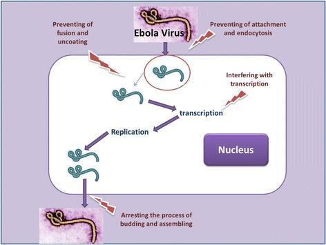
A pathophysiology diagram illustrating the Ebola virus (EBOV) life cycle within a host cell and identifying potential therapeutic targets. The diagram utilizes clinical micrographs of the filamentous, pleomorphic Ebola virus to represent entry and exit. The intracellular viral replication stages are mapped sequentially: attachment and endocytosis (entry), fusion and uncoating (releasing the viral genome), transcription, replication, and budding and assembly (exit). Potential pharmacological intervention points are highlighted by red lightning bolt icons, indicating strategies such as preventing attachment and endocytosis, preventing fusion and uncoating, interfering with transcription, and arresting the process of budding and assembling. The host cell is depicted with a distinct nucleus, and arrows trace the progression of the viral cycle from cellular entry to the release of progeny virions. This educational graphic is designed for medical virology and drug discovery contexts, emphasizing sites of action for future anti-Ebola therapeutics.
![A pathophysiology diagram illustrating the replication cycle of the SARS-CoV-2 virus (2019-nCoV) within a host cell and identifying potential therapeutic intervention points. The schematic depicts the virus engaging cell surface receptors ACE2 and CD147, followed by entry via endosomal or non-endosomal pathways. Once inside, the process of uncoating releases genomic positive-strand RNA [(+) RNA], which undergoes translation and transcription. Key viral components are shown being synthesized in the rough endoplasmic reticulum (ER), including accessory protein (AP), membrane protein (M), envelope protein (E), spike glycoprotein (S), and nucleocapsid protein (N). The diagram highlights the assembly of new virions within the endoplasmic reticulum-Golgi intermediate compartment (ERGIC) and the Golgi apparatus, followed by vesicle transport and viral release. Critical therapeutic targets are identified through the inclusion of entry inhibitors, protease inhibitors (targeting PLpro and 3CLpro), and replication inhibitors. The visualization serves as an educational tool for understanding coronavirus virology and pharmacotherapy.](https://cdn.orris.care/cdss_images/pmc_clinical_VQA_c35e92c4f4a5e28b4364f80ef93e2fa38f52483741d51efe2804ea4eac1386c6.jpg)
A pathophysiology diagram illustrating the replication cycle of the SARS-CoV-2 virus (2019-nCoV) within a host cell and identifying potential therapeutic intervention points. The schematic depicts the virus engaging cell surface receptors ACE2 and CD147, followed by entry via endosomal or non-endosomal pathways. Once inside, the process of uncoating releases genomic positive-strand RNA [(+) RNA], which undergoes translation and transcription. Key viral components are shown being synthesized in the rough endoplasmic reticulum (ER), including accessory protein (AP), membrane protein (M), envelope protein (E), spike glycoprotein (S), and nucleocapsid protein (N). The diagram highlights the assembly of new virions within the endoplasmic reticulum-Golgi intermediate compartment (ERGIC) and the Golgi apparatus, followed by vesicle transport and viral release. Critical therapeutic targets are identified through the inclusion of entry inhibitors, protease inhibitors (targeting PLpro and 3CLpro), and replication inhibitors. The visualization serves as an educational tool for understanding coronavirus virology and pharmacotherapy.
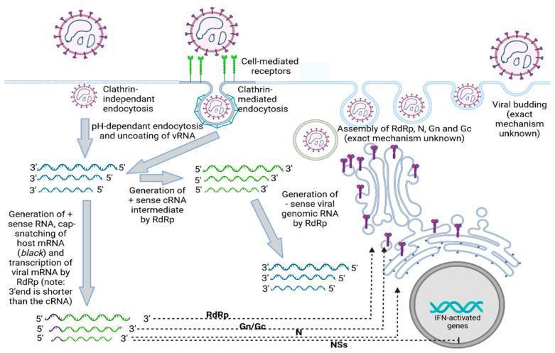
A pathophysiology diagram illustrating the replication cycle of a typical Bunyavirus within a host cell. The schematic details viral entry via two pathways: clathrin-independent endocytosis and receptor-mediated clathrin-dependent endocytosis. Following entry, the diagram shows pH-dependent uncoating and release of negative-sense viral RNA (vRNA) into the cytoplasm. The replication phase demonstrates the role of RNA-dependent RNA polymerase (RdRp) in synthesizing a positive-sense cRNA intermediate. Two divergent genetic processes are shown: (1) the generation of new negative-sense genomic vRNA and (2) 'cap-snatching' of host mRNA to produce translatable viral mRNA. The translation section highlights the synthesis of structural proteins (RdRp, Gn/Gc, N) and non-structural proteins (NSs), the latter shown inhibiting interferon (IFN)-activated genes in the nucleus. The cycle concludes with viral assembly at the Golgi apparatus and subsequent viral budding from the host membrane. This diagram is intended for advanced virology and microbiology education, focusing on negative-sense RNA virus replication and host-immune evasion mechanisms.
🦠 Medical Microbiology - Exam-Efficient Study Notes
UNIT 1 - What is Medical Microbiology?
| Pillar | What It Asks |
|---|---|
| Pathogen Biology | What is the microbe? How does it work? |
| Pathogenesis | How does it cause disease? |
| Diagnosis | How do we identify it? |
| Prevention/Treatment | How do we stop it? |
- Bacteriology
- Virology
- Mycology
- Parasitology
- Immunology
UNIT 2 - History Timeline (High-Yield for MCQs)
| Era | Person | Year | Contribution | Exam Hook |
|---|---|---|---|---|
| 17th C | Leeuwenhoek | 1674 | First to SEE microorganisms (simple microscope) | "Father of Microbiology" |
| 19th C | Pasteur | 1822-95 | Disproved spontaneous generation; germ theory; rabies & anthrax vaccines | "Father of Immunology" |
| 19th C | Koch | 1843-1910 | Koch's Postulates; discovered TB, anthrax, cholera agents; pure culture technique | "Father of Medical Microbiology" |
| 19th C | Gram | 1884 | Gram stain developed | Still used clinically today |
| 1909 | Ehrlich | 1909 | Salvarsan = first antimicrobial drug (for syphilis) | First "magic bullet" |
| 1928 | Fleming | 1928 | Discovered penicillin (first antibiotic) | Accidental discovery from mold |
| 21st C | mRNA vaccines | 2020 | COVID-19 vaccines - new vaccine platform | Still relevant clinically |
UNIT 3 - Koch's Postulates (VERY HIGH YIELD)
Classic Koch's Postulates (1884)
- The microorganism must be found in all cases of the disease (and its distribution matches the lesions).
- It must be isolated and grown in pure culture outside the host.
- When the pure culture is inoculated into a healthy susceptible host, it must reproduce the disease.
- The microorganism must be re-isolated from the experimentally infected host and shown to be the same organism.
Why They Sometimes Fail (Exam Favourite!)
- Treponema pallidum (syphilis) and Mycobacterium leprae (leprosy) - cannot be grown in vitro
- Neisseria gonorrhoeae - no animal model exists
- Some pathogens are found in healthy people too (asymptomatic carriers)
- Viruses and prions don't fully fit the classical model
Molecular Koch's Postulates (modern update)
- The virulence gene should be present in pathogenic but NOT non-pathogenic strains.
- Inactivating (knocking out) the gene should reduce virulence.
- Restoring the gene should restore virulence.
Exam tip: If a question says "which pathogen CANNOT satisfy Koch's postulates because it can't be cultured?" - the answer is M. leprae or T. pallidum.
UNIT 4 - Bacteria
4A - Size and Shape
| Shape | Name | Example |
|---|---|---|
| Sphere | Cocci | Staphylococcus, Streptococcus |
| Rod | Bacilli | E. coli, Bacillus |
| Spiral (rigid) | Spirilla | Helicobacter pylori |
| Spiral (flexible) | Spirochetes | Treponema, Borrelia |
| Comma | Vibrio | Vibrio cholerae |
4B - Bacterial Cell Structure (Memorize Layer by Layer)
OUTERMOST → INNERMOST
[Capsule] → [Cell Wall (peptidoglycan)] → [Cell Membrane] → [Cytoplasm + Nucleoid]
| Structure | Function | Clinical Relevance |
|---|---|---|
| Capsule | Anti-phagocytic, protects from host immunity | Major virulence factor - e.g., S. pneumoniae |
| Cell wall (peptidoglycan) | Structural support | Target of penicillin and cephalosporins |
| Pili / Fimbriae | Attachment to host cells | Critical for infection initiation |
| Flagella | Motility | Also antigenic (H antigen) |
| Plasmids | Extra-chromosomal DNA | Carry antibiotic resistance genes |
| 70S Ribosomes | Protein synthesis | Target of aminoglycosides, macrolides, tetracyclines |
| Nucleoid | Contains chromosomal DNA (NO nuclear membrane) | Prokaryote = no true nucleus |
Key point: Bacteria are PROKARYOTES - no true nucleus, no membrane-bound organelles.
4C - Gram Staining (Extremely High-Yield)
| Feature | Gram-Positive | Gram-Negative |
|---|---|---|
| Peptidoglycan | THICK (multiple layers) | THIN (single layer) |
| Outer membrane | ABSENT | PRESENT (contains LPS) |
| Stain color | PURPLE (retains crystal violet) | PINK/RED (takes up safranin counterstain) |
| Endotoxin (LPS) | None | YES - causes septic shock |
| Examples | Staphylococcus, Streptococcus, Bacillus | E. coli, Salmonella, Neisseria |
4D - Bacterial Metabolism and Oxygen Requirements
| Type | Oxygen Relationship | Example |
|---|---|---|
| Obligate aerobe | NEEDS oxygen to survive | Mycobacterium tuberculosis |
| Obligate anaerobe | KILLED by oxygen | Clostridium, Bacteroides |
| Facultative anaerobe | Can grow with OR without oxygen | E. coli, Staphylococcus |
| Microaerophile | Needs LOW oxygen levels | Campylobacter, H. pylori |
Exam tip: Most clinically important pathogens are facultative anaerobes (can infect tissues with varying O₂ levels).
4E - Bacterial Reproduction and Genetic Exchange
- Main method: Binary fission (one cell divides into two)
- DNA exchange mechanisms (give bacteria new traits like resistance):
| Method | Mechanism | Key Point |
|---|---|---|
| Conjugation | Direct cell-to-cell contact via pilus; plasmid transferred | Most common mechanism of antibiotic resistance spread |
| Transformation | Bacterium picks up free DNA from environment | S. pneumoniae classic example |
| Transduction | Bacteriophage (virus) moves DNA between bacteria | Virus acts as DNA carrier |
4F - How Bacteria Cause Disease (Pathogenesis)
- Producing toxins:
- Endotoxins = LPS from Gram-NEGATIVE cell wall; released on cell death; causes fever, septic shock
- Exotoxins = proteins actively secreted by bacteria; more potent; specific actions (e.g., cholera toxin, botulinum toxin, tetanus toxin)
- Direct tissue invasion - Salmonella, Shigella
- Evading immunity - capsule (anti-phagocytic), antigenic variation, enzyme production
UNIT 5 - Viruses
5A - Fundamental Properties
| Property | Details |
|---|---|
| Structure | Acellular - NOT made of cells |
| Genome | Either DNA OR RNA - never both |
| Size | 20 - 300 nanometers (need electron microscope) |
| Reproduction | Cannot reproduce independently - obligate intracellular parasites |
| Cell wall | NONE |
Key concept: Viruses are NOT alive in the classical sense. They are genetic material wrapped in protein. They hijack host cell machinery to replicate.
5B - Viral Replication Cycle (6 Steps)
1. ATTACHMENT → Virus binds to specific receptor on host cell surface
2. ENTRY (Penetration) → Virus enters cell (endocytosis or fusion)
3. UNCOATING → Viral genome is released from its capsid
4. REPLICATION & SYNTHESIS → Host cell machinery makes viral proteins and nucleic acid
5. ASSEMBLY → New viral particles are assembled
6. RELEASE → Virions exit by LYSIS (cell bursts) or BUDDING (enveloped viruses)

Exam tip for antiviral drug targets: Each step of replication can be targeted:
- Attachment inhibitors (e.g., entry inhibitors for HIV)
- Protease inhibitors (prevent assembly)
- Polymerase inhibitors (prevent replication)
5C - Viral Classification
| Basis | Options |
|---|---|
| Nucleic acid | DNA virus vs. RNA virus |
| Shape | Helical / Icosahedral / Complex |
| Envelope | Enveloped (e.g., HIV, Influenza) vs. Non-enveloped/Naked (e.g., Poliovirus) |
5D - Clinical Disease by Virus Type (High-Yield)
| System | Virus | Disease |
|---|---|---|
| Respiratory | Influenza, SARS-CoV-2 | Flu, COVID-19 |
| Neurological | Rabies, Poliovirus | Rabies, Polio |
| Hepatic | HBV, HCV, HAV | Hepatitis B, C, A |
| Oncogenic | HPV | Cervical cancer (types 16, 18) |
| Oncogenic | EBV | Burkitt lymphoma, nasopharyngeal carcinoma |
| Immunodeficiency | HIV | AIDS |
UNIT 6 - Fungi
| Feature | Details |
|---|---|
| Cell type | Eukaryotic (have true nucleus) |
| Cell wall | Made of CHITIN (NOT peptidoglycan) |
| Cell membrane | Contains ERGOSTEROL (NOT cholesterol like humans) |
| Morphology | Yeasts (unicellular), Molds (multicellular, filamentous), or Dimorphic (both) |
Antifungal Targets
- Ergosterol in the fungal membrane is the target of azoles (fluconazole) and amphotericin B
- Chitin synthesis in cell wall is targeted by echinocandins (caspofungin)
Exam tip: Ergosterol is specific to fungi - that's why antifungals can kill fungi without harming human cells (we use cholesterol, not ergosterol).
Dimorphic Fungi - "Mold in Cold, Yeast in Heat"
UNIT 7 - Parasites
Classification
| Type | Structure | Examples | Disease |
|---|---|---|---|
| Protozoa | Unicellular eukaryotes | Plasmodium, Giardia, Entamoeba | Malaria, Giardiasis, Amoebiasis |
| Helminths | Multicellular worms | Roundworms, tapeworms, flukes | Helminthiasis |
| Ectoparasites | Live ON body surface | Lice, ticks, fleas | Pediculosis, Lyme disease (tick vector) |
Key Points
- All parasites are eukaryotes (like fungi and human cells)
- This makes them harder to target with drugs - fewer unique targets
- Many have complex life cycles with intermediate and definitive hosts
- Plasmodium (malaria) uses the mosquito (Anopheles) as its vector
UNIT 8 - Sterilization and Disinfection
Definitions - Know These Precisely!
| Term | Definition | Kills Spores? |
|---|---|---|
| Sterilization | Complete elimination of ALL microorganisms including spores | YES |
| Disinfection | Kills most pathogens but NOT all (especially not spores) | Usually NO |
| Antisepsis | Disinfection applied to LIVING tissue (skin, mucosa) | NO |
| Pasteurization | Heat treatment to kill pathogens in food/liquid - NOT full sterilization | NO |
| Asepsis | Working systems designed to PREVENT contamination (e.g., sterile surgical technique) | - |
| Sanitization | Between disinfection and cleanliness - used in food service | NO |
Exam tip: "Sterilization" is the ONLY absolute term. Everything else (disinfection, antisepsis) is relative.
Physical Methods of Sterilization
HEAT (Most Common)
| Method | Conditions | What It Kills | Use |
|---|---|---|---|
| Autoclaving (moist heat) | 121°C, 15 psi, 15-20 min | EVERYTHING including spores | Surgical instruments, culture media - GOLD STANDARD |
| Boiling | 100°C | Most microbes; NOT spores | Emergency use only |
| Pasteurization | 74°C for 3-5 sec OR 62°C for 30 min | Vegetative pathogens | Milk, food - NOT sterilization |
| Hot air oven (dry heat) | 160-170°C for 2 hours | All forms | Glassware, oils, powders |
| Incineration | Direct burning | All forms | Contaminated waste disposal |
RADIATION
| Type | Mechanism | Use |
|---|---|---|
| Ionizing (X-ray, gamma rays) | Damages DNA | Disposable medical supplies (syringes, catheters) |
| Non-ionizing (UV light) | Creates thymine dimers in DNA | Surface and air disinfection; LIMITED penetration |
FILTRATION
- Uses membrane filters (pore size 0.22 µm)
- Physically removes bacteria by size
- Used for heat-sensitive liquids: antibiotics, vaccines, serum
- Does NOT remove viruses (too small to be filtered by standard membranes)
Chemical Methods of Sterilization
| Agent | Form | Use | Special Notes |
|---|---|---|---|
| Ethylene oxide | Gas | Heat-sensitive instruments (plastics, scopes) | Toxic, carcinogenic - requires aeration |
| Glutaraldehyde | Liquid | Endoscopes, surgical instruments | High-level disinfectant; prolonged contact = sterilization |
| Formaldehyde/Formalin | Liquid/Gas | Lab fixation, sterilization | Toxic; carcinogen |
| Peracetic acid | Liquid | Endoscopes, instruments | Fast-acting, eco-friendly |
| Alcohols (70% ethanol/isopropanol) | Liquid | Skin antisepsis, surface disinfection | NOT effective against spores |
| Chlorine compounds | Liquid | Water treatment, surface disinfection | Effective but corrosive |
Exam tips:
- "Which method sterilizes heat-sensitive items?" → Ethylene oxide gas or filtration
- "What is the MOST reliable sterilization method?" → Autoclaving
- "What does pasteurization NOT kill?" → Bacterial spores
UNIT 9 - Comparison Table: The Four Microorganism Types
| Feature | Bacteria | Viruses | Fungi | Parasites |
|---|---|---|---|---|
| Cell type | Prokaryote | Acellular | Eukaryote | Eukaryote |
| Size | 0.2-10 µm | 20-300 nm | Variable | Variable |
| Nucleus | No | No (no cells) | Yes | Yes |
| Cell wall | Peptidoglycan | None | Chitin | None (protozoa) |
| Reproduction | Binary fission | Host cell machinery | Spores, budding | Binary fission / complex cycles |
| Microscope | Light | Electron | Light | Light |
| Drug targets | Peptidoglycan, 70S ribosome | Viral enzymes | Ergosterol, chitin | Varies |
HIGH-YIELD EXAM QUICK FACTS
- First to SEE microorganisms = Leeuwenhoek (1674)
- First to PROVE germ theory = Pasteur
- First to link specific microbe to specific disease = Koch
- First antibiotic = Penicillin (Fleming, 1928)
- First antimicrobial drug ever = Salvarsan/Arsphenamine (Ehrlich, 1909) - for syphilis
- Gram+ = purple = thick peptidoglycan, no outer membrane
- Gram- = pink = thin peptidoglycan + outer membrane with LPS (endotoxin)
- Acid-fast organisms = waxy cell wall, resist standard stains - use ZN or fluorescent stain
- Only DNA OR RNA = viruses (never both in the same virion)
- Ergosterol = fungal membrane target (azoles, amphotericin B)
- Autoclaving = 121°C, 15 psi, 15-20 min = gold standard sterilization
- 0.22 µm filter = removes bacteria, NOT viruses
- Binary fission = main bacterial reproduction method
- Conjugation = main method of antibiotic resistance gene transfer between bacteria
- "Mold in cold, yeast in heat" = dimorphic fungi rule
MEMORY AIDS
SECTION 1 fundamentals of microbiology chapter 1,2,3,4,5,6, (Jawetz, Melnick, & Adelberg's) Basic bacteriology chapter 1 , 2, 3, 4, 5, 6, ( WARREN LEVINSON) Gram positive bacteria (staphylococci) SECTION3 chapter 13 (Jawetz, Melnick, & Adelberg's) Gram positive bacteria (streptococci) SECTION 3 chapter 14 (Jawetz, Melnick, & Adelberg's)