
Ludwig’s angina
Ludwig's angina management airway
Ludwig's Angina
Definition
- Rosen's Emergency Medicine, p. 3714
- Bailey and Love's Surgery, p. 1111
Anatomy of the Spaces Involved
- Sublingual space (above mylohyoid) - contains the tongue, sublingual gland, and Wharton's duct
- Submylohyoid (submaxillary) space (below mylohyoid) - contains the submandibular gland
- Tintinalli's Emergency Medicine, p. 1906
- Rosen's Emergency Medicine, p. 3714
Aetiology and Microbiology
-
Odontogenic origin in ~80% of cases - most commonly periapical abscesses of the lower second and third mandibular molars, whose roots lie below the mylohyoid line, allowing direct spread into the submandibular space
-
Other causes: salivary calculi, oral lacerations, mandibular fractures, and in rare cases, underlying oral cavity cancer
-
Infection is polymicrobial (mixed oral flora):
- Aerobic: Streptococcus species (especially hemolytic streptococci), Staphylococcus aureus (including MRSA), Gram-negative rods
- Anaerobic: Bacteroides fragilis, Fusobacterium, gas-producing organisms; anaerobes produce hyaluronidase, collagenase, and fibrinolysin that facilitate fascial spread
-
Spread is direct through fascial planes, NOT via lymphatics - this explains the bilateral, non-fluctuant, boardlike character without lymphadenopathy
-
Rosen's Emergency Medicine, p. 3714
-
Cummings Otolaryngology, p. 3317
-
Rosen's Emergency Medicine (Pediatric), p. 1906
Pathophysiology
- Along deep fascial layers to the larynx - causing glottic oedema
- Posteriorly via the parapharyngeal space to the retropharyngeal space
- Into the "danger space" - and then down to the superior mediastinum
- Along the carotid sheath (the "Lincoln Highway") to the aortic arch level
Clinical Features
| Feature | Details |
|---|---|
| Brawny, "woody" neck swelling | Bilateral submandibular induration - characteristically non-fluctuant |
| Tongue elevation/protrusion | Displaced upward and backward by sublingual swelling |
| Trismus | Due to masticator space involvement |
| Drooling / odynophagia | Cannot swallow, floor of mouth too swollen |
| Putrid halitosis | Characteristic of mixed anaerobic infection |
| "Sniffing position" | Patient leans forward, jaw jutting, neck extended to maximise airway |
| Stridor | Late finding indicating impending airway obstruction |
| Fever, rigors, toxicity | Systemic signs of severe infection |
| Anxiety | Hallmark of impending airway compromise |
CT Scan Appearance
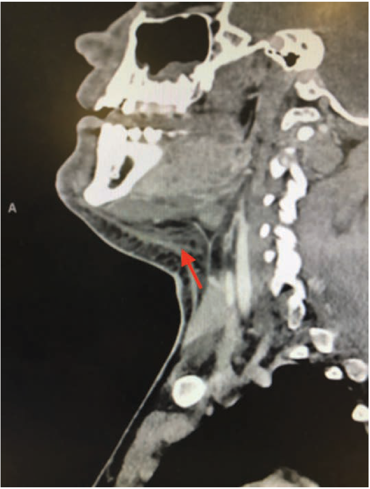
Management
1. Airway - FIRST PRIORITY
"Control the airway early, because intubation can be extremely difficult late in the clinical course of the disease." - Tintinalli's Emergency Medicine, p. 1908
- Awake nasal fiberoptic intubation - preferred by most experts; nasal route needed because trismus limits mouth opening
- Awake oral fiberoptic intubation - if trismus is not severe
- Surgical airway (tracheostomy/cricothyrotomy) - if above fail or anatomy is distorted; elective tracheostomy before incision and drainage is a classic (though now less common) approach
- Patient must be kept upright - supine positioning worsens tongue prolapse and can cause sudden complete obstruction
2. Antibiotics - Immediately
| Antibiotic | Regimen |
|---|---|
| Ampicillin-sulbactam | First-line (covers aerobic and anaerobic oral flora) |
| Penicillin G + Metronidazole | Alternative combination |
| Clindamycin | If penicillin allergy |
| Add vancomycin | If MRSA colonisation is suspected |
- Tintinalli's Emergency Medicine, p. 1908
3. Surgical Drainage
- Cases not rapidly responding to antibiotics
- Evidence of abscess formation on CT
- Progressive worsening
-
Curved submental incision to drain both submandibular triangles
-
The mylohyoid muscle may be incised to decompress the floor of the mouth
-
Both intraoral and external approaches used to ensure drainage of submaxillary and sublingual spaces
-
Corrugated drains placed, wound lightly sutured
-
General anaesthesia preferred over local for better access and patient comfort
-
Bailey and Love's Surgery, p. 1128-1136
-
Scott-Brown's Otorhinolaryngology, p. 8130
4. Admission
Complications
| Complication | Mechanism |
|---|---|
| Airway obstruction / asphyxia | Tongue displacement, glottic oedema - leading cause of death |
| Descending necrotising mediastinitis | Via danger space/carotid sheath spread; mortality 10-40% |
| Aspiration pneumonia / lung abscess | Abscess rupture into hypopharynx |
| Septicaemia / septic shock | Bacteraemia from mixed oral flora |
| Jugular vein thrombosis (Lemierre) | Via parapharyngeal space spread |
| Cavernous sinus thrombosis | Retrograde spread via valveless ophthalmic veins |
| Pericardial effusion / tamponade | Mediastinal extension |
| Great vessel erosion | From mediastinal extension |
- Tintinalli's Emergency Medicine (Complications section), p. 2509
Key Summary Points (for exam)
- Definition: Bilateral cellulitis of sublingual + submandibular + submental spaces
- Source: Odontogenic (~80%) - lower molar periapical abscesses
- Spread: Direct through fascial planes (NOT lymphatics) - so bilateral, no adenopathy
- Organism: Polymicrobial (streptococci + anaerobes)
- Classic sign: "Woody", non-fluctuant bilateral submandibular swelling with tongue elevation
- Priority: Airway first - awake fiberoptic (nasal) intubation preferred
- Antibiotics: Ampicillin-sulbactam (or Penicillin G + Metronidazole)
- Surgery: Submental incision with mylohyoid division for drainage
- Mortality: Primarily from asphyxia; mediastinal spread carries 10-40% mortality
Audiology Audiometry & hearing tests: • Otoacoustic emission — 2016, Oct 2020, 2021 • Speech audiometry — 2018, 2021 • Impedance audiometry — 2020; Tympanogram — 2020 • BERA — Oct 2020 • ASSR (auditory steady-state response) — 2019 • Bekesy audiometry — Oct 2025 • Masking in pure tone audiometry — 2023; Role of masking in audiology — 2018 • Tuning fork tests for malingering — 2018, 2019 • Cochlear microphonics — 2017, Oct 2025 Hearing screening & rehabilitation: • Universal neonatal hearing screening — 2016 (asked in both Paper I and II) • Neonatal hearing screening — 2020 • Issuing a hearing disability certificate — 2021 • BAHA / BAHA & indications — 2019, Oct 2020 Related (hearing loss & vestibular electrophysiology): • Genetics of non-syndromic hearing loss — 2021; Autosomal dominant syndromic hearing loss — 2023 • VEMP — 2017, 2022 • Caloric test — 2021 • Electroneurography — 2016 Give a short notes of the questions that I have given now
AUDIOLOGY - SHORT NOTES
1. Otoacoustic Emissions (OAE)
| Type | Stimulus | Details |
|---|---|---|
| TEOAE (Transient-Evoked) | Click/transient at 80-86 dB SPL | Responses averaged over 260 stimuli in first 20 ms; present if amplitude >4 dB above noise floor in 3 octave bands |
| DPOAE (Distortion Product) | Two pure tones f1 and f2 | Most robust product at 2f1-f2; tests restricted cochlear region; more frequency-specific than TEOAE |
| SOAE (Spontaneous) | No stimulus | Present in ~70% of normal ears |
- OAEs test only cochlear (OHC) function - retrocochlear lesions leave OAEs intact
- Presence of TEOAE means cochlear hearing cannot be worse than ~30-40 dB HL
- Absent in: cochlear hearing loss >30-40 dB HL, middle ear pathology (OAEs require intact middle ear transmission both ways)
- Key use in auditory neuropathy: OAEs are present (OHCs intact) but ABR is absent - the hallmark dissociation
- Used extensively in universal neonatal hearing screening
- Requires only 5-10 minutes per ear; sedation rarely needed
2. Speech Audiometry
Components:
- Lowest level (dB) at which the presence of speech is perceived (does not require word recognition)
- Should be within 10 dB of pure-tone threshold anywhere from 250-8000 Hz
- Used when SRT cannot be obtained (young children, language barriers)
- Lowest level at which the patient correctly identifies 50% of spondee words
- Spondees = two-syllable words with equal stress on both syllables (e.g., railroad, eardrum, sidewalk)
- Should agree within ±6-10 dB of the PTA (average of air-conduction thresholds at 500, 1000, and 2000 Hz)
- Primary purpose: cross-check of pure-tone thresholds
- Percentage of monosyllabic phonetically balanced (PB) words correctly repeated
- Presented at 25-35 dB SL above SRT (suprathreshold)
- Uses PB word lists (phonetic content mirrors normal English discourse)
| Score | Interpretation |
|---|---|
| 90-100% | Normal |
| 76-88% | Slight difficulty |
| 60-74% | Moderate difficulty |
| 40-58% | Poor |
| ≤40% | Very poor |
- Cochlear loss: WRS depressed in proportion to degree of loss; normal PI function shape
- Retrocochlear (8th nerve): WRS disproportionately poor; rollover of the PI (performance-intensity) function (score worsens at high intensities) - pathognomonic of retrocochlear lesion
- Brainstem lesion: poor in competition tasks, ipsilateral ear
- Temporal lobe lesion: dichotic deficits contralateral to lesion; quiet speech may be near normal
3. Impedance Audiometry & Tympanometry
- Tympanometry
- Static immittance (static compliance)
- Acoustic (stapedial) reflex thresholds
Tympanometry
Tympanogram Types (Jerger Classification):
| Type | Description | Clinical correlation |
|---|---|---|
| A | Normal peak near 0 daPa, normal compliance | Normal middle ear |
| As ("shallow") | Normal peak pressure but reduced height (stiff) | Otosclerosis, tympanosclerosis, malleus fixation |
| Ad ("deep") | Normal peak pressure but greatly increased height (flaccid) | Ossicular chain discontinuity, flaccid TM |
| B | Flat, no identifiable peak | Otitis media with effusion (fluid); large ECV = perforation or PE tube |
| C | Normal shape but peak shifted to negative pressure | Eustachian tube dysfunction, retracted TM |
- Peak pressure expressed in dekapascals (daPa); compliance in mMho or cm³
- In infants <6 months: use 1000 Hz probe tone (220 Hz gives false normals due to compliant canal walls)
Acoustic Reflex
- Contraction of stapedius muscle to loud sound (85 dB above threshold in normal ear)
- Cochlear loss: reflex present at reduced sensation level (recruitment)
- Retrocochlear (8th nerve): reflex absent or at elevated level; reflex decay
- Facial nerve paralysis: absent reflex on side of lesion
- Helps distinguish cochlear from retrocochlear pathology (SPAR test)
4. BERA (Brainstem Evoked Response Audiometry)
Waves and Generator Sites:
| Wave | Generator Site |
|---|---|
| I | Distal (cochlear) end of 8th nerve |
| II | Proximal 8th nerve (near brainstem) |
| III | Cochlear nucleus + trapezoid body/SOC |
| IV | Superior olivary complex, lateral lemniscus |
| V | Lateral lemniscus / inferior colliculus |
- Most reliable waves: I, III, and V
- Most clinically important: Wave V (last to disappear with decreasing intensity - used for threshold estimation)
- Stimulus: Click (tests predominantly 2000-4000 Hz basal cochlea region)
- Not significantly affected by sedation, anaesthesia, sleep state, or arousal
Clinical Uses:
- Threshold estimation - especially in infants, difficult-to-test patients, suspected non-organic hearing loss; Wave V threshold = estimated audiometric threshold
- Retrocochlear lesion detection - acoustic neuroma/vestibular schwannoma: 90% hit rate, ~80% specificity with click ABR; prolonged I-V interpeak interval
- Neonatal hearing screening (automated ABR)
- Intraoperative monitoring during posterior fossa surgery
ABR Interpretation by Type of Loss:
| Hearing Loss Type | ABR Finding |
|---|---|
| Normal | All parameters within normal limits |
| Conductive | Delayed absolute latencies (especially wave I); normal interpeak intervals |
| Sensory (cochlear) | Wave I diminished/absent; delayed absolutes; normal interpeak intervals; poor morphology |
| Neural (retrocochlear) | Wave I latency normal; delayed interpeak intervals (I-III, III-V, I-V); prolonged I-V interval |
5. ASSR (Auditory Steady-State Response)
- When a 1000 Hz tone is modulated at 90 Hz, the brain generates an electrical response at 90 Hz - this is detected and analysed statistically (phase-coherence algorithm)
- Response is objective - detected by automated statistical analysis without subjective interpretation
- Frequency-specific threshold estimation at 500, 1000, 2000, 4000 Hz - primary advantage over click ABR
- Assessment of severe-to-profound hearing loss (ABR cannot estimate thresholds beyond 70-80 dB nHL accurately; ASSR can)
- Hearing aid fitting guidance, especially in infants
- Predicting hearing sensitivity in those who cannot participate in behavioural testing
- Frequency-specific (click ABR gives only high-frequency average)
- Objective automated detection (no subjective wave identification)
- Can estimate profound hearing loss thresholds
- Multiple frequencies tested simultaneously (multi-frequency ASSR)
6. Bekesy Audiometry
Bekesy Tracing Types (Jerger Classification):
| Type | Description | Interpretation |
|---|---|---|
| Type I | Continuous and pulsed traces overlap (interweave) | Normal; conductive hearing loss |
| Type II | Pulsed trace = continuous trace up to ~1000 Hz; continuous drops below pulsed by <20 dB at high freq | Cochlear (sensory) loss |
| Type III | Continuous trace drops far below pulsed (>20-45 dB) throughout all frequencies | Retrocochlear (8th nerve/brainstem) - severe pathology |
| Type IV | Continuous drops below pulsed by >20 dB across all frequencies (less severe than III) | Retrocochlear (8th nerve lesion) |
| Type V | Pulsed trace lower than continuous trace (reverse of normal) | Non-organic (functional/malingering) hearing loss |
7. Masking in Pure Tone Audiometry
- Air conduction: ~40 dB (insert earphones: ~70 dB)
- Bone conduction: ~0-10 dB (essentially zero; always requires masking)
- AC: when the difference between AC threshold of test ear and BC threshold of non-test ear exceeds 40 dB (or 50 dB with supra-aural phones, 70 dB with insert phones)
- BC: always mask the non-test ear unless both ears have equal AC thresholds
- Speech audiometry: mask when AC difference >40 dB
- Narrow-band noise (NBN) - used for pure tone masking; centred around the test frequency
- Speech-spectrum noise (broadband) - used for masking during speech audiometry
- Undermasking: inadequate masking; non-test ear still participates
- Overmasking: mask level so high it crosses back to test ear and masks it
- Central masking: small threshold shift (~5 dB) in test ear even with proper masking (due to central auditory pathways)
8. Tuning Fork Tests for Malingering
Key Tests:
- Based on the Stenger principle: when two tones of the same frequency are presented simultaneously to both ears, the person is aware of only the louder tone
- Method: Present a tone to the "good" ear at 10 dB above its threshold; simultaneously present the same frequency to the "bad" ear at 10 dB below its claimed threshold
- If the patient has a true hearing loss in the bad ear, they will hear only the tone in the good ear and respond
- Positive Stenger (malingering): Patient does not respond - because they actually hear the louder tone in the supposedly deaf ear but deny it
- Can be done with tuning fork (tuning fork Stenger) or audiometer
- Modification of Rinne test for total unilateral deafness
- A vibrating tuning fork is placed on the vertex (midline bone conduction)
- Normally heard in both ears or the better ear
- In true unilateral deafness: patient hears it in the good ear
- In malingering (claiming unilateral deafness): patient claims not to hear it at all (which is impossible if the "good" ear is truly normal)
- Broadband noise masking is applied to the supposedly deaf ear
- In true normal hearing in that ear, the patient unconsciously raises their voice (Lombard effect)
- Malingerer with feigned unilateral deafness will raise their voice, betraying hearing in the masked ear
- Patient reads aloud; their voice is played back with a delay
- In persons with intact hearing, DAF causes characteristic speech disruption, slowing, and stuttering
- Malingerer with feigned hearing loss will show DAF disruption - proving hearing
- Weber in claimed unilateral deafness should lateralise to good ear; if patient says "both ears equally" or to the "deaf" side, suspect malingering
9. Cochlear Microphonics (CM)
- Generated by OHC receptor potentials (mechanotransduction)
- Recorded via electrocochleography (ECochG) electrodes (transtympanic or extratympanic)
- AC voltage (follows stimulus frequency)
- No latency (follows acoustic stimulus instantaneously)
- Polarity reverses when stimulus polarity is reversed (unlike neural responses)
- This polarity reversal is the key to distinguishing CM from stimulus artifact - by alternating rarefaction and condensation clicks and averaging, CM is preserved while neural responses cancel
- CM is preserved in auditory neuropathy (AN/ANSD) - OHCs are intact
- In AN: CM present + ABR absent + OAEs present = diagnostic triad
- CM is absent/reduced in cochlear (OHC) damage (e.g., ototoxicity, noise-induced HL)
- Increasing focus for diagnosing auditory neuropathy via ECochG
- Three potentials recorded: CM, Summating Potential (SP), and Compound Action Potential (CAP)
- Enlarged SP/AP ratio (>0.4-0.5) = diagnostic of Meniere's disease / endolymphatic hydrops
10. Universal Neonatal Hearing Screening (UNHS)
- Screen by 1 month
- Diagnose by 3 months
- Enroll in intervention by 6 months
| Test | Advantage | Limitation |
|---|---|---|
| TEOAE | Fast (5-10 min), objective, no sedation | Tests only OHC (cochlear) function; misses auditory neuropathy; fails with middle ear fluid |
| Automated ABR (AABR) | Tests entire auditory pathway including 8th nerve and brainstem | Takes longer; more expensive |
| DPOAE | Frequency-specific OHC testing | Same limitations as TEOAE |
- Stage 1: TEOAE/DPOAE (in well-baby nursery)
- Stage 2 (if refer): AABR
- Refer rate Stage 1: ~5-10%; after Stage 2: ~1-2%
- NICU admission >5 days
- Family history of congenital hearing loss
- Craniofacial anomalies
- CMV, rubella, toxoplasmosis infection
- Hyperbilirubinemia (kernicterus)
- Ototoxic drugs, aminoglycosides
11. Issuing a Hearing Disability Certificate
- Hearing loss >60 dB HL in the better ear in conversational frequencies (500, 1000, 2000, 4000 Hz) averaged
- Percentage disability is calculated using the PTA at 500, 1000, 2000, 4000 Hz
- Calculate monaural hearing impairment (MHI) for each ear using PTA
- BHI = (5 × better ear MHI + 1 × worse ear MHI) / 6
- MHI = 0% if PTA ≤25 dB; 100% if PTA ≥92 dB
- Formula: MHI (%) = (PTA - 25) × 1.5
12. BAHA (Bone-Anchored Hearing Aid)
- Titanium fixture (implanted in mastoid bone)
- Abutment (transcutaneous connector)
- External sound processor (detachable)
- Softband/Softband devices available for young infants (<age 5) without surgery
Indications:
- Chronic otitis media with hearing loss (when surgery not feasible or failed)
- Congenital aural atresia / microtia - unilateral or bilateral
- Otosclerosis (when stapedectomy not suitable)
- Draining ears where conventional hearing aid cannot be worn
- Inability to wear conventional hearing aids (canal problems)
- Unilateral profound SNHL (acoustic neuroma removal, sudden deafness)
- BAHA routes sound from the deaf side to the functioning cochlea via bone conduction (CROS-type function)
- Insufficient bone thickness (<2.5-3 mm) - relative (especially paediatric)
- Active infection at implant site
- Active bone disease
- BC thresholds in implanted ear ≤45 dB HL (for classic BAHA)
- Newer, more powerful processors: BC thresholds up to 55-65 dB HL
- No occlusion effect
- Better sound quality in conductive/mixed HL
- Useful when ear canal cannot accommodate hearing aid
13. Genetics of Non-Syndromic Hearing Loss
- DFNA = Autosomal Dominant (AD) deafness loci
- DFNB = Autosomal Recessive (AR) deafness loci
- DFNX = X-linked deafness loci
- Numbers = order of discovery (e.g., DFNB1, DFNA2)
- Encodes Connexin 26 - a gap junction protein essential for K+ recycling in the cochlea
- Accounts for up to 50% of all autosomal recessive non-syndromic hearing loss
- Most common mutation in Caucasians: 35delG (deletion of single guanine at position 35)
- In India: common mutations include W24X and 35delG
- Autosomal recessive; homozygous/compound heterozygous mutations cause SNHL
- GJB6 (Connexin 30) - adjacent gene, deletions cause similar phenotype
| Gene | Locus | Inheritance | Notes |
|---|---|---|---|
| GJB2 (Cx26) | DFNB1 | AR | Most common; K+ recycling |
| GJB6 (Cx30) | DFNB1 | AR | Often co-deleted with GJB2 |
| SLC26A4 (Pendrin) | DFNB4 | AR | Associated with enlarged vestibular aqueduct; also causes Pendred syndrome (syndromic - with goitre) |
| OTOF (Otoferlin) | DFNB9 | AR | Causes auditory neuropathy |
| MYO7A | DFNB2/DFNA11 | AR/AD | Also causes Usher syndrome type 1B |
| KCNQ4 | DFNA2 | AD | Progressive high-frequency SNHL |
| COCH | DFNA9 | AD | Progressive SNHL + vestibular dysfunction |
| POU3F4 | DFNX2 | X-linked | Perilymph gusher on stapedectomy |
Autosomal Dominant Syndromic Hearing Loss:
| Syndrome | Gene / Locus | Features |
|---|---|---|
| Waardenburg | PAX3, MITF, EDN3, EDNRB | White forelock, heterochromia iridis, dystopia canthorum (Type I); SNHL |
| Branchio-Oto-Renal (BOR) | EYA1 | Preauricular pits, branchial cysts, renal anomalies, mixed HL |
| Treacher Collins | TCOF1 | Mandibulofacial dysostosis, conductive HL, malar hypoplasia |
| Stickler syndrome | COL2A1 | Myopia, retinal detachment, cleft palate, SNHL |
| Neurofibromatosis type 2 | NF2 (merlin) | Bilateral vestibular schwannomas, SNHL |
14. VEMP (Vestibular Evoked Myogenic Potential)
Cervical VEMP (cVEMP):
- Tests the saccule - inferior vestibular nerve - vestibulospinal tract - SCM pathway
- Stimulus: Clicks or tone bursts at 500 Hz or 1000 Hz, high intensity (~90-100 dB nHL)
- Recording: Surface EMG from the ipsilateral tonically contracted SCM
- Response: Biphasic waveform - initial positive peak P1 (~13 ms) followed by negative N1 (~23 ms)
- Ipsilateral response
- SCM must be actively contracted (patient turns head away from stimulus side while seated)
- Not affected by sensorineural hearing loss
- Abolished by even small conductive hearing losses (5 dB air-bone gap can obliterate response)
- May be absent normally in patients >60 years
Ocular VEMP (oVEMP):
- Tests utricle - superior vestibular nerve pathway
- Recorded from inferior oblique muscle (under eye contralateral to stimulus)
- Contralateral response
Clinical Applications:
| Condition | cVEMP finding |
|---|---|
| Meniere's disease | Enhanced amplitude; lower thresholds; tuning shift toward 1000 Hz |
| Superior Canal Dehiscence (SCD) | Abnormally low threshold (<85 dB); enhanced amplitude |
| Vestibular neuritis | Absent or reduced amplitude (if inferior vestibular nerve affected) |
| Acoustic neuroma | Absent or reduced cVEMPs |
| Multiple sclerosis | Prolonged latencies |
| Otosclerosis | Absent (due to conductive component) |
15. Caloric Test
- Cold water: nystagmus fast phase to Opposite ear
- Warm water: nystagmus fast phase to Same ear
- 30°C (cold) and 44°C (warm) water - 250 mL over 40 seconds; or 20°C / 49°C air
- Patient positioned supine with head at 30° (horizontal canal becomes vertical - most sensitive position)
- Nystagmus measured by electronystagmography (ENG) or videonystagmography (VNG)
- Canal paresis (CP): reduced response from one ear to both warm and cold irrigation = unilateral peripheral vestibular hypofunction. CP% = (R warm + R cold) - (L warm + L cold) / total × 100. >25% asymmetry = significant
- Directional preponderance (DP): nystagmus beats more to one side regardless of which ear is irrigated = central pathology or otolith asymmetry. >30% = significant
- Bilateral weakness: reduced response to all four irrigations = bilateral peripheral vestibular failure
- Demonstrates peripheral vestibular function of each labyrinth separately (unlike rotation tests which test both together)
- Used in vestibular neuritis, Meniere's disease, acoustic neuroma assessment
- Reduced/absent caloric response on one side = confirmed unilateral vestibular hypofunction
16. Electroneurography (ENoG / Electroneuronography)
- Determine the degree of neural degeneration (Wallerian degeneration)
- Guide surgical decision-making (decompression)
- Stimulating electrode at stylomastoid foramen
- Recording electrodes over nasolabial fold
- Compare amplitude of affected side vs. normal side
- Expressed as % degeneration: (1 - affected amplitude / normal amplitude) × 100
-
90% degeneration within 14 days of onset in Bell's palsy + absent voluntary EMG = poor prognosis for spontaneous recovery = indication for surgical decompression
- <90% degeneration: good prognosis for recovery without surgery
Quick Revision Summary Table
| Test | What it measures | Key clinical use |
|---|---|---|
| TEOAE / DPOAE | OHC function (cochlear) | Neonatal screening; auditory neuropathy diagnosis |
| Speech audiometry | SRT, WRS | Cross-check PTA; cochlear vs retrocochlear |
| Tympanometry | Middle ear compliance | OME (Type B), ETD (Type C), otosclerosis (As) |
| Acoustic reflex | Stapedius reflex | Cochlear vs retrocochlear; facial nerve |
| BERA/ABR | 8th nerve to brainstem | Threshold (infants), acoustic neuroma, neonatal screening |
| ASSR | Frequency-specific thresholds | Severe-profound HL in infants; hearing aid fitting |
| Bekesy | Self-recorded thresholds | Cochlear vs retrocochlear; Type V = malingering |
| Masking | Prevent crosshearing | Accurate threshold in unilateral/asymmetric HL |
| Stenger test | Functional hearing loss | Unilateral malingering - most reliable TF test |
| Cochlear microphonic | OHC receptor potential | Auditory neuropathy; electrocochleography |
| UNHS | Neonatal hearing | Screen by 1 month, diagnose by 3, intervene by 6 |
| BAHA | Bone conduction hearing | Atresia, CHL, SSD - bypasses outer/middle ear |
| GJB2 genetics | Cx26 mutation | Most common cause of AR non-syndromic SNHL |
| cVEMP | Saccule - inferior VN | SCD (low threshold), Meniere's, vestibular neuritis |
| Caloric test | Horizontal SCC per side | Unilateral vestibular hypofunction (CP >25%) |
| ENoG | Facial nerve degeneration % | Bell's palsy: surgical decision at >90% degeneration |
Continue
AUDIOLOGY - DETAILED SHORT NOTES (Continued & Expanded)
1. MASKING IN PURE TONE AUDIOMETRY (Expanded)
Why Masking is Needed
Interaural Attenuation Values:
| Transducer | Air Conduction IA | Bone Conduction IA |
|---|---|---|
| Supra-aural headphones | 35-50 dB (conservative: 40 dB) | ~0 dB (negligible) |
| Insert earphones | 60-70 dB (up to 90 dB) | ~0 dB |
| Bone oscillator (mastoid/forehead) | N/A | 0 dB (always masks) |
Rules for When to Mask:
- Mask the non-test ear when:
AC threshold (test ear) minus BC threshold (non-test ear) ≥ 40 dB(supra-aural phones)AC threshold (test ear) minus BC threshold (non-test ear) ≥ 65-70 dB(insert phones)
- Mask the non-test ear whenever there is an air-bone gap > 10 dB in the test ear (i.e., whenever there is suspicion of a conductive component)
- Mask when SRT/presentation level of test ear differs from SRT or PTA of non-test ear by ≥ 45 dB
Types of Masking Noise Used:
| Test | Masking Noise |
|---|---|
| Pure-tone AC/BC | Narrow-band noise (NBN) centred at test frequency |
| Speech audiometry | Speech-spectrum (broadband) noise |
Plateau Method (Hood's Method) - Standard Procedure:
- Present initial masking noise to non-test ear at a level just audible (usually SRT of non-test ear + 10 dB EM)
- Increase masking in 10 dB steps and recheck threshold of test ear
- When 3 successive 10 dB increments of masking do not shift the test ear threshold = masking plateau reached = true threshold identified
- The stable zone represents: masking is sufficient to occupy the non-test ear but not yet loud enough to cross back to the test ear
Masking Dilemma (Overmasking):
- Occurs in bilateral conductive/mixed hearing loss
- Masking noise must be raised so high to overcome the conductive component in the non-test ear that it "crosses" and masks the test ear as well
- Solution: use insert earphones (higher IA); or accept unmasked thresholds with explanation
Central Masking:
- Even with proper masking, noise in the non-test ear can cause a small (~5 dB) threshold shift in the test ear through central auditory pathways
- Must be accounted for when interpreting masked thresholds
2. BEKESY AUDIOMETRY (Detailed)
Principle
- Continuous tone (fixed, uninterrupted)
- Pulsed (interrupted) tone - typically pulsing at 2.5 Hz with 50% duty cycle
Jerger's Five Types of Bekesy Tracings:
| Type | Description | Width of Tracing | Interpretation |
|---|---|---|---|
| Type I | Pulsed and continuous tracings interweave/overlap throughout all frequencies | ~10 dB | Normal; conductive hearing loss |
| Type II | Tracings overlap up to 1000 Hz; continuous drops below pulsed by <20 dB at high frequencies | Continuous widens | Cochlear (sensory) hearing loss |
| Type III | Continuous drops far below pulsed from low frequencies onward (>45 dB separation) | Very wide continuous | Retrocochlear (severe) - acoustic neuroma, brainstem lesion |
| Type IV | Continuous drops below pulsed throughout all frequencies by >20 dB | Wide | Retrocochlear (8th nerve) - moderate |
| Type V | Pulsed tone tracing is below the continuous tracing (reversed pattern) | Pulsed lower | Non-organic/Functional (malingering) hearing loss |
Physiological Basis:
- In a normal cochlea: continuous and pulsed tones produce the same threshold (outer hair cells adapt slightly to continuous tone but not enough to matter)
- In cochlear damage: outer hair cells adapt more rapidly to continuous tones (tone decay / auditory fatigue) - continuous tracing drops below pulsed especially at high frequencies (Type II)
- In retrocochlear lesions: severe abnormal auditory adaptation (pathological tone decay) - continuous tone fades out rapidly (Types III/IV)
- In malingering: patient deliberately adjusts the button inconsistently; pulsed tone (intermittent) is easier to fake as "not heard" - pulsed tracing is lower than continuous (Type V - pathognomonic of pseudohypacusis)
Clinical Notes:
- Type V Bekesy: sensitivity and specificity are fair; Bekesy equipment is now less commonly available
- Fixed-frequency Bekesy audiometry: tones presented at fixed frequencies rather than frequency sweep - useful for measuring tone decay at specific frequencies
- Superseded largely by ABR for retrocochlear diagnosis and by ABR/ASSR for functional hearing loss evaluation
3. TUNING FORK TESTS FOR MALINGERING (Expanded)
Overview of Non-Organic Hearing Loss (NOHL)
- Patient claims hearing loss that is either absent or less severe than claimed
- Common in: medicolegal claims, compensation cases, military exemptions
A. Stenger Test (Most Reliable for Unilateral Malingering)
- Present tone to good ear at 10 dB ABOVE its threshold
- Simultaneously present same frequency to claimed deaf ear at 10 dB BELOW its claimed threshold
- Ask patient: "Do you hear anything?"
- Negative Stenger (normal/organic): Patient responds - they genuinely cannot hear the tone in the "bad" ear, so they hear only the good ear tone and respond normally
- Positive Stenger (malingering): Patient does not respond - because they DO hear the louder tone in the "bad" ear (proving hearing is present) and refuse to acknowledge it
B. Lombard Test (Malingering / Functional)
C. Chimani-Moos Test (Claimed Total Unilateral Deafness)
- A vibrating tuning fork placed on the vertex (midline skull) should be heard by the better-hearing ear
- In true unilateral deafness: patient correctly reports hearing in the good ear
- In malingering (feigned unilateral deafness): patient says "I don't hear it at all" - which is anatomically impossible if the good ear is truly normal
D. Delayed Auditory Feedback (DAF)
- Patient reads aloud; their voice is replayed through headphones with a short delay (~200 ms)
- In persons with intact hearing, DAF causes characteristic speech disruption, slowing, repetitions, and stuttering
- A malingerer with feigned hearing loss will show DAF disturbance - proving hearing in the "deaf" ear
E. Doerfler-Stewart Test (Bilateral Functional Loss)
- Uses competing noise to unmask inconsistencies in threshold for speech
- If patient's SRT shifts to better levels when noise is introduced (paradoxical improvement) = functional loss
Other Useful Audiological Tests for NOHL:
| Test | Basis |
|---|---|
| Bekesy Type V | Pulsed tracing lower than continuous |
| ABR / ASSR | Objective threshold; cannot be voluntarily manipulated |
| OAE present + claimed severe HL | OHCs intact; hearing loss claimed is false |
| Acoustic reflex at normal SL | Reflex at 70-85 dBHL rules out severe HL |
| SRT-PTA discrepancy > 12 dB | Speech threshold unexpectedly better than PTA |
4. IMPEDANCE AUDIOMETRY - ACOUSTIC REFLEX (Expanded)
Acoustic Stapedial Reflex
- Acoustic nerve → Cochlear nucleus → Superior olivary complex → Facial nerve motor nucleus → Stapedius
Interpretation:
| Condition | Reflex |
|---|---|
| Normal | Present at 70-100 dB HL bilaterally |
| Cochlear SNHL | Present but at reduced SL (Metz recruitment: reflex at <60 dB SL = recruitment = cochlear) |
| Retrocochlear (8th nerve) | Absent or elevated; pathological reflex decay (>50% in 10 sec at 500, 1000 Hz) |
| Middle ear effusion | Absent (due to stiffness / mass) |
| Ossicular disruption | Absent |
| Otosclerosis | Absent (stapes fixation) |
| Facial nerve palsy (proximal to stapedius branch) | Absent ipsilaterally |
| VIIth nerve palsy distal to stapedius | Present |
Reflex Decay Test:
- Sustained tone at 500 or 1000 Hz presented at 10 dB above reflex threshold for 10 seconds
- Normal: amplitude maintained (< 50% decay)
- Retrocochlear: > 50% amplitude decay in 10 seconds = positive decay = retrocochlear pathology
5. BAHA - DETAILED (Bone-Anchored Hearing Aid)
History & Components:
- First implanted by Tjellstrom in Sweden in 1977; commercially available 1987
- Three components:
- Titanium fixture - surgically inserted into mastoid bone; osseointegrates over 3-6 months (adults), 3 months (children)
- Abutment - percutaneous connector (or transcutaneous magnet in BAHA Attract)
- External sound processor - detachable; converts sound to vibrations
Surgical Technique:
- Single-stage procedure using linear incision technique (preserves soft tissue)
- 4 mm fixture preferred (3 mm associated with higher fixture loss rate)
- Recommended minimum skull thickness: 2.5-3 mm
- Minimum age: FDA recommends ≥5 years for surgery; internationally debated (successful cases in 14 months reported)
- Children < 5 years: use Softband (headband holding processor against skin - non-surgical bone conduction)
Types of BAHA Systems:
| System | Type | Details |
|---|---|---|
| BAHA Connect (Cochlear) | Percutaneous abutment | Direct coupling; best sound transmission |
| BAHA Attract (Cochlear) | Transcutaneous magnet | No skin penetration; lower infection risk; MRI safe to 1.5T; slight signal dampening |
| Ponto (Oticon Medical) | Percutaneous abutment | Similar to BAHA Connect |
| Sophono / Osia | Transcutaneous | MRI safe up to 3T |
Audiological Criteria:
| Indication | BC threshold requirement |
|---|---|
| Conductive/Mixed HL | BC PTA ≤ 45 dB HL in implanted ear (standard processors) |
| More powerful processors | BC PTA up to 55-65 dB HL |
| Single-Sided Deafness (SSD) | Better ear PTA ≤ 20 dB HL |
BAHA in Single-Sided Deafness (SSD):
- Routes sound from deaf side to the contralateral functioning cochlea via bone conduction
- Benefits: eliminates head shadow effect; improves speech understanding in noise
- Does NOT restore sound localisation
- Alternative for SSD: CROS hearing aid, Bi-CROS, cochlear implant
Complications:
- Soft tissue reactions (Holger grading 0-4; Grade 4 = infection requiring removal)
- Fixture loss (up to 14% over 15 years in paediatric patients)
- Skin overgrowth around abutment
- Higher complication rate in children vs adults
6. ELECTRONEUROGRAPHY (ENoG) - Detailed
Principle:
Method:
- Stimulating electrode: placed over stylomastoid foramen
- Recording electrode: placed over nasolabial fold (orbicularis oris)
- Both sides tested
- Percentage degeneration =
[1 - (affected amplitude / normal side amplitude)] × 100
Timing of Degeneration:
- After nerve injury, degeneration does NOT show immediately
- Wallerian degeneration begins at 72 hours after injury
- Degeneration is complete by 14-21 days
- ENoG is most useful: 3-21 days after onset (too early = underestimates; too late = irreversible)
Clinical Application (Bell's Palsy / Temporal Bone Fracture):
| ENoG finding | Prognosis |
|---|---|
| <90% degeneration | Good spontaneous recovery expected |
| ≥90% degeneration within 14 days + absent voluntary EMG | Poor prognosis; consider surgical decompression |
-
90% degeneration on ENoG within 6 days to 14 days of onset PLUS
- Absent voluntary motor unit potentials on EMG
- Decision must be made within 14 days (irreversible degeneration after that)
Comparison with Other Facial Nerve Tests:
| Test | What it measures | Clinical use |
|---|---|---|
| Nerve Excitability Test (NET) | Threshold for facial twitch | Simple; less accurate |
| Maximum Stimulation Test (MST) | Compares maximal twitch both sides | Qualitative; useful early |
| ENoG | % axonal degeneration | Most accurate prognostic test; used for surgical decision |
| EMG | Voluntary motor units / fibrillations | Distinguishes neuropraxia from axonotmesis; useful after 3 weeks |
| Intraoperative EMG | Real-time nerve monitoring | Best for acoustic neuroma surgery, parotid surgery |
Limitations:
- Cannot distinguish degrees of axonotmesis
- Cannot predict outcome once degeneration >90% without EMG
- Cannot image the nerve (MRI needed if tumour suspected)
- Technical errors if electrode placement is inconsistent
7. GENETICS OF NON-SYNDROMIC HEARING LOSS (Expanded)
Epidemiology:
- 1-3/1000 live births: permanent bilateral congenital SNHL
- 60% genetic, 40% environmental/unknown
- Of genetic SNHL: 70% non-syndromic (NSHL), 30% syndromic
Inheritance patterns in NSHL:
- Autosomal recessive (ARNSHL): ~80% - most common; typically prelingual, severe-profound
- Autosomal dominant (ADNSHL): ~15% - often postlingual, progressive, mild-moderate
- X-linked: ~2-3%
- Mitochondrial: ~1% (maternal inheritance)
Connexin genes - the most common cause:
- Located on chromosome 13q12
- Encodes gap junction protein Connexin 26 - essential for K+ recycling in the cochlea (K+ flows from hair cells back to stria vascularis via gap junction network)
- Disruption leads to K+ accumulation, hair cell damage, SNHL
- Accounts for 50% of all ARNSHL in many populations
- Common mutations by ethnicity:
- Caucasians/Ashkenazi Jews: 35delG (c.35delG)
- East Asians (Japan, Korea): 235delC
- Ashkenazi Jews also: 167delT
- India: W24X (p.Trp24X) most common; also 35delG
- Phenotype: congenital, bilateral, severe-profound SNHL; CT temporal bone - normal
- No syndromic features
- del(GJB6-D13S1830) deletion - often compound heterozygous with GJB2 mutation
- Similar phenotype to GJB2
Other important NSHL genes:
| Gene | Locus | Type | Key feature |
|---|---|---|---|
| SLC26A4 (Pendrin) | DFNB4 | AR | Enlarged vestibular aqueduct (EVA); penduloscopy shows dilated EV; also causes Pendred syndrome (with thyroid goitre) - most common syndromic ARSNHL |
| OTOF (Otoferlin) | DFNB9 | AR | Auditory neuropathy; otoferlin is essential for synaptic vesicle exocytosis at inner hair cell ribbon synapse |
| MYO15A | DFNB3 | AR | Severe-profound prelingual SNHL |
| TMPRSS3 | DFNB8/10 | AR | Progressive or congenital SNHL |
| MYO7A | DFNB2/DFNA11 | AR or AD | Also causes Usher type 1B (syndromic: RP + SNHL) |
| KCNQ4 | DFNA2 | AD | Progressive high-frequency SNHL; adult onset |
| COCH | DFNA9 | AD | Progressive SNHL with episodic vertigo; "cochleovestibular degeneration" |
| WFS1 (Wolframin) | DFNA6/14 | AD | Low-frequency SNHL |
| TECTA (Tectorin) | DFNA8/12 | AD | Mid-frequency SNHL |
| POU3F4 | DFNX2 | X-linked | Perilymph gusher on stapedectomy (dilated internal auditory canal on CT); deep notch at 2000-4000 Hz |
| PRPS1 | DFNX1 | X-linked | Severe prelingual SNHL in males |
Mitochondrial hearing loss:
- A1555G mutation in 12S rRNA - aminoglycoside-induced SNHL (even normal doses); maternal inheritance
- A3243G mutation - associated with MELAS syndrome; SNHL component
Autosomal Dominant Syndromic Hearing Loss - Revision Table:
| Syndrome | Gene(s) | Key Features | Type of HL |
|---|---|---|---|
| Waardenburg Type I | PAX3 | Dystopia canthorum, white forelock, heterochromia, premature greying | SNHL |
| Waardenburg Type II | MITF, SNAI2 | No dystopia canthorum; more severe HL | SNHL |
| Waardenburg Type III | PAX3 | Type I + limb anomalies (Klein-Waardenburg) | SNHL |
| Waardenburg Type IV | EDNRB, EDN3, SOX10 | Type II + Hirschsprung disease | SNHL |
| Branchio-Oto-Renal (BOR) | EYA1 | Preauricular pits, branchial cysts, renal anomalies | Mixed/CHL/SNHL |
| Treacher Collins | TCOF1, POLR1C/D | Malar/mandibular hypoplasia, coloboma, absent external ear | CHL |
| CHARGE | CHD7 | Coloboma, Heart, Atresia choanae, Retarded growth, Genitourinary, Ear (cup-shaped pinna) | Mixed |
| Neurofibromatosis Type 2 | NF2 (merlin) | Bilateral vestibular schwannomas | SNHL (progressive) |
| Stickler | COL2A1 | Myopia, retinal detachment, cleft palate, arthropathy | SNHL |
8. CALORIC TEST (Detailed)
Principle:
- Warm water: endolymph currents move toward ampulla (ampullopetal) → stimulates hair cells → nystagmus fast phase toward same side (WARM SAME)
- Cold water: endolymph currents move away from ampulla (ampullofugal) → inhibits hair cells → nystagmus fast phase toward opposite side (COLD OPPOSITE)
Test Setup (Fitzgerald-Hallpike Bithermal Caloric Test):
- Patient supine with head at 30° (brings horizontal canal into vertical plane - maximum convection effect)
- Water temperatures: 30°C (7°C below body temp) and 44°C (7°C above body temp)
- Volume: 250 mL over 40 seconds (some protocols: 5 mL ice water = monothermal)
- Nystagmus recorded by ENG/VNG for 2-3 minutes after each irrigation
- 5-minute rest between each irrigation (4 irrigations total: R44, L44, R30, L30)
- Nystagmus measured in slow component velocity (°/sec)
Analysis:
CP% = [(RW + RC) - (LW + LC)] / (RW + RC + LW + LC) × 100
- R=right, L=left, W=warm, C=cold
- > 25% asymmetry = canal paresis (reduced response in one ear = unilateral peripheral vestibular hypofunction)
DP% = [(RW + LC) - (LW + RC)] / (RW + RC + LW + LC) × 100
- Nystagmus beats more in one direction regardless of which ear is stimulated
- > 30% = directional preponderance
- Indicates: central pathology, or compensation after peripheral lesion
- All four responses reduced below normal (<12°/sec SCV)
- Causes: bilateral vestibular failure, aminoglycoside ototoxicity, meningitis, bilateral Meniere's
Clinical Interpretation:
| Finding | Meaning |
|---|---|
| Unilateral canal paresis | Peripheral vestibular hypofunction (affected ear) - vestibular neuritis, acoustic neuroma |
| Bilateral weakness | Bilateral vestibular failure |
| Directional preponderance | Central pathology OR recovery phase of peripheral lesion |
| Perverted nystagmus (vertical on horizontal test) | Central lesion |
| Normal caloric + positive BPPV | Posterior canal pathology (caloric only tests horizontal canal) |
Comparison with rotation test:
- Caloric test: tests each labyrinth separately - cannot be done with rotation tests
- Rotation test: tests both together but more sensitive for bilateral loss
9. UNIVERSAL NEONATAL HEARING SCREENING - Protocol Details
India's National Programme:
- RBSK (Rashtriya Bal Swasthya Karyakram) includes early detection of hearing loss as one of the "4 Ds" (Defects, Deficiencies, Diseases, Developmental delays)
- Guidelines by Ministry of Health & Family Welfare and AIIMS New Delhi recommend UNHS in all babies
Two-Tier Protocol:
BIRTH → TEOAE (Day 1-2 in hospital)
         ↓              ↓
       PASS           REFER
         ↓              ↓
      Discharge   Repeat TEOAE (4-6 weeks)
                       ↓              ↓
                     PASS           REFER
                       ↓              ↓
                  Discharge     AABR (by 3 months)
                                     ↓              ↓
                                   PASS           REFER
                                                    ↓
                                            Diagnostic ABR/ASSR
                                            + Impedance (by 3 months)
                                                    ↓
                                            Confirm diagnosis → Intervention by 6 months
Neonatal Indicators (High Risk for HL - JCIH 2019 criteria):
- NICU admission ≥5 days
- In utero infections: CMV, rubella, herpes, toxoplasma, syphilis
- Craniofacial anomalies (including ear anomalies)
- Family history of permanent childhood SNHL
- Syndromes associated with HL (Down, Waardenburg, Pendred, etc.)
- Hyperbilirubinemia requiring exchange transfusion
- Ototoxic medications (aminoglycosides, loop diuretics)
- Bacterial meningitis
- Birth asphyxia (low Apgar scores)
- Mechanical ventilation >5 days
Auditory Neuropathy Detection - the gap in OAE-only screening:
- TEOAE/DPOAE will PASS in auditory neuropathy (OHCs are intact)
- AABR will REFER (neural pathway disrupted)
- This is why AABR (not OAE alone) should be used in NICU babies
10. HEARING DISABILITY CERTIFICATE - Indian Guidelines
Legal Framework:
- Rights of Persons with Disabilities (RPWD) Act, 2016 (replaces PWD Act 1995)
- Benchmark disability for hearing: ≥40% disability in better ear
Assessment Method:
- 0% if PTA ≤ 25 dB HL (normal hearing)
- 100% if PTA ≥ 91 dB HL
- For intermediate values: MHI (%) = (PTA - 25) × 1.5
Example Calculation:
- Right ear PTA = 75 dB → MHI = (75-25) × 1.5 = 75%
- Left ear PTA = 90 dB → MHI = (90-25) × 1.5 = 97.5% (cap at 100%)
- BHI = (5×75 + 1×97.5)/6 = (375+97.5)/6 = 78.75% → eligible for certificate
Disability % Thresholds:
| BHI % | Classification |
|---|---|
| 0-25% | No disability benefit |
| 26-39% | Hearing impairment (below benchmark; some state schemes) |
| ≥40% | Benchmark disability - eligible for all RPWD Act benefits |
| ≥80% | High support needs |
Who can issue the certificate:
- ENT Surgeon / Audiologist at a Government Medical College or District Hospital
- Medical Board at AIIMS or State Medical Boards for complex cases
- Certificate issued on government-prescribed form (Form VI under RPWD Act 2016)
What certificate provides:
- 4% reservation in government jobs
- Concessions in education, transport, income tax (80DD/80U)
- Assistive devices subsidy (ADIP scheme - Assistance to Disabled Persons)
- Access to cochlear implant programme under RBSK/NHM
QUICK EXAM SUMMARY - High-Yield Points
| Topic | Key Exam Point |
|---|---|
| OAE | Reflects OHC function; absent in cochlear loss >30 dB; present in auditory neuropathy |
| TEOAE | 260 stimuli, 20 ms window, 80-86 dB SPL; present if ≥4 dB above noise floor |
| DPOAE | 2f1-f2 is the key distortion product |
| Speech audiometry | SRT within ±6 dB of PTA; rollover = retrocochlear sign |
| Tympanogram B | Flat; OME (normal ECV) or perforation (large ECV) |
| Tympanogram As | Shallow peak; otosclerosis |
| Tympanogram Ad | Deep peak; ossicular discontinuity |
| Tympanogram C | Negative peak; ETD |
| ABR Wave V | Last to disappear; used for threshold; generator = lateral lemniscus/IC |
| I-V interpeak interval | >4.4 ms = retrocochlear (prolonged in acoustic neuroma) |
| ASSR | 80-90 Hz modulation; frequency-specific; best for severe-profound HL |
| Bekesy Type V | Pulsed LOWER than continuous = malingering |
| Stenger test | Positive (no response) = malingering confirmed |
| Masking plateau | Stable threshold across 3×10 dB masking steps |
| Cochlear microphonic | AC voltage, mirrors stimulus, from OHCs; polarity reverses with stimulus reversal |
| VEMP | cVEMP = saccule; low threshold <85 dB = SCD; absent = vestibular neuritis |
| Caloric COWS | Cold Opposite Warm Same; CP >25% = canal paresis |
| ENoG | >90% degeneration within 14 days + absent EMG = surgical decompression |
| GJB2/Cx26 | Most common ARNSHL gene; 35delG (Caucasian), W24X (India) |
| SLC26A4 | Enlarged vestibular aqueduct; Pendred syndrome |
| OTOF | Auditory neuropathy |
| UNHS | 1-3-6 rule: screen by 1 month, diagnose by 3, intervene by 6 |
| BAHA criteria | BC ≤45 dB HL; min age 5 yrs (surgery); Softband from 3 months |
| Disability certificate | BHI ≥40%; formula: (5×better + 1×worse)/6; PTA at 500,1000,2000,4000 Hz |
Saliva
Saliva
1. Overview
- Guyton & Hall Medical Physiology, p. 800
- Scott-Brown's Otorhinolaryngology Head & Neck Surgery, p. 3236
2. Salivary Glands and Their Secretions
Major Glands:
| Gland | Type of Secretion | Contribution (Unstimulated) | Contribution (Stimulated) | Key Features |
|---|---|---|---|---|
| Parotid | Purely serous (watery, proteinaceous) | ~20-25% | 69% | Rich in amylase; secretion increases most with stimulation (gustatory/olfactory); drained by Stensen's duct |
| Submandibular | Mixed (serous > mucous) | 60-70% | 26% | Predominant source of resting/basal saliva; higher mucin and Ca²⁺ content; drained by Wharton's duct |
| Sublingual | Mixed (mucous > serous) | ~5% | 5% | Most viscous; predominantly mucinous; drained by ducts of Rivinus |
| Minor glands | Purely mucous | Remaining 10% | - | Rich in mucin; maintain oral mucosal lubrication |
-
Saliva becomes more viscous in order: parotid → submandibular → sublingual → minor gland
-
Ca²⁺ concentration is twice as high in submandibular gland saliva
-
Gustatory and olfactory stimulation predominantly increase parotid secretion
-
Alpha-amylase is the most abundant salivary protein; 40% of total body amylase is produced by salivary glands
-
K.J. Lee's Essential Otolaryngology, p. 11920
3. Microanatomy of Salivary Glands
Acinus → Intercalated duct → Striated duct → Excretory duct → Oral cavity
- Serous acini: Spherical cells with abundant zymogen granules rich in amylase
- Mucous acini: Tubular cells with mucin granules - washed out on H&E (appear pale/empty)
- Mixed acini: Serous demilunes cap mucous acini in submandibular/sublingual glands
- Scott-Brown's, p. 3163-3222
4. Mechanism of Salivary Secretion (Two-Stage Process)
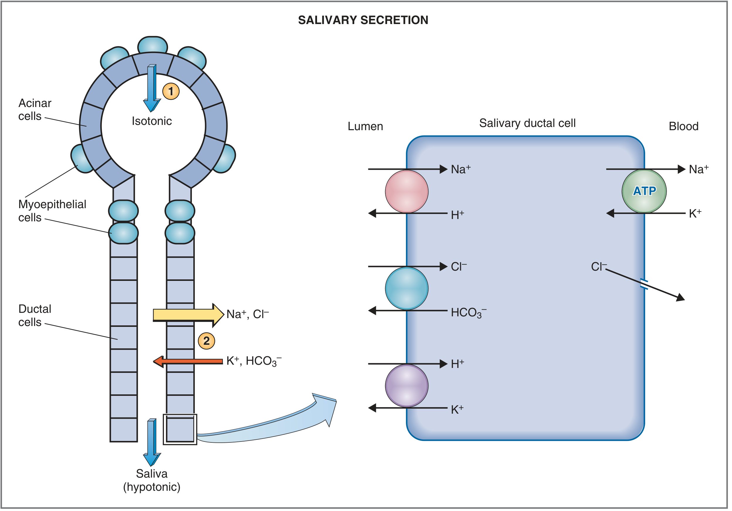
Stage 1 - Acinar secretion (Primary saliva):
- Acinar cells secrete an isotonic, plasma-like primary secretion containing water, electrolytes (Na⁺, K⁺, Cl⁻, HCO₃⁻ at plasma concentrations), enzymes (amylase, lipase), and mucins
- Driven by: parasympathetic stimulation → muscarinic M3 receptors → IP₃ → ↑ intracellular Ca²⁺ → exocytosis of granules + Cl⁻ secretion into acinar lumen → water follows osmotically
Stage 2 - Ductal modification (Final saliva):
| Ion | Transport | Net effect |
|---|---|---|
| Na⁺ | Actively reabsorbed from lumen (Na⁺/H⁺ exchange; Na⁺-K⁺-ATPase on basolateral side) | Final saliva Na⁺ = ~15 mEq/L (1/7 to 1/10 of plasma) |
| Cl⁻ | Passively reabsorbed following Na⁺ electrochemical gradient; also Cl⁻/HCO₃⁻ exchange | Final saliva Cl⁻ = very low |
| K⁺ | Actively secreted into lumen | Final saliva K⁺ = ~30 mEq/L (7× plasma) |
| HCO₃⁻ | Secreted into lumen (especially stimulated; selectively increased at high flow rates) | Final saliva HCO₃⁻ = 50-70 mEq/L (2-3× plasma) |
- Guyton, p. 800; Costanzo Physiology, p. 362-363
5. Composition of Saliva
Electrolyte Comparison with Plasma:
| Electrolyte | Saliva | Plasma | Ratio |
|---|---|---|---|
| Na⁺ | ~15 mEq/L | ~140 mEq/L | 1/10 plasma |
| K⁺ | ~30 mEq/L | ~4 mEq/L | 7× plasma |
| Cl⁻ | ~15 mEq/L | ~105 mEq/L | Low |
| HCO₃⁻ | 50-70 mEq/L | ~25 mEq/L | 2-3× plasma |
| Ca²⁺ | Present | Present | Higher in submandibular |
Organic Composition (Unstimulated Saliva):
| Component | Concentration (mg/mL) | Function |
|---|---|---|
| Total protein | 220 | Multiple |
| α-Amylase (ptyalin) | 38 | Starch digestion |
| Mucin | 2.7 | Lubrication |
| Lysozyme (muramidase) | 22 | Antibacterial |
| Secretory IgA (sIgA) | 19 | Immune defence |
| EGF | 3.4 | Wound healing |
| IgG | 1.4 | Immune |
| Lactoferrin | 0.03 | Antimicrobial |
| Glucose | 1.0 | Trace (diagnostic) |
| Urea | 20 | Excretory |
| Uric acid | 1.5 | Antioxidant |
- Histology: A Text and Atlas, Table 16.1, p. 1484
6. Effect of Flow Rate on Composition
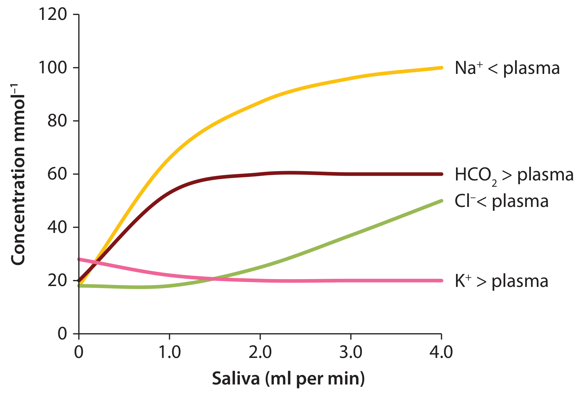
- Ductal cells have maximum contact time with saliva
- Na⁺ and Cl⁻ are maximally reabsorbed → lowest concentrations
- K⁺ is maximally secreted → highest concentration
- Saliva is most hypotonic
- Less ductal modification time
- Na⁺ and Cl⁻ rise toward plasma levels
- HCO₃⁻ rises (exception to contact-time rule - selectively stimulated by parasympathetic activity)
- Saliva osmolality increases (less hypotonic)
7. Functions of Saliva
A. Digestive Functions:
- Carbohydrate digestion: α-amylase (ptyalin) cleaves α-1,4 glycosidic bonds of starch → maltose, maltotriose, α-dextrins. Begins in mouth; continues in oesophagus and stomach until gastric acid (pH <4) denatures it
- Lipid digestion: Lingual lipase (from von Ebner glands on tongue) begins fat digestion; active even at low gastric pH
- Food dissolution: Dissolves food particles to stimulate taste buds (saliva is the medium for taste perception)
- Moistening bolus: Mucin glycoproteins lubricate the food bolus for easy mastication and deglutition
B. Protective / Oral Hygiene Functions:
- Buffering: HCO₃⁻ (primary buffer) neutralises acid produced by oral bacteria and acid from gastric reflux - prevents dental caries and oesophageal mucosal damage. Resting pH ~6.7; stimulated pH ~7.4-7.8
- Antimicrobial:
- Lysozyme - cleaves muramic acid in bacterial cell walls (especially staphylococci)
- Lactoferrin - chelates iron, depriving bacteria of essential nutrient
- sIgA - prevents bacterial adhesion to oral surfaces; opsonisation
- Thiocyanate ions + lysozyme - bactericidal combination
- Peroxidase system - antimicrobial oxidative mechanism
- Dental protection:
- Salivary Ca²⁺ and PO₄³⁻ are essential for remineralisation of early enamel carious lesions and mineralisation of newly erupted teeth
- Proline-rich proteins and statherins form the acquired pellicle - a protective protein coat on tooth surfaces
- Prevents dental plaque formation
- Washing action: Flow of saliva physically washes away food debris, bacteria, and toxins
C. Other Functions:
- Speech: Saliva lubricates the oral mucosa, tongue, and lips, enabling normal articulation
- Wound healing: EGF (Epidermal Growth Factor) in saliva promotes oral mucosal healing - loss of salivary function impedes wound healing
- Thirst regulation: Moistens the oral mucosa; when salivary flow falls, dryness signals thirst
- Excretion: Viruses (HIV, EBV, CMV), drugs, heavy metals (lead), and metabolic waste products (urea, uric acid) are excreted in saliva - the basis of salivary diagnostics
- Kallikrein-kinin vasodilation: Acinar cells secrete kallikrein → cleaves kininogen → bradykinin → vasodilation → ↑ salivary gland blood flow during secretion
- Taste solvent: Water in saliva dissolves sapid molecules to interact with taste receptor cells
8. Nervous Regulation of Salivation
- Under exclusively neural control (unlike other GI secretions which have both neural and hormonal regulation)
- Stimulated by BOTH parasympathetic AND sympathetic systems (unusual - most organs have opposing actions from the two systems)
- Parasympathetic dominance - interruption → gland atrophy
Parasympathetic Control (dominant):
| Gland | Nucleus of Origin | Preganglionic Nerve | Ganglion | Postganglionic Nerve |
|---|---|---|---|---|
| Parotid | Inferior salivatory nucleus (medulla) | CN IX (glossopharyngeal) → lesser petrosal nerve | Otic ganglion | Auriculotemporal nerve (CN V₃) |
| Submandibular & Sublingual | Superior salivatory nucleus (pons) | CN VII (facial) → chorda tympani | Submandibular ganglion | Short postganglionic fibres |
- Large volume of watery, enzyme-rich saliva
- Vasodilation of salivary gland blood vessels (facilitates high secretion)
- Contraction of myoepithelial cells (eject saliva)
Sympathetic Control (secondary):
- Origin: Thoracic segments T1-T3 → superior cervical ganglion → postganglionic fibres along external carotid artery branches
- Neurotransmitter: Norepinephrine → β-adrenergic receptors → cAMP
- Effect: Produces small volume of thick, viscous, protein-rich saliva
- Vasoconstriction (reduces gland blood flow)
Stimuli for Salivation:
-
Increase: Taste (especially sour/acid = 8-20× basal rate), smell of food, smooth objects in mouth, nausea, conditioned reflexes (Pavlov), stomach/intestinal irritation
-
Decrease: Fear, dehydration, sleep, anticholinergic drugs, aging (resting flow)
-
Guyton, p. 800-801; Costanzo Physiology, p. 363-364
9. Salivary Flow Rates
| State | Flow Rate | Dominant Gland |
|---|---|---|
| Basal/resting (unstimulated) | 0.001-0.2 mL/min/gland; total ~0.5 mL/min | Submandibular (60-70%) |
| Stimulated (maximal) | Up to 4-5 mL/min; total 1-2.5 L/day | Parotid (69%) |
| During sleep | Near zero | - |
10. Xerostomia (Dry Mouth)
Causes:
| Category | Examples |
|---|---|
| Drugs (most common) | Antihistamines, anticholinergics, antidepressants (TCAs), antipsychotics, antihypertensives, diuretics |
| Autoimmune | Sjögren's syndrome (lymphocytic destruction of acini) |
| Radiation | Head and neck radiotherapy - irreversible acinar damage |
| Dehydration | Any cause of systemic dehydration |
| Ageing | Decreased basal flow |
| Anxiety/fear | Sympathetic dominance inhibits parasympathetic secretion |
Consequences:
- Rampant dental caries (loss of buffering, antimicrobial, and remineralising functions)
- Oral candidiasis
- Oral ulceration, mucositis
- Dysphagia, dysarthria
- Impaired taste
- Poor denture retention
11. Salivary Diagnostics
- HIV antibodies - salivary ELISA approved for HIV screening
- Cortisol - reflects free serum cortisol; used in Cushing's diagnosis
- Drugs and metabolites - drug testing
- Hormones - testosterone, oestradiol, progesterone
- Blood group substances - ~80% of people are secretors (express ABO antigens in secretions including saliva) - used in forensic medicine
Quick Reference Summary
| Feature | Value |
|---|---|
| Daily volume | 800-1500 mL |
| pH | 5.6-7.4 (resting 6.7; stimulated 7.8) |
| Osmolality | Hypotonic to plasma |
| Na⁺ | 1/7-1/10 of plasma (~15 mEq/L) |
| K⁺ | 7× plasma (~30 mEq/L) |
| HCO₃⁻ | 2-3× plasma (50-70 mEq/L) |
| Water content | 99.5% |
| Most abundant protein | α-Amylase |
| Dominant immunoglobulin | sIgA |
| Dominant unstimulated gland | Submandibular (60-70%) |
| Dominant stimulated gland | Parotid (69%) |
| Primary nerve supply | Parasympathetic (CN VII & IX) |
| Ductal cell function | Na⁺/Cl⁻ absorption; K⁺/HCO₃⁻ secretion |
Tracheo oesophageal prosthesis
Tracheo-Oesophageal Prosthesis (TEP / Voice Prosthesis)
1. Background - Why It Is Needed
- Loss of voice (the primary issue)
- Pulmonary dysfunction - loss of the nasal airway means loss of humidification, filtration, and warming of inhaled air
- Loss of olfaction (anosmia) - nasal airflow is disrupted
- Cummings Otolaryngology, p. 2147
2. History
| Year | Milestone |
|---|---|
| 1873 | Billroth performed the first total laryngectomy; Gussenbauer fitted the patient with a mechanical "artificial larynx" - a tracheotomy tube with pharyngeal extension, reed vibrator, and aspiration valve |
| 1972 | Mozolewski (Poland) published the first modern prosthetic device for 24 patients (in Polish - largely unnoticed) |
| 1980 | Singer and Blom published their landmark paper on tracheoesophageal voice restoration using the Blom-Singer prosthesis - the true beginning of the modern era |
| 1983 | Panje prosthesis introduced (added retention flange and safety strap) |
| 1988 | Provox (indwelling, low-resistance) introduced by Hilgers and Schouwenburg - became the most widely used VP |
| 2003 | Provox 2 (improved) |
| 2011 | Provox Vega (Seldinger technique, pre-mounted on dilator) - current standard |
- Cummings Otolaryngology, p. 2145-2146
3. Principle of Tracheo-Oesophageal Speech
- A small, surgically created fistula called a tracheo-oesophageal puncture (TEP) connects the posterior tracheal wall to the anterior oesophageal wall
- A one-way voice prosthesis (VP) is inserted into this fistula
- When the patient occludes the stoma (with a thumb, finger, or hands-free valve), pulmonary air is diverted through the VP into the pharynx/oesophagus
- This air causes the mucosa of the PE segment to vibrate, generating sound
- The sound is then articulated in the vocal tract (lips, tongue, teeth) into intelligible speech
"TE speech is pulmonary driven and thus closest to normal laryngeal speech." - Cummings, p. 2149
- Opens when patient occludes the stoma and blows air → air flows from trachea to oesophagus
- Closes when patient stops → prevents saliva/food from entering the trachea (aspiration)
4. Alaryngeal Speech Methods - Comparison
| Method | Mechanism | Advantages | Disadvantages |
|---|---|---|---|
| TEP with Voice Prosthesis (Gold Standard) | Pulmonary air via TEP fistula vibrates PE segment mucosa | Most natural sound; 90% success; pulmonary powered; fast recovery (2 weeks); applicable even after extensive pharyngeal reconstruction | Requires surgery; stoma occlusion needed; prosthesis maintenance required |
| Oesophageal speech | Patient swallows/traps air into oesophagus and expels it to vibrate PE segment | No device; no surgery needed | Difficult to learn (30-50% success); limited air supply; short phonation time; poor volume; NOT applicable after pharyngeal flap repair |
| Electrolarynx (Electrolaryngeal speech) | Battery-powered buzzing device placed against neck; sound transmitted through neck tissues and articulated | Easy to use immediately; no surgery | Robotic/mechanical sound; one hand occupied; skin contact needed; poor intelligibility |
5. Tracheo-Oesophageal Puncture (TEP) - Technique
Primary vs Secondary TEP:
| Timing | Description | Preferred? |
|---|---|---|
| Primary TEP | Performed at the same time as total laryngectomy | YES - method of choice; patient wakes from surgery with VP already in place; no second procedure; stenting with NGT not needed |
| Secondary TEP | Performed weeks to months after total laryngectomy as a separate procedure | When primary not possible (e.g., gastric pull-up where proximal oesophagus is mobilised - delay 4-5 weeks) |
Primary TEP Technique (Provox Vega Puncture Set - PVPS):
- At conclusion of total laryngectomy, the posterior tracheal wall and anterior oesophageal wall are identified (the "party wall")
- A Seldinger-type PVPS instrument is used: guidewire introduced through the party wall, followed by a dilator with the VP pre-mounted on it
- The dilator is withdrawn, leaving the VP in situ - stenting the TEP fistula immediately
- No nasogastric tube stent needed
- The device both creates and stents the puncture in a single step
Surgical Refinements to Optimise Results:
- Short myotomy of the upper oesophageal sphincter (cricopharyngeal myotomy) - prevents hypertonicity of PE segment (the most common cause of TEP failure)
- Suturing trachea in a separate fenestra in inferior skin flap - creates a stable stoma
- Sectioning of sternal heads of SCM muscles - prevents a "deep/recessed" stoma
- T-shaped pharyngeal mucosa closure without tension - prevents pseudo-vallecula formation
- Cummings Otolaryngology, p. 2150
6. Types of Voice Prostheses
A. Non-Indwelling Prostheses:
- Patient can self-remove and self-replace
- Requires good manual dexterity and vision
- Example: Original Blom-Singer duckbill prosthesis (Fig. 111.7A), Panje prosthesis
- Increasingly replaced by indwelling devices
B. Indwelling Prostheses:
- Cannot be self-replaced - must be replaced by trained clinician/nurse/speech therapist
- Longer in-situ lifetime (months)
- Lower maintenance burden on patient
- Examples:
| Prosthesis | Developer | Key Feature |
|---|---|---|
| Groningen button | Nijdam (Netherlands) | First indwelling VP; replaced by Provox |
| Provox (1988) | Hilgers & Schouwenburg | Low resistance; self-retaining flanges; became world standard |
| Provox 2 | Hilgers | Improved; softer; rotation-independent |
| Provox Vega (2011) | Atos Medical | Adjustable diameter (16, 20, 22.5 Fr); pre-mounted on PVPS dilator; current standard |
| Provox NID | - | Non-indwelling version |
| VoiceMaster | Schouwenburg | Valveless, self-retaining |
| Blom-Singer Indwelling | Blom-Singer/InHealth | Widely used in USA |
Structure of a Typical VP (Provox Vega):
- Shaft length: 4 mm to 12.5 mm (various sizes - matched to party wall thickness)
- Diameter: 16 Fr, 20 Fr, or 22.5 Fr
- Material: Silicone
- Components:
- Tracheal flange - prevents anterior displacement into trachea
- Oesophageal flange - prevents posterior displacement into oesophagus
- One-way flap valve at oesophageal end - opens inward (trachea→oesophagus) for speech; closes to prevent aspiration
7. Indications for TEP Voice Prosthesis
- Patient has undergone (or will undergo) total laryngectomy
- Intact oesophagus at the level of TEP (not divided, as in total pharyngolaryngooesophagectomy)
- Patient is medically fit for the procedure
- Motivated patient who desires and can manage a prosthesis
- Good hand dexterity and vision (for non-indwelling)
- Good pulmonary reserve
- No significant PE segment hypertonicity
- Pharyngeal reconstruction cases (free flap, pedicled flap) - TEP still possible if oesophagus intact
- Post-chemoradiotherapy laryngectomy (higher complication rate but still feasible)
- TEP at the level where oesophagus has been resected (gastric pull-up)
- Severe pulmonary insufficiency
- Dementia/inability to manage prosthesis
- Significant reflux disease (relative - can be managed)
8. Voice Prosthesis Sizing and Fitting
Measuring Shaft Length:
- The party wall thickness (distance from tracheal to oesophageal lumen) is measured before inserting the VP
- Measured with a sizing gauge - the correct shaft length ensures both flanges seat properly
- Too short: VP leaks around flanges; too long: flanges don't engage properly
- Common lengths: 4, 6, 8, 10, 12.5 mm
In-Situ Lifetime:
- Indwelling VP: average 4-6 months (Provox 2, Provox Vega)
- Replaced when leakage through the prosthesis occurs (valve failure from Candida colonisation)
- Daily antifungal prophylaxis (e.g., yoghurt, antifungal drops) can extend lifetime
9. How the Patient Speaks
Manual Occlusion (most common):
- Patient places thumb or finger over the stoma
- Exhales - air passes through VP into pharynx → PE segment vibrates → speech
- One hand is occupied while speaking
Hands-Free Speaking Valves (HME with speaking valve):
- Heat and Moisture Exchanger (HME) is an adhesive housing attached over the stoma
- An automatic speaking valve within the HME closes when the patient speaks (air pressure during speech closes the valve, diverting air through VP)
- Opened by sniffing or quiet breathing
- Allows hands-free speech - a major quality-of-life improvement
- Also provides pulmonary rehabilitation (humidifies and warms inspired air - compensating for the lost nasal airway)
10. Complications and Management
A. Leakage THROUGH the Prosthesis (most common reason for replacement)
- Replace the VP (primary management)
- Antifungal prophylaxis: oral antifungals (fluconazole), local antifungal drops; dairy products (yoghurt) may reduce Candida load
- Low-resistance VPs or modified valve designs may have longer lifetimes
B. Leakage AROUND the Prosthesis (periprosthetic leakage)
- Gastro-oesophageal/laryngopharyngeal reflux (most important cause - up to 6× higher risk with severe reflux; refluxate disrupts E-cadherin/β-catenin tight junctions → epithelial-mesenchymal transition → fistula enlargement)
- Pressure necrosis from oversized VP
- Radiation damage to party wall tissues
- Yeast/bacterial infection
- Insert a larger diameter VP (e.g., upgrade from 16 Fr to 20 Fr, or 20 Fr to 22.5 Fr)
- Treat reflux aggressively: proton pump inhibitors, dietary modification, anti-reflux measures
- If party wall too thin for any VP: injection of bulking agents around TEP (e.g., collagen, fat)
- Surgical closure of TEP and re-puncture in severe cases
- In refractory cases: permanent TEP closure (<1% of patients)
C. Hypertonicity of the Pharyngo-Oesophageal Segment
- Cricopharyngeal myotomy (surgical) - performed at time of TL or secondarily
- Botulinum toxin A injection into the PE segment - most effective non-surgical treatment; injected under EMG guidance; effects last 3-6 months
- Pharyngeal neurectomy - sectioning of the pharyngeal branches of CN X
D. Granuloma/Pseudo-Vallecula
- Granulation tissue around TEP opening → may block air entry from oesophageal side
- Treated by surgical excision or silver nitrate cauterisation
E. TEP Closure/Stenosis
- Spontaneous shrinkage of the fistula if VP is removed and not immediately replaced
- Always keep a stent or catheter in the TEP if the VP must be removed temporarily
- Re-dilation and re-insertion usually possible
F. Stoma Problems
- Stenosis of the tracheostoma → must be managed before VP fitting
- Stoma button or stoma protector prostheses may help
Summary of Complications Table:
| Complication | Common Cause | Management |
|---|---|---|
| Leakage through VP | Candida on valve | Replace VP; antifungals |
| Leakage around VP | GORD/reflux; fistula widening | PPI; larger VP; bulking agent |
| No voice / failure to phonate | PE hypertonicity | Myotomy; Botox injection |
| VP dislodgement | Flanges fail | Replacement |
| Aspiration | VP incompetence | Replace VP; check sizing |
| TEP stenosis | VP removed without stenting | Re-dilation |
| Granuloma at TEP | Tissue reaction | Cauterise/excise |
| Candida biofilm | Retained proteins in VP | Antifungal prophylaxis; yoghurt |
11. Post-Laryngectomy Rehabilitation: Three-Component Programme
1. Voice Rehabilitation (TEP + VP - as above)
2. Pulmonary Rehabilitation (HME):
- After TL, the upper airway (nose, sinuses, pharynx) is bypassed → inspired air reaches the tracheobronchial tree cold, dry, and unfiltered
- HME (Heat and Moisture Exchanger): A foam/paper-cassette device mounted over the stoma
- Absorbs heat and moisture during exhalation; releases it back during inhalation
- Reduces coughing, sputum production, and pulmonary complications
- Obligatory part of post-TL care
- Integrated with speaking valves for hands-free TEP speech
3. Olfaction Rehabilitation:
- After TL, nasal airflow ceases → anosmia in the vast majority of patients
- Nasal Airflow Inducing Maneuver (NAIM) / "Polite yawning" technique:
- Patient learns to lower the jaw while sealing the lips → creates negative pressure → draws air passively through the nose
- Can restore smell in the vast majority of TL patients with training
12. Outcomes
| Outcome Measure | Data |
|---|---|
| TEP speech success rate | ~90% |
| Time to useful voice | ~2 weeks after TEP |
| Mean maximum phonation time | 16-17 seconds (approaching normal) |
| Average VP in-situ lifetime (Provox Vega) | 4-6 months |
| TEP permanent closure rate | <1% of patients |
| Effect of radiotherapy | Higher complication rate; does NOT significantly reduce overall voice success |
13. Key Exam Points
- TEP = the gold standard for post-laryngectomy voice rehabilitation; 90% success
- Singer and Blom (1980) - credited with modern TEP voice restoration
- Pharyngo-oesophageal segment (PE segment) = the new sound source (vibrating mucosa)
- VP is a one-way valve: trachea → oesophagus (for speech); prevents aspiration
- Primary TEP (at time of TL) is preferred over secondary TEP
- Hypertonicity of PE segment = most common cause of failure to phonate → treat with Botox or myotomy
- Leakage through VP = Candida on valve → replace VP + antifungals
- Leakage around VP = reflux-induced fistula widening → PPI + larger diameter VP
- HME is mandatory for pulmonary rehabilitation after TL
- Indwelling VPs replaced by clinician; non-indwelling replaced by patient
- TE voice has no gender difference in fundamental frequency (~100 Hz in both sexes)
Rigid bronchoscopy
rigid bronchoscopy instrument technique airway
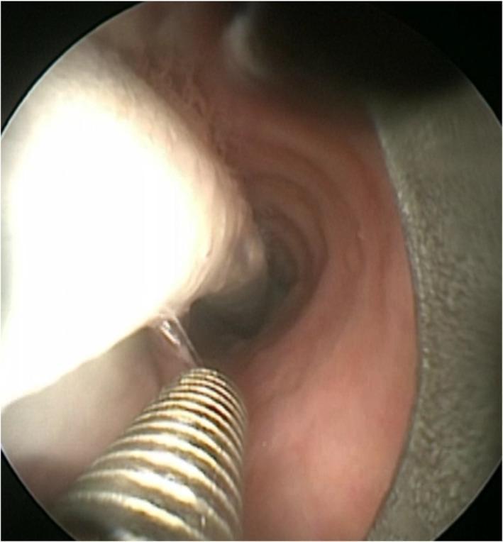
This endoscopic clinical photograph displays an intraoperative view of the human airway during a rigid bronchoscopy procedure. The image depicts the pink, mucosal lining of the trachea or bronchus, providing a central lumen for navigation. Two primary medical instruments are visible: a white, cylindrical laser-resistant tracheal tube (laser tube) positioned along the left and a metallic, corrugated rigid bronchoscope sliding parallel to it. The rigid bronchoscope is being advanced under direct endoscopic visualization to guide its positioning relative to the laser tube. This technique is utilized in interventional pulmonology and thoracic surgery for managing severe central airway obstructions, such as tumor debulking, while ensuring stable ventilation through the laser tube. The educational focus is on the spatial relationship and simultaneous use of ventilatory and surgical instrumentation within the restricted airway lumen.
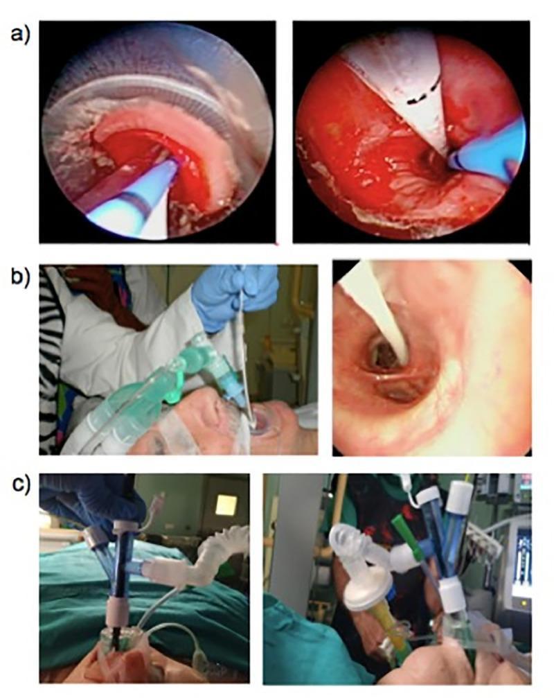
This composite of clinical and intraoperative images demonstrates three different airway management techniques used during transbronchial cryobiopsy (TBCB). Section (a) features endoscopic views showing a flexible bronchoscope and cryoprobe positioned within the trachea. The left view shows the instrument tip nearing a bronchial segment, while the right view illustrates the cryoprobe and an occlusion balloon deployed to manage potential post-biopsy bleeding. Section (b) includes a clinical photograph and an endoscopic view of rigid bronchoscopy. The clinical photo shows the metal rigid bronchoscope being manipulated by a clinician, while the endoscopic view shows the distal end of the rigid tube within the trachea alongside an occlusion balloon catheter. Section (c) displays external clinical photographs of a laryngeal mask airway (LMA) being used to facilitate the procedure, showing the connections for the ventilator circuit and the port through which the flexible bronchoscope is inserted. These images contrast the visual access and instrumentation differences between flexible intubation, rigid bronchoscopy, and supraglottic airway management in interventional pulmonology.
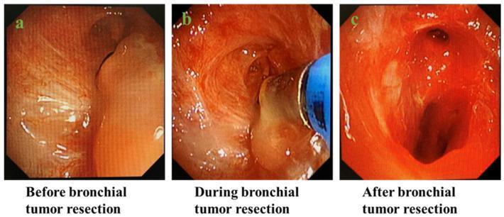
This series of three clinical endoscopic photographs demonstrates the process of an endobronchial tumor resection using rigid bronchoscopy. Panel (a), labeled 'Before bronchial tumor resection,' shows a significant, smooth-surfaced, pale pinkish-orange mass nearly occluding the bronchial lumen, leaving only a narrow, slit-like airway passage. Panel (b), 'During bronchial tumor resection,' illustrates the intraoperative stage where a blue-colored endoscopic instrument is actively debulking or manipulating the tumor tissue. This stage highlights the mechanical intervention within the airway. Panel (c), 'After bronchial tumor resection,' reveals a widely patent bronchial lumen with the primary obstruction successfully removed. The airway now exhibits a clear, oval-shaped opening, allowing for restored ventilation. The surrounding mucosa appears erythematous but intact. These images provide a comparison of pre-operative airway obstruction, the procedural application of rigid bronchoscopy, and the post-operative restoration of airway patency, serving as an educational resource for interventional pulmonology and thoracic surgery.