
Shigellosis tell me in details
shigellosis treatment management
Shigella dysentery pathology histology
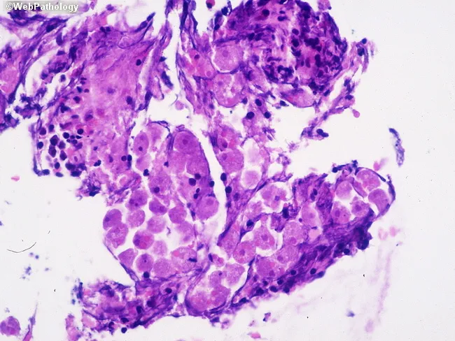
This is a bright-field light microscopy image of a colonic mucosal biopsy stained with Hematoxylin and Eosin (H&E), at high magnification (approximately 400x total). The histology shows inflamed colonic mucosa with an inflammatory exudate in the lamina propria and within the ulcerated mucosal surface. Within this exudate, round to oval trophozoites with granular cytoplasm and a small, centrally placed karyosome are evident, consistent with Entamoeba histolytica. These trophozoites may contain ingested erythrocytes, a characteristic diagnostic clue for invasive amebiasis. Alveolated borders (pseudopodia) are not readily visible in all fields but may be inferred by amoeboid cytoplasm. The surrounding tissue demonstrates acute and chronic inflammatory infiltrates, with neutrophils, macrophages, and occasional eosinophils; focal mucosal necrosis and ulceration are possible. The image highlights tissue invasion by protozoa within a colitis framework, supporting a diagnosis of amebic colitis rather than noninvasive Entamoeba species. Clinically, this histopathologic pattern correlates with dysentery, abdominal pain, and potential systemic spread if untreated. Diagnostic significance lies in identifying Entamoeba histolytica trophozoites in tissue, guiding antiparasitic therapy (metronidazole or tinidazole) followed by luminal eradication therapy. This image is relevant for educational pathology teaching, differential diagnosis of colitis, and research on invasive enteric protozoa. This educational image aids clinical correlation.
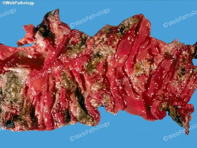
This gross pathology image depicts a colectomy specimen from a patient with Entamoeba histolytica-associated amebic colitis. The colon shows extensive mucosal ulceration with coalescing, serpiginous lesions and an irregular, shaggy fibrinopurulent exudate adherent to the mucosal surface. The involved segments include the cecum and ascending colon, consistent with the common distribution of amebic infection; in severe disease the ileum may be involved though not evident here. The mucosa is friable, erythematous, and eroded with necrotic debris; hemorrhagic areas reflect microvascular injury. This gross pattern can mimic other ulcerative colitides, particularly inflammatory bowel disease, but coarse ulcer margins and exudate are suggestive of amoebic colitis. Clinically, amebiasis presents with abdominal pain, dysentery, weight loss, and may progress to fulminant colitis, megacolon, or perforation. Definitive diagnosis requires histological demonstration of Entamoeba histolytica trophozoites, often with ingested red blood cells, in colonic tissue, or molecular/serologic evidence. Management hinges on anti-amoebic therapy (metronidazole followed by luminal agents) and surgical intervention in complications such as perforation or necrosis. This image highlights a severe surgical specimen with coalescent ulcers and a shaggy exudate that correlates with profound inflammatory destruction and potential life-threatening complications. Correlation with serology or tissue histology confirms amoebic etiology and guides therapy.
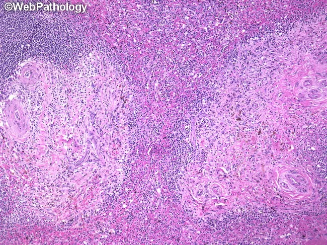
Systemic pathology panel: Histology of a splenectomy specimen from a patient with systemic mastocytosis. Modality: light microscopy on hematoxylin and eosin (H&E) stained sections. Gross features include a thickened splenic capsule with prominent fibrosis and a cut surface showing multiple 1–2 mm nodular foci scattered throughout the parenchyma. Microscopically, these nodules comprise mast cell infiltrates that are spindle-shaped and round, dispersed through both red pulp and white pulp, with a granulomatous appearance. Eosinophils and plasma cells accompany the infiltrate. Immunophenotype aligns with systemic mastocytosis: flow cytometry positive for CD2, CD25, CD117 and tryptase; negative for CD3, CD4, CD20, and CD34. The architectural disruption reflects involvement of the splenic microenvironment, often with hypersplenism and cytopenias as clinical correlates. In this case, patient history includes flushing, dermatologic lesions, diarrhea, hepatosplenomegaly, anemia, and thrombocytopenia; the final diagnosis is systemic mastocytosis with splenic involvement. Radiology is not applicable in this histology image. Clinically, such splenic involvement can be massive in aggressive systemic mastocytosis or systemic mastocytosis with associated hematologic neoplasm. This image illustrates diagnostic clues such as mast cells in fibrotic granulomas within both splenic pulp compartments and provides a reference for differential considerations and therapeutic implications. Useful for education and research instruction purposes.
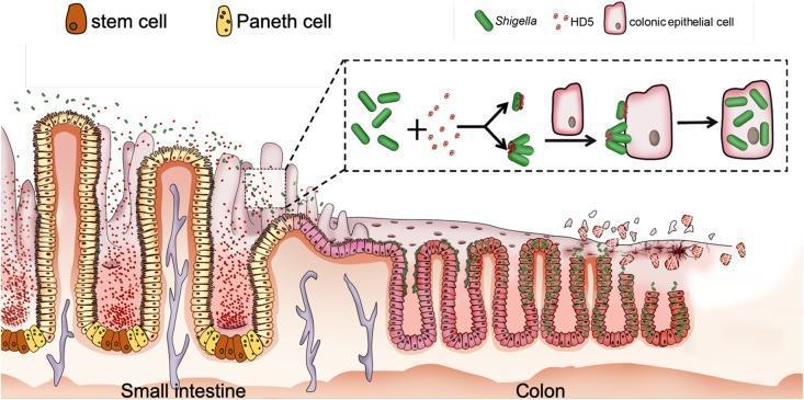
This pathophysiology diagram illustrates the proposed mechanism by which human alpha-defensin 5 (HD5) promotes Shigella infection in the colonic epithelium. The visual is divided into two main sections: the small intestine and the colon. In the small intestine section, the epithelium is characterized by long villi containing stem cells (brown) and Paneth cells (yellow) at the crypt bases. Paneth cells are shown secreting HD5 (red dots) into the intestinal lumen, where they encounter rod-shaped Shigella bacteria (green). A detailed inset shows the molecular interaction: HD5 binds to the bacterial surface, acting as a molecular bridge or causing bacterial clustering to enhance adhesion. In the colon section, the epithelium transitions to a flatter structure with shorter crypts. The HD5-coated Shigella are depicted adhering to the apical surface of colonic epithelial cells (pink), followed by cellular invasion and intracellular replication. The rightmost portion of the diagram shows resulting tissue damage, epithelial sloughing, and loss of mucosal integrity, highlighting the clinical significance of defensins as unintended facilitators of bacterial pathogenicity in certain gastrointestinal infections.
Shigellosis - Comprehensive Overview
Definition
- Goldman-Cecil Medicine, p. 3155
The Pathogen
- Gram-negative, nonmotile, facultatively anaerobic bacilli (rods)
- Oxidase negative, fermenters
- Lipopolysaccharide (LPS) has three components: somatic O polysaccharide, core polysaccharide (common antigen), and lipid A (endotoxin)
- The four species are differentiated by the O-antigen of LPS
Four Species & Clinical Significance
| Species | Serotypes | Geographic Distribution | Notes |
|---|---|---|---|
| S. flexneri | 15 serotypes | Low/middle-income countries (developing world) | Most common globally (66% of isolates); S. flexneri 2a predominates |
| S. sonnei | 1 serotype | High-income countries (developed world) | Most common in Western nations (24% of isolates) |
| S. dysenteriae | 15 serotypes | Epidemic outbreaks | Most severe; produces Shiga toxin; only 5% of isolates |
| S. boydii | 19 serotypes | Mostly South Asia | Relatively uncommon (5%) |
- Medical Microbiology 9e, p. 1974
- Goldman-Cecil Medicine, p. 3155
Epidemiology
- Reservoir: Humans are the ONLY known reservoir - no animal reservoir
- Transmission: Predominantly fecal-oral route (person-to-person contact, fomites, contaminated food/water)
- Very low infective dose: as few as 10 organisms for S. dysenteriae, 180 organisms for S. flexneri and S. sonnei - this makes it highly contagious
- House flies facilitate transmission in settings with open defecation
- Increasingly recognized as a sexually transmitted infection, particularly among men who have sex with men (MSM)
- No seasonal incidence (consistent with person-to-person spread at low inoculum)
Global Burden
- Second most common cause of diarrheal death worldwide (after rotavirus) and the most common bacterial cause
- Main pathogen associated with dysentery (attributable fraction 64%)
- ~150,000 deaths/year globally (GBD 2019); ~95,000 in children under 5
- ~10.5 million disability-adjusted life years (DALYs) lost annually
- Up to 165 million cases per year worldwide
High-Risk Groups
-
Children under 5 years (especially in daycare centers, nurseries)
-
Adults in crowded, poor-sanitation environments
-
Elderly patients
-
Travelers from high-income to low-income countries
-
Siblings and parents of affected children
-
Institutionalized individuals
-
MSM
-
Goldman-Cecil Medicine, p. 3155-3156; Robbins Pathologic Basis of Disease, p. 736
Pathogenesis
- Acid resistance: Shigella is resistant to gastric acid, which partially explains the very low infective dose
- M cell entry: After passing the stomach, bacteria are taken up by M (microfold) cells in the intestinal epithelium - specialized antigen-sampling cells
- Lamina propria escape: Bacteria proliferate within M cells, then escape into the lamina propria
- Macrophage interaction: Bacteria are phagocytosed by macrophages and induce apoptosis of those macrophages
- Inflammatory damage: The inflammatory response damages the surface epithelium
- Basolateral invasion: Both luminal and lamina propria bacteria gain access to the basolateral membranes of colonic epithelial cells - bacteria invade the cytoplasm more efficiently from the basolateral (not apical) side
- Intercellular spread: Bacteria spread from cell to cell, evading the immune response
Key Virulence Factors
- All Shigella spp. carry virulence plasmids, some encoding a Type III Secretion System (T3SS) that directly injects bacterial proteins into host cytoplasm
- S. dysenteriae serotype 1 also releases Shiga toxin (Stx), which inhibits eukaryotic protein synthesis, causing host cell damage and death - this toxin also damages vascular endothelium (explaining HUS)
- Robbins Pathologic Basis of Disease, p. 736-737
Morphology / Pathology
-
Infection is most prominent in the left (distal) colon, though the ileum may be involved (due to M cells overlying Peyer's patches)
-
Gross: Mucosa is hemorrhagic and ulcerated; pseudomembranes may form
-
Histology (early): Similar to other acute self-limited colitides (Campylobacter) - neutrophils in surface and crypt epithelia, crypt abscesses
-
Distinctive feature: Aphthous ulcers overlying Peyer's patches (due to tropism for M cells)
-
As disease progresses: ulceration extends, pseudomembrane formation
-
Robbins Pathologic Basis of Disease, p. 737
Clinical Features
Two Main Presentations
- Onset 1-3 days after ingestion
- Nonbloody, watery stools
- Often self-limiting
- Caused by enterotoxin effect on small bowel
- Develops 1-2 days after onset of watery diarrhea
- Hallmarks: Bloody/mucoid stools, fever, abdominal cramps, tenesmus (painful urge to defecate with passage of little stool)
- Frequent small-volume bloody stools (may be >30/day in severe cases)
- Represents colonic invasion
Complications
-
Toxic megacolon: Life-threatening dilation of the colon
-
Intestinal perforation: Risk with severe disease
-
Rectal prolapse: Especially in malnourished children
-
Hemolytic Uremic Syndrome (HUS): Predominantly with S. dysenteriae serotype 1; microangiopathic hemolytic anemia + thrombocytopenia + acute kidney injury (from Shiga toxin endothelial damage)
-
Bacteremia/Sepsis: More common in malnourished children and immunocompromised hosts; may lead to septic shock
-
Seizures: Seen in young children (febrile seizures or "Ekiri" syndrome in severe cases with cerebral edema)
-
Reactive Arthritis: 1-2% of cases, especially HLA-B27 individuals; seronegative spondyloarthropathy involving the joints, and potentially uveitis, urethritis, and erythema nodosum
-
Post-infectious IBS: Shigella infection increases the risk of subsequent irritable bowel syndrome
-
Malnutrition: Prolonged or recurrent infection contributes significantly in endemic areas
-
Goldman-Cecil Medicine, p. 3156-3157
Diagnosis
-
No clinical feature reliably distinguishes shigellosis from other watery diarrhea causes
-
Stool microscopy: Shows red blood cells and leukocytes (nonspecific)
-
PCR (qPCR): Modern gold standard - enteric multiplex NAATs are the current gold standard; more sensitive than culture
-
Stool culture: Uses selective media (e.g., MacConkey, Hektoen enteric agar, XLD agar); essential for determining antibiotic susceptibility
-
Rectal swab culture is an alternative to stool culture
-
Work ongoing to develop low-cost molecular diagnostics for field use in LMICs
-
Medical Microbiology 9e, p. 1994-1995; Goldman-Cecil Medicine, p. 3157
Treatment
Supportive Care (All Cases)
- Oral Rehydration Solution (ORS): For dehydrated patients; IV fluids (glucose + saline) only for severe dehydration or inability to tolerate oral fluids
- Zinc sulfate 20 mg: Supplement alongside ORS - reduces duration of illness
- Antimotility agents (e.g., loperamide) are CONTRAINDICATED - they prolong symptoms and bacterial shedding by increasing intestinal contact time
When to Use Antibiotics
- Antibiotics reduce duration of fever, diarrhea, and fecal shedding
- Indicated for:
- Dysentery (bloody diarrhea)
- Severe illness in immunocompromised patients
- Epidemic settings
- Not routinely indicated for non-febrile, uncomplicated watery diarrhea (self-limiting in high-income countries)
- Treatment should always be guided by local susceptibility data first, then individual sensitivity profiles
Antibiotic Regimens
| Scenario | First Choice | Alternatives |
|---|---|---|
| Empirical (oral) | Ciprofloxacin 500 mg once daily x 3 days | - |
| Ciprofloxacin-resistant | Azithromycin 500 mg twice daily x 1 day | Cefixime 400 mg once daily x 3 days, or TMP-SMX 160/800 mg twice daily x 5 days |
| Severe/vomiting/unconscious | IV/IM Ceftriaxone 1 g daily x 3 days | - |
- First line oral: Ciprofloxacin 15 mg/kg twice daily x 3 days
- Severe illness (parenteral): Ceftriaxone 50-100 mg/kg once daily x 3 days IV or IM
Antimicrobial Resistance - Growing Crisis
-
Resistance to ampicillin, tetracycline, and TMP-SMX is now widespread
-
Fluoroquinolone-resistant strains are emerging globally
-
A recent systematic review and meta-analysis (2025) documented widespread ESBL-producing and carbapenemase-producing Shigella in Africa, highlighting the growing threat of extensively drug-resistant (XDR) strains
-
Goldman-Cecil Medicine, p. 3157-3158; Medical Microbiology 9e, p. 2000-2003
Prevention & Control
- Hand washing is the single most important measure
- Proper disposal of soiled materials
- Access to clean water and sanitation
- Proper food handling; supervision of food handlers
- Infection control measures to prevent institutional spread
- No licensed vaccine is currently available - this is a major unmet global health need; multiple candidate vaccines are in development
Summary Table
| Feature | Details |
|---|---|
| Organism | Shigella spp. (Gram-negative, nonmotile rod) |
| Most common species (global) | S. flexneri (developing world) |
| Most common species (developed) | S. sonnei |
| Most severe | S. dysenteriae type 1 (Shiga toxin) |
| Infective dose | As low as 10-180 organisms |
| Reservoir | Humans only |
| Transmission | Fecal-oral; also STI in MSM |
| Key virulence | T3SS; Shiga toxin (S. dysenteriae) |
| Site of invasion | Basolateral colonic epithelium via M cells |
| Classic presentation | Dysentery: fever + bloody stools + tenesmus |
| Life-threatening complication | HUS, toxic megacolon, sepsis |
| Gold-standard diagnosis | Multiplex PCR + culture with susceptibility |
| First-line antibiotic (adult) | Ciprofloxacin 500 mg once daily x 3 days |
| Contraindicated drug | Antimotility agents (loperamide) |
| Vaccine | None currently available |