
explain arterial supply of mouth in detail for presentation
Reading File
Now let me search the medical library for arterial supply of the mouth in parallel.
Finding Sources
Finding Sources
Finding Sources
Gray's Anatomy for Students has relevant content. Let me read those sections and also search for more specific content.
Reading File
Finding Sources
Finding Sources
Excellent content found. Let me now read the detailed sections on facial artery, maxillary artery, and the Scott-Brown's lingual artery sections in parallel.
Reading File
Reading File
Reading File
Searching Images
arterial supply oral cavity mouth lingual facial maxillary artery diagram
Good content gathered. Let me now read the maxillary artery branches in detail (for palate and teeth supply) and fetch the key diagram image.
Reading File
Reading File
Loading Image
Searching Images
lingual artery tongue floor of mouth sublingual deep lingual branches anatomy
Excellent - I have a great anatomy diagram. Now let me get the maxillary artery branches for the palate and alveolar regions (pterygopalatine fossa branches), and also read the Scott-Brown's detailed section on lingual artery vascular territory.
Finding Sources
Finding Sources
Now let me read the Scott-Brown's detailed arteries section for the palate and teeth supply:
Reading File
Loading Image
I now have comprehensive information and two excellent anatomy diagrams. Let me compile the full detailed presentation-ready answer.
Arterial Supply of the Mouth (Oral Cavity) - Detailed Presentation Notes
Overview
The oral cavity receives its arterial supply predominantly from the external carotid artery (ECA) via three major branches:
- Facial artery
- Lingual artery
- Maxillary artery
A minor contribution also comes from the internal carotid artery (via the ophthalmic artery).
Diagram 1: Vasculature of the Face and Maxillary Artery Branches
(Gray's Anatomy for Students)
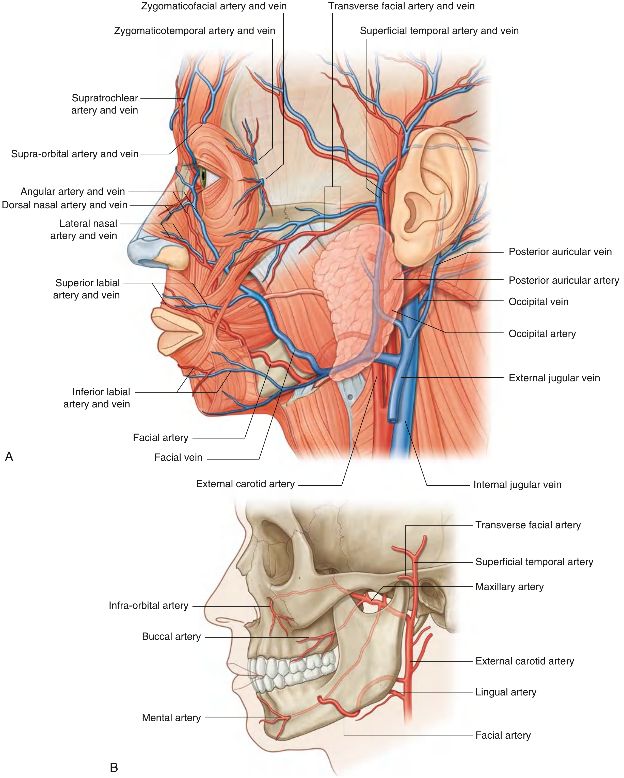
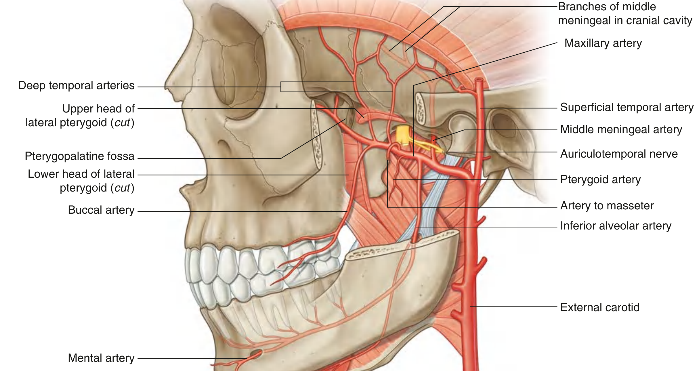
Diagram 2: Maxillary Artery and Its Branches
(Gray's Anatomy for Students)
1. FACIAL ARTERY
Origin: Anterior surface of the external carotid artery in the neck.
Course:
- Passes deep through the neck structures
- Appears at the lower border of the mandible, posterior to the submandibular gland
- Curves around the inferior border of the mandible just anterior to the masseter (pulse palpable here)
- Runs upward and medially in a tortuous course across the face
- Passes deep to platysma, risorius, zygomaticus major and minor; superficial to buccinator and levator anguli oris
- Terminates as the angular artery at the medial corner of the eye
Branches supplying the mouth:
| Branch | Territory |
|---|---|
| Inferior labial artery | Lower lip |
| Superior labial artery | Upper lip + branch to nasal septum |
| Submental artery (in the neck) | Floor of mouth, chin, submandibular region |
| Tonsillar branch | Tonsil, root of tongue |
| Ascending palatine artery | Soft palate, palatine tonsil |
- The superior and inferior labial branches anastomose across the midline with their counterparts from the opposite facial artery, creating an important connection between the two external carotid arteries.
- The platysma and mylohyoid muscles also receive branches from the facial artery.
2. LINGUAL ARTERY
Origin: External carotid artery, at the level of the tip of the greater horn of the hyoid bone.
Course:
- Forms an upward loop, then passes deep to the hyoglossus muscle
- Enters the floor of the oral cavity through the oropharyngeal triangle (bounded by mylohyoid, superior constrictor, and middle constrictor muscles)
- Travels forward in the plane between hyoglossus and genioglossus to the apex of the tongue
- Near the tip of the tongue, it anastomoses with its fellow from the opposite side
Branches (from proximal to distal):
A. Dorsal Lingual Arteries (2-3 branches)
- Arise medial to the hyoglossus
- Ascend to the posterior dorsum of the tongue
- Supply: mucous membrane of the dorsum, palatoglossal arch, soft palate, tonsil, and epiglottis
B. Sublingual Artery
- Arises at the anterior margin of the hyoglossus
- Passes forward between genioglossus and mylohyoid
- Supplies: sublingual gland, floor of mouth mucosa
- Sends a branch through the lingual foramen on the posterior aspect of the mandibular symphysis (important for implant surgery)
C. Deep Lingual Artery
- The terminal branch of the lingual artery
- Runs on the inferior surface of the tongue, near the lingual frenum
- Visible through mucosa on the underside of the tongue
Additional supply to the tongue root: Tonsillar and ascending palatine branches of the facial artery + ascending pharyngeal artery also supply the root of the tongue. In the valleculae region, epiglottic branches of the superior laryngeal artery anastomose with inferior dorsal lingual branches.
3. MAXILLARY ARTERY
Origin: Larger of the two terminal branches of the external carotid artery.
Course: Originates within the parotid gland → passes forward between the neck of the mandible and sphenomandibular ligament → traverses the infratemporal fossa → enters the pterygopalatine fossa through the pterygomaxillary fissure.
The maxillary artery is divided into 3 parts based on its relationship to the lateral pterygoid muscle:
Part 1 (Mandibular/Petrous Part) - Key oral branches:
Inferior Alveolar Artery
- Descends with the inferior alveolar nerve into the mandibular foramen
- Runs in the mandibular canal supplying mandibular molars and premolars
- Divides into:
- Incisive branch - continues to supply mandibular incisors
- Mental artery - exits the mental foramen to supply the chin and lower lip
- Before entering the mandibular foramen, gives off the mylohyoid artery (to mylohyoid and anterior belly of digastric)
Part 2 (Pterygoid Part) - Key oral branches:
Buccal Artery
- Runs with the buccal nerve on the superficial surface of the buccinator
- Supplies: skin, buccinator muscle, and oral mucosa of the cheek
- Also supplies buccal gingiva of lower posterior teeth
Part 3 (Pterygopalatine Part) - Key oral branches:
Greater Palatine Artery
- Descends through the palatine canal (with the greater palatine nerve)
- Gives off 2-3 lesser palatine arteries through lesser palatine canals
- Greater palatine artery supplies: hard palate (major supply) and palatal gingiva of maxillary teeth
- Lesser palatine arteries supply: soft palate and tonsil (anastomose with ascending palatine branch of facial artery)
- Terminal branch passes through the incisive canal to anastomose with the sphenopalatine artery
Nasopalatine Artery (terminal branch of sphenopalatine artery)
- Passes through the incisive canal
- Supplies the anterior palate (incisive/canine region)
- Palatal gingiva: nasopalatine supplies incisors and canines; greater palatine supplies premolars and molars
Superior Alveolar Arteries (supply upper jaw teeth):
| Artery | Origin | Territory |
|---|---|---|
| Posterior superior alveolar | 3rd part of maxillary artery (pterygopalatine fossa) | Upper molars, premolars, maxillary sinus |
| Middle superior alveolar | Infraorbital artery | Upper premolars (variable) |
| Anterior superior alveolar | Infraorbital artery (in infraorbital canal) | Upper incisors, canines, maxillary sinus mucosa |
These three form the superior dental plexus.
Infra-orbital Artery
- Enters the face through the infraorbital foramen
- Supplies: lower eyelid, upper lip, cheek between them
- Gives off anterior (and middle) superior alveolar branches
4. CONTRIBUTIONS FROM ASCENDING PHARYNGEAL ARTERY
- A direct branch of the ECA
- Supplies tissue at the root of the tongue and tonsillar region
- Anastomoses with dorsal lingual arteries in the valleculae
5. TRANSVERSE FACIAL ARTERY
- Branch of the superficial temporal artery (terminal branch of ECA)
- Arises within the parotid gland
- Crosses the face transversely, between the zygomatic arch and parotid duct
- Lies on the superficial surface of masseter
- Minor contribution to cheek vascularity
Summary Table: Regional Arterial Supply
| Region | Primary Artery | Source |
|---|---|---|
| Upper lip | Superior labial (facial a.) | ECA |
| Lower lip | Inferior labial (facial a.) + mental a. | ECA |
| Tongue (body/apex) | Deep lingual a. | Lingual a. (ECA) |
| Tongue (dorsum/posterior) | Dorsal lingual aa. | Lingual a. (ECA) |
| Floor of mouth | Sublingual a. | Lingual a. (ECA) |
| Hard palate (post.) | Greater palatine a. | Maxillary a. (ECA) |
| Hard palate (ant.) | Nasopalatine (sphenopalatine) a. | Maxillary a. (ECA) |
| Soft palate | Lesser palatine + ascending palatine a. | Maxillary a. + Facial a. (ECA) |
| Tonsil/tongue root | Ascending palatine + tonsillar + dorsal lingual | Facial a. + Lingual a. (ECA) |
| Upper teeth | Post/Mid/Ant superior alveolar aa. | Maxillary a. (ECA) |
| Lower teeth | Inferior alveolar a. (incisive branch) | Maxillary a. (ECA) |
| Cheek/buccal mucosa | Buccal a. | Maxillary a. (ECA) |
| Chin | Mental a. | Inferior alveolar a. → Maxillary a. (ECA) |
| Gingiva (upper buccal, molar) | Post. superior alveolar + buccal a. | Maxillary a. (ECA) |
| Gingiva (lower lingual) | Inferior alveolar + lingual a. | Maxillary a. + Lingual a. (ECA) |
Key Anastomoses (Clinically Important)
- Labial arteries anastomose across the midline - connects both external carotid circulations
- Greater palatine artery anastomoses with sphenopalatine/nasopalatine via incisive canal
- Deep lingual arteries anastomose at the tongue tip bilaterally
- Lesser palatine anastomoses with ascending palatine (facial artery)
- Superior laryngeal (internal carotid tributary) anastomoses with dorsal lingual arteries near valleculae
Clinical Relevance
- Mandibular block anesthesia targets the inferior alveolar nerve/artery at the mandibular foramen
- Lingual foramen bleeding during anterior mandibular implant placement can cause life-threatening floor-of-mouth hemorrhage (sublingual artery branch)
- Epistaxis (posterior) involves sphenopalatine artery which anastomoses with greater palatine via incisive canal
- Tongue flaps for reconstruction exploit the reliable lingual artery territory
- Radial forearm free flap used for oral reconstruction brings its own arterial supply
Sources: Gray's Anatomy for Students (9780323934237); Scott-Brown's Otorhinolaryngology Head & Neck Surgery (9781138094642)
venous supply and lymphatic drainage for oral cavity
The venous drainage of the oral cavity follows the arterial supply (facial vein, lingual vein, pterygoid plexus), while lymphatics drain primarily to submental, submandibular, and deep cervical nodes. Let me pull the full detail from the textbooks.
Searching sources...
Finding Sources
Finding Sources
Finding Sources
Reading File
Finding Sources
Reading File
Finding Sources
Finding Sources
Reading File
Finding Sources
Searching Images
lymphatic drainage oral cavity tongue cervical lymph nodes levels diagram
Reading File
Finding Sources
Reading File
Finding Sources
Loading Image
Reading File
I now have all the material needed for a complete, presentation-quality answer. Let me compile it.
Venous Supply and Lymphatic Drainage of the Oral Cavity
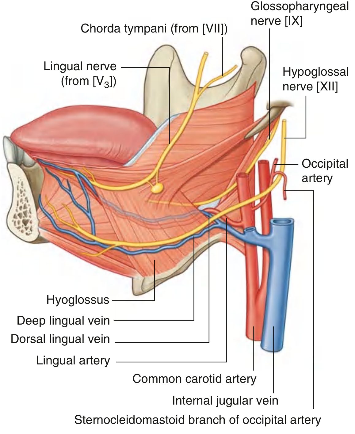
Diagram: Arteries, Veins, and Nerves of the Tongue
(Gray's Anatomy for Students)
PART 1: VENOUS DRAINAGE
The venous drainage of the oral cavity parallels its arterial supply but drains into two main channels: the facial vein and the pterygoid venous plexus, both of which ultimately empty into the internal jugular vein (IJV).
1. Veins of the Tongue
The tongue is drained by two sets of veins:
A. Deep Lingual Vein
- Begins near the apex of the tongue, runs back along the ventral (inferior) surface
- Visible through the mucosa on the underside of the tongue, on each side of the frenulum
- Anteriorly, it accompanies the lingual artery; posteriorly, it separates from it at the hyoglossus muscle
- Travels with the hypoglossal nerve [XII] on the external surface of the hyoglossus
- Joins the sublingual vein (from the sublingual gland) to form the vena comitans nervi hypoglossi (companion vein of the hypoglossal nerve)
- This vena comitans passes backward with the hypoglossal nerve and drains into the lingual, facial, or internal jugular vein
- Exits the floor of the oral cavity through the oropharyngeal triangle (bounded by mylohyoid, superior constrictor, middle constrictor)
B. Dorsal Lingual Veins
- Drain the dorsum and lateral sides of the tongue
- Follow the lingual artery between hyoglossus and genioglossus
- Join the lingual veins accompanying the lingual artery
- Drain into the internal jugular vein at the level of the hyoid bone
2. Veins of the Lips and Face
- Superior labial vein - drains the upper lip → facial vein
- Inferior labial vein - drains the lower lip → facial vein
- The facial vein (formed by supratrochlear + supra-orbital veins as the angular vein at the medial orbit) descends with the facial artery, collects these tributaries, and drains into the internal jugular vein in the neck
- The facial vein and lingual veins form a venous plexus in the submandibular triangle, which then drains as a single tributary (the common facial vein) into the IJV
- The common facial vein also receives some drainage from the retromandibular vein (through the parotid gland)
3. Veins of the Cheek
- The cheek is drained by the buccal vein
- This leads to the pterygoid venous plexus in the infratemporal fossa
4. Veins of the Palate
- The veins of the hard palate accompany the greater palatine and nasopalatine arteries
- Drain largely into the pterygoid venous plexus
5. Veins of the Teeth and Gingivae
| Region | Vein | Ultimate Drainage |
|---|---|---|
| Upper jaw/teeth (anterior) | Superior alveolar veins | Facial vein |
| Upper jaw/teeth (posterior) | Superior alveolar veins | Pterygoid plexus |
| Lower jaw/teeth | Inferior alveolar veins | Mental foramen → facial vein; OR mandibular foramen → pterygoid plexus |
| Gingiva (buccal, lingual, palatine) | Buccal, lingual, greater palatine, nasopalatine veins | Pterygoid plexus (except lingual veins which pass directly to IJV) |
6. The Pterygoid Venous Plexus
A major collecting hub for the oral cavity, located between the medial and lateral pterygoid muscles (and between lateral pterygoid and temporalis).
Receives drainage from:
- Nasal cavity, roof and lateral wall of oral cavity
- All teeth
- Muscles of the infratemporal fossa
- Paranasal sinuses, nasopharynx
- Inferior ophthalmic vein (from orbit, via inferior orbital fissure)
Connects to:
- Posteriorly: maxillary vein → joins superficial temporal vein to form the retromandibular vein → IJV
- Anteriorly: facial vein (via the deep facial vein in the cheek)
- Superiorly: cavernous sinus via emissary veins through foramen ovale, foramen lacerum, and a small sphenoidal foramen
Clinical note - "Danger area of the face": Because the pterygoid plexus connects to the cavernous sinus via valveless emissary veins, infections from the upper lip, nose, and oral cavity above the mouth can spread retrogradely into the cranial cavity. This is why the region above the mouth is called the "danger area." Inadvertent injection into the pterygoid plexus during dental anesthesia can also backflow intracranially due to the absence of venous valves.
Summary: Venous Drainage by Region
| Region | Primary Vein(s) | Drains Into |
|---|---|---|
| Tongue (ventral/apex) | Deep lingual vein → vena comitans n. hypoglossi | Lingual v. / facial v. / IJV |
| Tongue (dorsum/lateral) | Dorsal lingual veins | IJV (at hyoid level) |
| Lips | Superior & inferior labial veins | Facial vein → IJV |
| Cheek | Buccal vein | Pterygoid plexus → retromandibular v. → IJV |
| Hard palate | Palatine veins | Pterygoid plexus |
| Upper teeth (anterior) | Superior alveolar veins | Facial vein |
| Upper teeth (posterior) | Superior alveolar veins | Pterygoid plexus |
| Lower teeth | Inferior alveolar veins | Facial v. (mental) / pterygoid plexus (mandibular) |
| Gingiva | Buccal, lingual, palatine veins | Pterygoid plexus (lingual veins → IJV directly) |
PART 2: LYMPHATIC DRAINAGE
The principal nodes receiving lymphatics from the oral cavity are the submental (Level IA), submandibular (Level IB), and jugulodigastric (Level IIA) nodes. All ultimately drain to the deep cervical chain and then to the thoracic duct (left) or right lymphatic duct.
Note: Drainage patterns are highly variable. Many regions drain bilaterally, especially midline structures. This is critical in oncology.
1. Lymphatics of the Tongue
The tongue lymphatics are divided into three zones:
A. Marginal Vessels (Tip/Apex and Lateral Margins)
Tip of tongue / lingual frenum area:
- Vessels descend under the mucosa
- Some drain to submental nodes (Level IA) and also pass to jugulo-omohyoid nodes (Level III)
- Some vessels cross under the frenulum to drain contralateral nodes (important: bilateral drainage)
- Some drain to submandibular nodes (Level IB)
- One vessel often reaches the jugulo-omohyoid node directly
Lateral margin of tongue:
- Drain to submandibular nodes (Level IB)
- Others end in jugulodigastric (Level IIA) or jugulo-omohyoid (Level III) nodes
- Posterior lateral margin vessels traverse the pharyngeal wall to reach jugulodigastric nodes
B. Central Vessels (Body of Tongue)
- Drain to either or both sides into deep cervical nodes (jugulodigastric and jugulo-omohyoid)
- Some pierce the mylohyoid muscle to enter submandibular nodes
- Bilateral drainage is characteristic of the tongue body
C. Dorsal/Posterior Vessels (Circumvallate region and behind)
- Run posteroinferiorly
- Those near the median plane drain to both sides - jugulodigastric and jugulo-omohyoid nodes
Summary for tongue (K.J. Lee's Classification by neck levels):
| Region | Nodal Levels |
|---|---|
| Anterior (oral) tongue | Level I (submental + submandibular), Level II (jugulodigastric), III, IV; midline → bilateral |
| Posterior (oropharyngeal) tongue | Both ipsilateral + contralateral deep cervical (jugulodigastric) + retropharyngeal nodes |
2. Lymphatics of the Lips and Cheek
| Structure | Primary Nodes | Notes |
|---|---|---|
| Upper lip | Submandibular nodes (Level IB) | |
| Cheek | Submandibular nodes (Level IB) | |
| Lateral lower lip | Submandibular nodes (Level IB) | |
| Central/midline lower lip | Submental nodes (Level IA) | Bilateral drainage possible |
3. Lymphatics of the Floor of Mouth
| Region | Primary Nodes | Notes |
|---|---|---|
| Lateral floor of mouth | Ipsilateral submandibular nodes (Level IB) → deep cervical chain | |
| Anterior floor of mouth | Submental nodes (Level IA) → both left and right deep cervical chains | Bilateral; important for cancer management |
| Lingual lymph nodes | Located in the floor of mouth above the mylohyoid | Clinically relevant for floor-of-mouth tumours |
4. Lymphatics of the Teeth and Gingivae
| Region | Primary Nodes |
|---|---|
| Most teeth | Submandibular nodes (Level IB), ipsilateral |
| Mandibular incisors | Submental nodes (Level IA) |
| Mandibular molars (occasionally) | Jugulodigastric nodes (Level IIA) directly |
| Lingual and palatal gingiva | Jugulodigastric nodes, directly or via submandibular |
| Buccal oral mucosa (bilateral) | Level I submental and submandibular; asymmetrically enlarged nodes are suspicious |
5. Lymphatics of the Palate
| Region | Primary Nodes |
|---|---|
| Bulk of palate | Jugulodigastric nodes (Level IIA) directly |
| Soft palate and tonsil | Jugulodigastric nodes; may also involve retropharyngeal nodes |
| Palatal gingiva | Jugulodigastric nodes, directly or via submandibular |
Nodal Level Reference
| Level | Node Group | Oral Cavity Drainage |
|---|---|---|
| IA | Submental | Tip of tongue, anterior floor of mouth, midline lower lip, mandibular incisors |
| IB | Submandibular | Lateral tongue, lateral floor of mouth, cheek, upper lip, lateral lower lip, most teeth |
| IIA | Upper deep cervical (jugulodigastric) | Posterior/lateral tongue, palate, gingiva, molars; major secondary node for whole oral cavity |
| III | Middle deep cervical (jugulo-omohyoid) | Tongue tip (direct), secondary from Level I and II |
| IV | Lower deep cervical | Tongue (secondary drainage) |
| Retropharyngeal | Posterior oral tongue, soft palate |
Key surgical point: For oral cavity cancer, the standard neck dissection is Selective Neck Dissection of Levels I-III (supraomohyoid neck dissection), covering submental, submandibular, and upper jugular nodes. The submandibular gland is included in the specimen when Level IB nodes are removed. Any asymmetrically enlarged node in the bilateral drainage zones of midline structures is suspicious for metastasis.
Sources: Scott-Brown's Otorhinolaryngology Head & Neck Surgery (9781138094642); Gray's Anatomy for Students (9780323934237); K.J. Lee's Essential Otolaryngology (9781260122237); Cummings Otolaryngology (9780323612173)
Make a presentation combining arterial, venous, and lymphatic anatomy
Reading File
Reading File
Reading File
Good. I have everything I need. Let me now build the full presentation.
Running Command
ls -la /tmp/workspace/ 2>/dev/null || echo "workspace empty or missing"
Running Command
mkdir -p /tmp/workspace/oral-cavity-pptx
Writing File
~/oral-cavity-pptx/build_pptx.py
"""
Oral Cavity: Arterial, Venous & Lymphatic Anatomy
Presentation Builder
"""
import json, subprocess, base64
from io import BytesIO
from pptx import Presentation
from pptx.util import Inches, Pt, Emu
from pptx.dml.color import RGBColor
from pptx.enum.text import PP_ALIGN, MSO_ANCHOR
from pptx.enum.shapes import MSO_SHAPE
# ── Color Palette ──────────────────────────────────────────────────────────────
C_BG = RGBColor(0x0D, 0x1B, 0x2A) # deep navy background
C_ACCENT = RGBColor(0xC8, 0x3B, 0x3B) # arterial red
C_VENOUS = RGBColor(0x3A, 0x6B, 0xC4) # venous blue
C_LYMPH = RGBColor(0x2E, 0xA6, 0x6A) # lymph green
C_GOLD = RGBColor(0xF5, 0xC5, 0x42) # section header gold
C_WHITE = RGBColor(0xFF, 0xFF, 0xFF)
C_LIGHT = RGBColor(0xE8, 0xEE, 0xF4) # light text
C_DARK_CARD = RGBColor(0x16, 0x2A, 0x40) # card background
C_MID = RGBColor(0xA0, 0xB4, 0xCC) # muted text
C_DIVIDER = RGBColor(0x2A, 0x3F, 0x5A)
FONT_MAIN = "Calibri"
FONT_TITLE = "Calibri"
# ── Helper: slide background ───────────────────────────────────────────────────
def set_bg(slide, color=C_BG):
bg = slide.shapes.add_shape(MSO_SHAPE.RECTANGLE,
Inches(0), Inches(0),
Inches(13.333), Inches(7.5))
bg.fill.solid()
bg.fill.fore_color.rgb = color
bg.line.fill.background()
return bg
# ── Helper: add text box ───────────────────────────────────────────────────────
def tb(slide, text, x, y, w, h,
font=FONT_MAIN, size=18, bold=False, italic=False,
color=C_WHITE, align=PP_ALIGN.LEFT, wrap=True,
valign=MSO_ANCHOR.TOP):
shape = slide.shapes.add_textbox(Inches(x), Inches(y), Inches(w), Inches(h))
tf = shape.text_frame
tf.word_wrap = wrap
tf.vertical_anchor = valign
tf.margin_left = Pt(4)
tf.margin_right = Pt(4)
tf.margin_top = Pt(2)
tf.margin_bottom= Pt(2)
p = tf.paragraphs[0]
p.alignment = align
r = p.add_run()
r.text = text
r.font.name = font
r.font.size = Pt(size)
r.font.bold = bold
r.font.italic= italic
r.font.color.rgb = color
return shape
# ── Helper: coloured pill/badge ────────────────────────────────────────────────
def pill(slide, x, y, w, h, fill_color, radius=None):
shp = slide.shapes.add_shape(MSO_SHAPE.ROUNDED_RECTANGLE,
Inches(x), Inches(y), Inches(w), Inches(h))
shp.fill.solid()
shp.fill.fore_color.rgb = fill_color
shp.line.fill.background()
return shp
def rect(slide, x, y, w, h, fill_color, line_color=None, line_w=1):
shp = slide.shapes.add_shape(MSO_SHAPE.RECTANGLE,
Inches(x), Inches(y), Inches(w), Inches(h))
shp.fill.solid()
shp.fill.fore_color.rgb = fill_color
if line_color:
shp.line.color.rgb = line_color
shp.line.width = Pt(line_w)
else:
shp.line.fill.background()
return shp
# ── Helper: horizontal rule ───────────────────────────────────────────────────
def hline(slide, x, y, w, color=C_DIVIDER, width=1.5):
from pptx.enum.shapes import MSO_CONNECTOR
ln = slide.shapes.add_connector(MSO_CONNECTOR.STRAIGHT,
Inches(x), Inches(y),
Inches(x+w), Inches(y))
ln.line.color.rgb = color
ln.line.width = Pt(width)
# ── Helper: multi-line text box with separate paragraphs ─────────────────────
def tb_lines(slide, lines, x, y, w, h,
font=FONT_MAIN, size=16, bold=False,
color=C_LIGHT, spacing=1.15, wrap=True,
align=PP_ALIGN.LEFT):
"""lines = list of (text, indent_level, bold_override, color_override)"""
shape = slide.shapes.add_textbox(Inches(x), Inches(y), Inches(w), Inches(h))
tf = shape.text_frame
tf.word_wrap = wrap
tf.margin_left = Pt(4)
tf.margin_right = Pt(4)
tf.margin_top = Pt(2)
tf.margin_bottom= Pt(2)
first = True
from pptx.oxml.ns import qn
from lxml import etree
for (text, indent, bold_o, color_o) in lines:
if first:
p = tf.paragraphs[0]
first = False
else:
p = tf.add_paragraph()
p.alignment = align
# space after
pPr = p._p.get_or_add_pPr()
spcAft = etree.SubElement(pPr, qn('a:spcAft'))
spcPct = etree.SubElement(spcAft, qn('a:spcPct'))
spcPct.set('val', '0')
r = p.add_run()
r.text = text
r.font.name = font
r.font.size = Pt(size)
r.font.bold = bold_o if bold_o is not None else bold
r.font.color.rgb = color_o if color_o is not None else color
return shape
# ── Helper: fetch remote image → BytesIO ─────────────────────────────────────
def fetch_image(url):
result = json.loads(subprocess.check_output(
["python", "/tmp/skills/shared/scripts/fetch_images.py", url]
))
item = result[0]
if item.get("base64"):
raw = base64.b64decode(item["base64"].split(",", 1)[1])
return BytesIO(raw)
return None
# ══════════════════════════════════════════════════════════════════════════════
# PRESENTATION
# ══════════════════════════════════════════════════════════════════════════════
prs = Presentation()
prs.slide_width = Inches(13.333)
prs.slide_height = Inches(7.5)
blank = prs.slide_layouts[6]
IMAGE_FACE_ARTERY = "https://cdn.orris.care/cdss_images/335aae4b6a630052e093bc8abd9359cb78e9be1bdfebc0bace6bccb4513fbf98.png"
IMAGE_MAX_ARTERY = "https://cdn.orris.care/cdss_images/de85d349043c8d6194fbc6aad09287fd42863c6f3192a4641e70514b3358d849.png"
IMAGE_LINGUAL_VEIN = "https://cdn.orris.care/cdss_images/4fb66bf6990b9f9f9b4c18a7171d35c5d5e934c6128ccae3c44f7cc001259d30.png"
print("Fetching images...")
img_face = fetch_image(IMAGE_FACE_ARTERY)
img_max = fetch_image(IMAGE_MAX_ARTERY)
img_lv = fetch_image(IMAGE_LINGUAL_VEIN)
print("Images fetched.")
# ─────────────────────────────────────────────────────────────────────────────
# SLIDE 1 ─ TITLE
# ─────────────────────────────────────────────────────────────────────────────
slide = prs.slides.add_slide(blank)
set_bg(slide)
# Top accent bar (red)
rect(slide, 0, 0, 13.333, 0.08, C_ACCENT)
# Bottom accent bar (green-blue gradient substitute)
rect(slide, 0, 7.42, 13.333, 0.08, C_LYMPH)
# Central title card
rect(slide, 1.2, 1.6, 10.9, 4.3, C_DARK_CARD)
# Left colour strip (3 colours = arterial / venous / lymph)
rect(slide, 1.2, 1.6, 0.22, 1.43, C_ACCENT)
rect(slide, 1.2, 3.03, 0.22, 1.43, C_VENOUS)
rect(slide, 1.2, 4.47, 0.22, 1.43, C_LYMPH)
tb(slide, "ORAL CAVITY", 2.0, 1.85, 10.0, 0.8,
font=FONT_TITLE, size=42, bold=True, color=C_WHITE, align=PP_ALIGN.LEFT)
tb(slide, "Vascular & Lymphatic Anatomy", 2.0, 2.55, 10.0, 0.7,
font=FONT_TITLE, size=28, bold=False, color=C_GOLD, align=PP_ALIGN.LEFT)
hline(slide, 2.0, 3.35, 9.1, C_DIVIDER, 1.0)
tb(slide, "Arterial Supply | Venous Drainage | Lymphatic Drainage", 2.0, 3.5, 10.0, 0.6,
font=FONT_MAIN, size=18, color=C_MID, align=PP_ALIGN.LEFT)
tb(slide, "Sources: Gray's Anatomy for Students | Scott-Brown's Otorhinolaryngology | K.J. Lee's Essential Otolaryngology",
2.0, 5.0, 9.5, 0.5, font=FONT_MAIN, size=11, color=C_MID, italic=True)
# ─────────────────────────────────────────────────────────────────────────────
# SLIDE 2 ─ OVERVIEW / OVERVIEW OF BLOOD SUPPLY
# ─────────────────────────────────────────────────────────────────────────────
slide = prs.slides.add_slide(blank)
set_bg(slide)
rect(slide, 0, 0, 13.333, 0.08, C_ACCENT)
# Slide header
rect(slide, 0, 0.08, 13.333, 0.75, C_DARK_CARD)
tb(slide, "Overview of Oral Cavity Blood Supply", 0.3, 0.1, 10.0, 0.65,
font=FONT_TITLE, size=26, bold=True, color=C_WHITE)
# Three column cards
cols = [
("ARTERIAL", C_ACCENT, 0.25,
["External Carotid Artery (ECA)", "is the dominant source",
"", "3 major branches:", " Facial Artery", " Lingual Artery", " Maxillary Artery",
"", "Minor: Internal Carotid (ophthalmic a.)"]),
("VENOUS", C_VENOUS, 4.7,
["2 main draining channels:", "", " Facial Vein", " Pterygoid Venous Plexus",
"", "Both ultimately drain to:", " Internal Jugular Vein (IJV)",
"", "Valveless connections to cavernous sinus"]),
("LYMPHATIC", C_LYMPH, 9.1,
["3 principal node groups:", "", " Submental (Level IA)", " Submandibular (Level IB)", " Jugulodigastric (Level IIA)",
"", "All drain to deep cervical chain",
"", "Midline structures drain bilaterally"]),
]
for (label, color, cx, lines) in cols:
rect(slide, cx, 1.05, 4.1, 5.9, C_DARK_CARD)
rect(slide, cx, 1.05, 4.1, 0.5, color)
tb(slide, label, cx+0.1, 1.08, 3.9, 0.42, font=FONT_TITLE,
size=18, bold=True, color=C_WHITE, align=PP_ALIGN.CENTER)
y = 1.7
for line in lines:
is_header = not line.startswith(" ") and line != "" and ":" in line
color_l = C_GOLD if is_header else (C_MID if line == "" else C_LIGHT)
bold_l = True if is_header else False
size_l = 13 if not is_header and line.startswith(" ") else 14
tb(slide, line, cx+0.15, y, 3.8, 0.36,
font=FONT_MAIN, size=size_l, bold=bold_l, color=color_l)
y += 0.33
# ─────────────────────────────────────────────────────────────────────────────
# SLIDE 3 ─ FACIAL ARTERY
# ─────────────────────────────────────────────────────────────────────────────
slide = prs.slides.add_slide(blank)
set_bg(slide)
rect(slide, 0, 0, 13.333, 0.08, C_ACCENT)
rect(slide, 0, 0.08, 13.333, 0.75, C_DARK_CARD)
tb(slide, "Facial Artery", 0.3, 0.1, 6.0, 0.65,
font=FONT_TITLE, size=26, bold=True, color=C_ACCENT)
tb(slide, "ARTERIAL SUPPLY", 8.0, 0.1, 5.0, 0.65,
font=FONT_TITLE, size=13, bold=True, color=C_MID, align=PP_ALIGN.RIGHT)
# Left panel - text
rect(slide, 0.2, 1.0, 6.0, 6.3, C_DARK_CARD)
pill(slide, 0.3, 1.1, 2.0, 0.38, C_ACCENT)
tb(slide, "ORIGIN & COURSE", 0.3, 1.13, 2.0, 0.32,
font=FONT_MAIN, size=12, bold=True, color=C_WHITE, align=PP_ALIGN.CENTER)
lines_fa_course = [
("Branch of External Carotid Artery (ECA)", 0, True, C_LIGHT),
("Passes deep through neck structures", 0, False, C_LIGHT),
("Appears at lower border of mandible", 0, False, C_LIGHT),
("(posterior to submandibular gland)", 0, False, C_MID),
("Pulse felt at inferior border of mandible", 0, False, C_LIGHT),
("(anterior to masseter)", 0, False, C_MID),
("Runs tortuous course upward + medially", 0, False, C_LIGHT),
("Terminates as angular artery at medial orbit", 0, False, C_LIGHT),
]
tb_lines(slide, lines_fa_course, 0.35, 1.55, 5.8, 2.5,
font=FONT_MAIN, size=13, color=C_LIGHT)
pill(slide, 0.3, 3.85, 2.5, 0.38, C_ACCENT)
tb(slide, "BRANCHES TO ORAL CAVITY", 0.3, 3.88, 2.5, 0.32,
font=FONT_MAIN, size=12, bold=True, color=C_WHITE, align=PP_ALIGN.CENTER)
branches_fa = [
("Inferior Labial Artery -> Lower lip", 0, True, C_GOLD),
("Superior Labial Artery -> Upper lip + nasal septum branch", 0, True, C_GOLD),
("Submental Artery -> Floor of mouth, chin, submandibular region", 0, False, C_LIGHT),
("Ascending Palatine Artery -> Soft palate, tonsil", 0, False, C_LIGHT),
("Tonsillar Branch -> Tonsil, root of tongue", 0, False, C_LIGHT),
("Labial branches anastomose across midline", 0, False, C_MID),
("(connects both ECA circulations)", 0, False, C_MID),
]
tb_lines(slide, branches_fa, 0.35, 4.28, 5.8, 2.8, font=FONT_MAIN, size=12)
# Right panel - diagram image
if img_face:
img_face_bytes = img_face
slide.shapes.add_picture(img_face_bytes, Inches(6.5), Inches(0.95), width=Inches(6.6))
# ─────────────────────────────────────────────────────────────────────────────
# SLIDE 4 ─ LINGUAL ARTERY
# ─────────────────────────────────────────────────────────────────────────────
slide = prs.slides.add_slide(blank)
set_bg(slide)
rect(slide, 0, 0, 13.333, 0.08, C_ACCENT)
rect(slide, 0, 0.08, 13.333, 0.75, C_DARK_CARD)
tb(slide, "Lingual Artery", 0.3, 0.1, 6.0, 0.65,
font=FONT_TITLE, size=26, bold=True, color=C_ACCENT)
tb(slide, "ARTERIAL SUPPLY", 8.0, 0.1, 5.0, 0.65,
font=FONT_TITLE, size=13, bold=True, color=C_MID, align=PP_ALIGN.RIGHT)
# Left col
rect(slide, 0.2, 1.0, 5.8, 6.3, C_DARK_CARD)
pill(slide, 0.3, 1.1, 2.0, 0.38, C_ACCENT)
tb(slide, "ORIGIN & COURSE", 0.3, 1.13, 2.0, 0.32,
font=FONT_MAIN, size=12, bold=True, color=C_WHITE, align=PP_ALIGN.CENTER)
la_course = [
("Origin: ECA at tip of greater horn of hyoid bone", 0, True, C_LIGHT),
("Forms upward loop, then passes deep to hyoglossus", 0, False, C_LIGHT),
("Enters floor of oral cavity via oropharyngeal triangle", 0, False, C_LIGHT),
("(bounded by mylohyoid + sup. + mid. constrictors)", 0, False, C_MID),
("Travels between hyoglossus and genioglossus", 0, False, C_LIGHT),
("Anastomoses with fellow at tongue apex (bilateral)", 0, True, C_GOLD),
]
tb_lines(slide, la_course, 0.35, 1.55, 5.5, 2.2, font=FONT_MAIN, size=13)
pill(slide, 0.3, 3.6, 1.8, 0.38, C_ACCENT)
tb(slide, "4 BRANCHES", 0.3, 3.63, 1.8, 0.32,
font=FONT_MAIN, size=12, bold=True, color=C_WHITE, align=PP_ALIGN.CENTER)
branch_cards = [
("Dorsal Lingual Aa. (2-3)", "Arise medial to hyoglossus\nPosterior dorsum, soft palate,\npalatoglossal arch, tonsil, epiglottis"),
("Sublingual Artery", "Anterior margin of hyoglossus\nSublingual gland, floor of mouth\nLingual foramen branch -> mandible"),
("Deep Lingual Artery", "Terminal branch\nInferior surface of tongue\nNear lingual frenum - visible through mucosa"),
]
bx = 0.28
for (bname, bdesc) in branch_cards:
rect(slide, bx, 4.1, 1.85, 2.7, RGBColor(0x1B, 0x32, 0x4A))
tb(slide, bname, bx+0.07, 4.15, 1.72, 0.42,
font=FONT_MAIN, size=12, bold=True, color=C_GOLD)
tb(slide, bdesc, bx+0.07, 4.6, 1.72, 2.1,
font=FONT_MAIN, size=11, color=C_LIGHT, wrap=True)
bx += 1.95
# Right col - image
if img_lv:
img_lv2 = fetch_image(IMAGE_LINGUAL_VEIN)
slide.shapes.add_picture(img_lv2, Inches(6.3), Inches(0.95), width=Inches(6.8))
# Additional supply to root
tb(slide, "Additional supply to tongue root: ascending palatine (facial a.) + ascending pharyngeal a. + superior laryngeal a. (epiglottic region)",
0.28, 7.05, 12.8, 0.4, font=FONT_MAIN, size=11, color=C_MID, italic=True)
# ─────────────────────────────────────────────────────────────────────────────
# SLIDE 5 ─ MAXILLARY ARTERY
# ─────────────────────────────────────────────────────────────────────────────
slide = prs.slides.add_slide(blank)
set_bg(slide)
rect(slide, 0, 0, 13.333, 0.08, C_ACCENT)
rect(slide, 0, 0.08, 13.333, 0.75, C_DARK_CARD)
tb(slide, "Maxillary Artery", 0.3, 0.1, 7.0, 0.65,
font=FONT_TITLE, size=26, bold=True, color=C_ACCENT)
tb(slide, "ARTERIAL SUPPLY | 3 PARTS", 7.0, 0.1, 6.0, 0.65,
font=FONT_TITLE, size=13, bold=True, color=C_MID, align=PP_ALIGN.RIGHT)
# 3 parts as columns
part_data = [
("PART 1\nMandibular",
RGBColor(0x8B, 0x20, 0x20),
["Inferior Alveolar Artery:",
" Into mandibular foramen",
" Mandibular molars + premolars",
" Splits into:",
" Incisive -> mandibular incisors",
" Mental -> chin + lower lip",
" Mylohyoid branch (before foramen)",
"",
"Between neck of mandible",
"and sphenomandibular ligament"]),
("PART 2\nPterygoid",
RGBColor(0x7A, 0x36, 0x10),
["Buccal Artery:",
" With buccal nerve on buccinator",
" Skin, muscle, oral mucosa of cheek",
" Buccal gingiva (lower posterior)",
"",
"Masseteric, Pterygoid,",
"Deep Temporal arteries",
"(muscles of mastication)",
"",
"Related to lateral pterygoid muscle"]),
("PART 3\nPterygopalatine",
RGBColor(0x1E, 0x5C, 0x3A),
["Greater Palatine Artery:",
" Hard palate (primary supply)",
" Palatal gingiva of maxillary teeth",
" -> Lesser palatine aa.",
" Soft palate + tonsil",
"Nasopalatine (sphenopalatine):",
" Anterior palate (incisors + canines)",
" via incisive canal",
"Superior Alveolar Arteries:",
" Post/Mid/Ant -> upper teeth plexus",
"Infra-orbital Artery:",
" Upper lip, lower eyelid, cheek"]),
]
px = 0.2
for (title, col, lines) in part_data:
rect(slide, px, 1.0, 4.2, 6.3, C_DARK_CARD)
rect(slide, px, 1.0, 4.2, 0.62, col)
tb(slide, title, px+0.1, 1.03, 4.0, 0.56,
font=FONT_TITLE, size=14, bold=True, color=C_WHITE, align=PP_ALIGN.CENTER)
y = 1.72
for line in lines:
is_sub = line.startswith(" ")
is_ind = line.startswith(" ")
is_bold_h = not is_sub and not is_ind and line != "" and not line.startswith("(")
col_l = C_GOLD if is_bold_h and ":" in line else (C_MID if line == "" else C_LIGHT)
sz = 11 if is_sub else (12 if is_ind else 12)
tb(slide, line, px+0.12, y, 3.9, 0.35,
font=FONT_MAIN, size=sz, bold=(is_bold_h and ":" in line), color=col_l)
y += 0.32
px += 4.35
# ─────────────────────────────────────────────────────────────────────────────
# SLIDE 6 ─ ARTERIAL SUMMARY TABLE
# ─────────────────────────────────────────────────────────────────────────────
slide = prs.slides.add_slide(blank)
set_bg(slide)
rect(slide, 0, 0, 13.333, 0.08, C_ACCENT)
rect(slide, 0, 0.08, 13.333, 0.75, C_DARK_CARD)
tb(slide, "Arterial Supply - Regional Summary", 0.3, 0.1, 10.0, 0.65,
font=FONT_TITLE, size=24, bold=True, color=C_ACCENT)
headers = ["Region", "Primary Artery", "Source"]
rows = [
("Upper lip", "Superior labial a. (facial a.)", "ECA"),
("Lower lip", "Inferior labial + mental a.", "ECA"),
("Tongue - body/apex", "Deep lingual a.", "Lingual a. (ECA)"),
("Tongue - dorsum/post", "Dorsal lingual aa.", "Lingual a. (ECA)"),
("Floor of mouth", "Sublingual a.", "Lingual a. (ECA)"),
("Hard palate (post.)", "Greater palatine a.", "Maxillary a. (ECA)"),
("Hard palate (ant.)", "Nasopalatine / sphenopalatine a.", "Maxillary a. (ECA)"),
("Soft palate", "Lesser palatine + ascending palatine", "Maxillary + Facial (ECA)"),
("Upper teeth", "Post./Mid./Ant. superior alveolar aa.","Maxillary a. (ECA)"),
("Lower teeth", "Inferior alveolar a. (incisive br.)", "Maxillary a. (ECA)"),
("Cheek / buccal mucosa","Buccal a.", "Maxillary a. (ECA)"),
("Chin", "Mental a.", "Inf. alveolar -> Maxillary"),
]
col_widths = [3.4, 5.5, 3.8]
col_starts = [0.2, 3.65, 9.2]
# Header row
hy = 0.97
for i, (hdr, cw, cs) in enumerate(zip(headers, col_widths, col_starts)):
rect(slide, cs, hy, cw-0.05, 0.42, C_ACCENT)
tb(slide, hdr, cs+0.1, hy+0.05, cw-0.15, 0.32,
font=FONT_MAIN, size=13, bold=True, color=C_WHITE, align=PP_ALIGN.LEFT)
ry = 1.42
for ri, row in enumerate(rows):
bg_col = C_DARK_CARD if ri % 2 == 0 else RGBColor(0x12, 0x22, 0x35)
rect(slide, 0.2, ry, 12.93, 0.40, bg_col)
for j, (val, cw, cs) in enumerate(zip(row, col_widths, col_starts)):
txt_col = C_GOLD if j == 1 else C_LIGHT
tb(slide, val, cs+0.1, ry+0.04, cw-0.15, 0.34,
font=FONT_MAIN, size=12, color=txt_col)
ry += 0.40
# ─────────────────────────────────────────────────────────────────────────────
# SLIDE 7 ─ VENOUS DRAINAGE TONGUE + LIPS
# ─────────────────────────────────────────────────────────────────────────────
slide = prs.slides.add_slide(blank)
set_bg(slide)
rect(slide, 0, 0, 13.333, 0.08, C_VENOUS)
rect(slide, 0, 0.08, 13.333, 0.75, C_DARK_CARD)
tb(slide, "Venous Drainage of the Oral Cavity", 0.3, 0.1, 10.0, 0.65,
font=FONT_TITLE, size=24, bold=True, color=C_VENOUS)
tb(slide, "VENOUS SUPPLY", 10.0, 0.1, 3.0, 0.65,
font=FONT_TITLE, size=13, bold=True, color=C_MID, align=PP_ALIGN.RIGHT)
# Left: tongue veins
rect(slide, 0.2, 1.0, 6.2, 5.9, C_DARK_CARD)
pill(slide, 0.3, 1.08, 2.5, 0.38, C_VENOUS)
tb(slide, "VEINS OF THE TONGUE", 0.3, 1.11, 2.5, 0.32,
font=FONT_MAIN, size=12, bold=True, color=C_WHITE, align=PP_ALIGN.CENTER)
tongue_veins = [
("Deep Lingual Vein", True, C_GOLD),
("Begins near apex, runs along ventral surface", False, C_LIGHT),
("Visible through mucosa beside frenulum", False, C_LIGHT),
("Travels with hypoglossal nerve [XII]", False, C_LIGHT),
("Joins sublingual vein from sublingual gland", False, C_LIGHT),
("-> Forms vena comitans nervi hypoglossi", False, C_MID),
("Drains to: lingual / facial / IJV", False, C_VENOUS),
("", False, C_MID),
("Dorsal Lingual Veins", True, C_GOLD),
("Drain dorsum + lateral sides of tongue", False, C_LIGHT),
("Follow lingual artery (hyoglossus / genioglossus)", False, C_LIGHT),
("Drain into IJV at level of hyoid bone", False, C_VENOUS),
]
y = 1.55
for (t, b, c) in tongue_veins:
tb(slide, t, 0.35, y, 5.9, 0.35, font=FONT_MAIN, size=12, bold=b, color=c)
y += 0.33
# Right: lips / cheek / palate / gingiva
rect(slide, 6.7, 1.0, 6.4, 5.9, C_DARK_CARD)
pill(slide, 6.8, 1.08, 3.2, 0.38, C_VENOUS)
tb(slide, "LIPS, CHEEK, PALATE, TEETH", 6.8, 1.11, 3.2, 0.32,
font=FONT_MAIN, size=12, bold=True, color=C_WHITE, align=PP_ALIGN.CENTER)
other_veins = [
("Lips", True, C_GOLD),
("Superior + inferior labial veins -> Facial vein", False, C_LIGHT),
("Facial vein drains to IJV in the neck", False, C_LIGHT),
("Common facial vein also receives retromandibular vein", False, C_MID),
("", False, None),
("Cheek", True, C_GOLD),
("Buccal vein -> Pterygoid venous plexus", False, C_LIGHT),
("", False, None),
("Hard Palate", True, C_GOLD),
("Palatine veins -> Pterygoid plexus", False, C_LIGHT),
("", False, None),
("Upper Teeth", True, C_GOLD),
("Ant. superior alveolar veins -> Facial vein", False, C_LIGHT),
("Post. superior alveolar veins -> Pterygoid plexus", False, C_LIGHT),
("", False, None),
("Lower Teeth", True, C_GOLD),
("Inf. alveolar veins: mental foramen -> Facial vein", False, C_LIGHT),
("OR mandibular foramen -> Pterygoid plexus", False, C_LIGHT),
("", False, None),
("Gingiva", True, C_GOLD),
("Buccal/lingual/palatine veins -> Pterygoid plexus", False, C_LIGHT),
("Lingual veins -> IJV (direct)", False, C_MID),
]
y = 1.55
for (t, b, c) in other_veins:
if c:
tb(slide, t, 6.85, y, 6.0, 0.33, font=FONT_MAIN, size=12, bold=b, color=c)
y += 0.28
# ─────────────────────────────────────────────────────────────────────────────
# SLIDE 8 ─ PTERYGOID PLEXUS (clinical)
# ─────────────────────────────────────────────────────────────────────────────
slide = prs.slides.add_slide(blank)
set_bg(slide)
rect(slide, 0, 0, 13.333, 0.08, C_VENOUS)
rect(slide, 0, 0.08, 13.333, 0.75, C_DARK_CARD)
tb(slide, "Pterygoid Venous Plexus", 0.3, 0.1, 8.0, 0.65,
font=FONT_TITLE, size=26, bold=True, color=C_VENOUS)
tb(slide, "VENOUS SUPPLY", 10.5, 0.1, 2.6, 0.65,
font=FONT_TITLE, size=13, bold=True, color=C_MID, align=PP_ALIGN.RIGHT)
# Main description
rect(slide, 0.2, 1.0, 8.3, 5.0, C_DARK_CARD)
pill(slide, 0.3, 1.08, 2.8, 0.38, C_VENOUS)
tb(slide, "LOCATION & TRIBUTARIES", 0.3, 1.11, 2.8, 0.32,
font=FONT_MAIN, size=12, bold=True, color=C_WHITE, align=PP_ALIGN.CENTER)
plexus_info = [
("Located between medial and lateral pterygoid muscles", False, C_LIGHT),
("Also between lateral pterygoid and temporalis", False, C_LIGHT),
("", False, None),
("Receives drainage from:", True, C_GOLD),
(" Nasal cavity, oral cavity (roof + lateral wall)", False, C_LIGHT),
(" All teeth", False, C_LIGHT),
(" Muscles of infratemporal fossa", False, C_LIGHT),
(" Paranasal sinuses + nasopharynx", False, C_LIGHT),
(" Inferior ophthalmic vein (via inf. orbital fissure)", False, C_LIGHT),
("", False, None),
("Connects posteriorly to maxillary vein", False, C_LIGHT),
("-> retromandibular vein -> IJV", False, C_MID),
("Connects anteriorly to facial vein", False, C_LIGHT),
("(via deep facial vein in the cheek)", False, C_MID),
("Connects superiorly to cavernous sinus", False, C_LIGHT),
("(via emissary veins - no valves!)", False, C_MID),
]
y = 1.55
for (t, b, c) in plexus_info:
if c:
tb(slide, t, 0.35, y, 8.0, 0.33, font=FONT_MAIN, size=13, bold=b, color=c)
y += 0.33
# Clinical alert box
rect(slide, 0.2, 6.1, 12.9, 1.2, RGBColor(0x3A, 0x10, 0x10))
rect(slide, 0.2, 6.1, 0.18, 1.2, C_ACCENT)
tb(slide, "CLINICAL: 'Danger Area of the Face'", 0.5, 6.15, 8.0, 0.42,
font=FONT_MAIN, size=14, bold=True, color=C_ACCENT)
tb(slide, "Because the pterygoid plexus connects to the cavernous sinus via VALVELESS emissary veins, "
"infections above the mouth can spread intracranially. Inadvertent injection into the plexus during "
"dental anesthesia can backflow intracranially.",
0.5, 6.58, 12.5, 0.65, font=FONT_MAIN, size=12, color=C_LIGHT, wrap=True)
# Right: summary table
rect(slide, 8.8, 1.0, 4.3, 5.0, C_DARK_CARD)
tb(slide, "VENOUS DRAINAGE SUMMARY", 8.9, 1.05, 4.1, 0.4,
font=FONT_MAIN, size=12, bold=True, color=C_VENOUS, align=PP_ALIGN.CENTER)
summary_rows = [
("Tongue", "Lingual veins / IJV"),
("Lips", "Facial vein -> IJV"),
("Cheek", "Buccal v. -> Pterygoid plexus"),
("Palate", "Pterygoid plexus"),
("Upper teeth", "Facial v. / Pterygoid plexus"),
("Lower teeth", "Facial v. / Pterygoid plexus"),
("Gingiva", "Pterygoid plexus (lingual->IJV)"),
]
ry2 = 1.52
for (region, drain) in summary_rows:
tb(slide, region, 8.85, ry2, 1.9, 0.38, font=FONT_MAIN, size=11, bold=True, color=C_GOLD)
tb(slide, drain, 10.75, ry2, 2.2, 0.38, font=FONT_MAIN, size=11, color=C_LIGHT)
ry2 += 0.42
# ─────────────────────────────────────────────────────────────────────────────
# SLIDE 9 ─ LYMPH OVERVIEW + TONGUE
# ─────────────────────────────────────────────────────────────────────────────
slide = prs.slides.add_slide(blank)
set_bg(slide)
rect(slide, 0, 0, 13.333, 0.08, C_LYMPH)
rect(slide, 0, 0.08, 13.333, 0.75, C_DARK_CARD)
tb(slide, "Lymphatic Drainage - Overview & Tongue", 0.3, 0.1, 10.0, 0.65,
font=FONT_TITLE, size=24, bold=True, color=C_LYMPH)
tb(slide, "LYMPHATIC DRAINAGE", 10.0, 0.1, 3.0, 0.65,
font=FONT_TITLE, size=13, bold=True, color=C_MID, align=PP_ALIGN.RIGHT)
# Left: overview card
rect(slide, 0.2, 1.0, 4.2, 6.0, C_DARK_CARD)
pill(slide, 0.3, 1.08, 2.5, 0.38, C_LYMPH)
tb(slide, "PRINCIPAL NODE GROUPS", 0.3, 1.11, 2.5, 0.32,
font=FONT_MAIN, size=11, bold=True, color=C_WHITE, align=PP_ALIGN.CENTER)
node_groups = [
("Level IA - Submental", True, C_GOLD),
("Tip of tongue, anterior floor of mouth,", False, C_LIGHT),
("midline lower lip, mandibular incisors", False, C_LIGHT),
("", False, None),
("Level IB - Submandibular", True, C_GOLD),
("Lateral tongue, lateral floor of mouth,", False, C_LIGHT),
("cheek, upper lip, lateral lower lip, most teeth", False, C_LIGHT),
("", False, None),
("Level IIA - Jugulodigastric", True, C_GOLD),
("Post./lateral tongue, palate, gingiva,", False, C_LIGHT),
("molars; secondary node for whole oral cavity", False, C_LIGHT),
("", False, None),
("Level III - Jugulo-omohyoid", True, C_GOLD),
("Tongue tip (direct), secondary from I+II", False, C_LIGHT),
("", False, None),
("Retropharyngeal nodes", True, C_GOLD),
("Posterior oral tongue + soft palate", False, C_LIGHT),
]
y = 1.55
for (t, b, c) in node_groups:
if c:
tb(slide, t, 0.35, y, 3.9, 0.33, font=FONT_MAIN, size=12, bold=b, color=c)
y += 0.32
# Right: tongue lymphatics
rect(slide, 4.65, 1.0, 8.4, 6.0, C_DARK_CARD)
pill(slide, 4.75, 1.08, 3.2, 0.38, C_LYMPH)
tb(slide, "LYMPHATICS OF THE TONGUE (3 Zones)", 4.75, 1.11, 3.2, 0.32,
font=FONT_MAIN, size=12, bold=True, color=C_WHITE, align=PP_ALIGN.CENTER)
tongue_lymph = [
("Zone 1: MARGINAL VESSELS (Tip + Lateral margins)", True, C_GOLD),
("Tip/frenum area: submental (IA) + jugulo-omohyoid (III)", False, C_LIGHT),
("Cross-midline drainage to CONTRALATERAL nodes", False, C_ACCENT),
("Lateral margin: submandibular (IB)", False, C_LIGHT),
("Also jugulodigastric (IIA) / jugulo-omohyoid (III)", False, C_LIGHT),
("Posterior lateral margin: traverses pharyngeal wall to IIA", False, C_MID),
("", False, None),
("Zone 2: CENTRAL VESSELS (Body of tongue)", True, C_GOLD),
("Drains BILATERALLY to deep cervical nodes", False, C_ACCENT),
("Especially IIA (jugulodigastric) + III (jugulo-omohyoid)", False, C_LIGHT),
("Some pierce mylohyoid to submandibular nodes (IB)", False, C_LIGHT),
("", False, None),
("Zone 3: DORSAL VESSELS (Circumvallate + posterior)", True, C_GOLD),
("Run posteroinferiorly", False, C_LIGHT),
("Median plane: BOTH sides -> IIA + III", False, C_ACCENT),
("", False, None),
("Anterior (oral) tongue: Levels I, II, III, IV", True, C_LYMPH),
("Posterior (oropharyngeal) tongue: bilateral deep cervical + retropharyngeal", True, C_LYMPH),
]
y = 1.55
for (t, b, c) in tongue_lymph:
if c:
tb(slide, t, 4.8, y, 8.0, 0.34, font=FONT_MAIN, size=12, bold=b, color=c)
y += 0.31
# ─────────────────────────────────────────────────────────────────────────────
# SLIDE 10 ─ LYMPH: REGION BY REGION
# ─────────────────────────────────────────────────────────────────────────────
slide = prs.slides.add_slide(blank)
set_bg(slide)
rect(slide, 0, 0, 13.333, 0.08, C_LYMPH)
rect(slide, 0, 0.08, 13.333, 0.75, C_DARK_CARD)
tb(slide, "Lymphatic Drainage - Region by Region", 0.3, 0.1, 10.0, 0.65,
font=FONT_TITLE, size=24, bold=True, color=C_LYMPH)
regions = [
("LIPS", [
("Upper lip + cheek", "Submandibular (Level IB)"),
("Lateral lower lip", "Submandibular (Level IB)"),
("Central/midline lower lip", "Submental (Level IA) - bilateral"),
]),
("FLOOR OF MOUTH", [
("Lateral floor of mouth", "Ipsilateral submandibular (IB) -> deep cervical"),
("Anterior floor of mouth", "Submental (IA) -> BOTH left + right deep cervical"),
("Lingual nodes (above mylohyoid)", "Clinically important for floor-of-mouth tumours"),
]),
("TEETH & GINGIVA", [
("Most teeth", "Submandibular (IB) - ipsilateral"),
("Mandibular incisors", "Submental (IA)"),
("Mandibular molars (occasionally)", "Jugulodigastric (IIA) directly"),
("Lingual + palatal gingiva", "Jugulodigastric (IIA), or via submandibular"),
]),
("PALATE", [
("Bulk of palate", "Jugulodigastric (IIA) directly"),
("Soft palate + tonsil", "Jugulodigastric (IIA) + retropharyngeal nodes"),
("Palatal gingiva", "Jugulodigastric (IIA) or via submandibular"),
]),
]
col_x = [0.2, 4.55, 9.0]
col_w = 4.25
for ci, (title, rows) in enumerate(regions):
x = col_x[ci] if ci < 3 else col_x[2]
# For 4 regions, do 2x2
if ci == 0:
x = 0.2; ry = 1.0
elif ci == 1:
x = 4.55; ry = 1.0
elif ci == 2:
x = 0.2; ry = 4.1
else:
x = 4.55; ry = 4.1
rect(slide, x, ry, 4.25, 3.2, C_DARK_CARD)
rect(slide, x, ry, 4.25, 0.45, C_LYMPH)
tb(slide, title, x+0.1, ry+0.06, 4.0, 0.35,
font=FONT_MAIN, size=13, bold=True, color=C_WHITE, align=PP_ALIGN.CENTER)
iy = ry + 0.53
for (region2, nodes) in rows:
tb(slide, region2, x+0.15, iy, 4.0, 0.32, font=FONT_MAIN, size=12, bold=True, color=C_GOLD)
iy += 0.3
tb(slide, nodes, x+0.25, iy, 3.9, 0.34, font=FONT_MAIN, size=11, color=C_LIGHT, wrap=True)
iy += 0.38
# Right column: neck level table
rect(slide, 9.0, 1.0, 4.1, 6.3, C_DARK_CARD)
tb(slide, "NECK LEVEL REFERENCE", 9.05, 1.05, 3.9, 0.42,
font=FONT_MAIN, size=12, bold=True, color=C_LYMPH, align=PP_ALIGN.CENTER)
level_data = [
("Level IA", "Submental", "Tongue tip, ant. floor of mouth, midline lower lip"),
("Level IB", "Submandibular", "Lat. tongue, floor mouth, cheek, upper lip, teeth"),
("Level IIA", "Jugulodigastric", "Post. tongue, palate, gingiva, whole oral cavity 2°"),
("Level III", "Jugulo-omohyoid", "Tongue tip (direct), 2° from I+II"),
("Level IV", "Lower jugular", "Tongue (secondary)"),
("Retropharyngeal", "–", "Post. oral tongue, soft palate"),
]
ly = 1.55
for (lvl, name, desc) in level_data:
rect(slide, 9.05, ly, 3.9, 0.9, RGBColor(0x12, 0x22, 0x35))
tb(slide, lvl, 9.1, ly+0.03, 1.2, 0.3, font=FONT_MAIN, size=11, bold=True, color=C_GOLD)
tb(slide, name, 10.35, ly+0.03, 2.55, 0.3, font=FONT_MAIN, size=11, bold=False, color=C_LYMPH)
tb(slide, desc, 9.1, ly+0.33, 3.8, 0.52, font=FONT_MAIN, size=10, color=C_MID, wrap=True)
ly += 0.95
# ─────────────────────────────────────────────────────────────────────────────
# SLIDE 11 ─ CLINICAL CORRELATIONS
# ─────────────────────────────────────────────────────────────────────────────
slide = prs.slides.add_slide(blank)
set_bg(slide)
rect(slide, 0, 0, 13.333, 0.08, C_GOLD)
rect(slide, 0, 0.08, 13.333, 0.75, C_DARK_CARD)
tb(slide, "Clinical Correlations", 0.3, 0.1, 10.0, 0.65,
font=FONT_TITLE, size=26, bold=True, color=C_GOLD)
clinical_points = [
(C_ACCENT, "Mandibular Block Anesthesia",
"Targets inferior alveolar nerve + artery at mandibular foramen; anesthetizes mandibular molars, premolars, and lower lip"),
(C_ACCENT, "Floor-of-Mouth Hemorrhage",
"Sublingual artery branch enters through lingual foramen on posterior mandibular symphysis; "
"anterior mandibular implant placement can cause life-threatening bleeding in the floor of the mouth"),
(C_ACCENT, "Tongue Vascularity",
"Deep lingual arteries anastomose at the tongue tip. Lingual artery ligation distal to the origin "
"of the dorsal lingual arteries is safer for tongue-base resections"),
(C_VENOUS, "Danger Area of Face / Dental Anesthesia",
"Valveless pterygoid plexus connects to cavernous sinus; facial infections above the mouth + "
"inadvertent intravenous injection during inferior alveolar block can spread intracranially"),
(C_VENOUS, "Thyroglossofacial Confluence (Surgical Anatomy)",
"During neck dissection / hypoglossal nerve dissection, a venous confluence of thyroid, face, and tongue veins "
"(venae comitantes nervi hypoglossi) empties into the IJV - division of the common facial vein improves access"),
(C_LYMPH, "Oral Cavity Cancer - Bilateral Lymph Node Risk",
"Midline structures (tongue body, floor of mouth, lower lip) drain bilaterally; "
"any asymmetrically enlarged node in bilateral drainage zones is suspicious for metastasis"),
(C_LYMPH, "Supraomohyoid Neck Dissection (SND I-III)",
"Standard for oral cavity cancer; removes submental (IA), submandibular (IB), and upper jugular (IIA) nodes; "
"submandibular gland is included in the Level IB specimen"),
(C_LYMPH, "Lingual Lymph Nodes in Floor of Mouth",
"Small nodes located above the mylohyoid in the floor of mouth; clinically important when managing "
"floor-of-mouth carcinoma (staging and surgical planning)"),
(C_LYMPH, "Posterior Epistaxis Anastomosis",
"Greater palatine artery anastomoses with sphenopalatine (nasopalatine) via incisive canal; "
"posterior epistaxis may respond to descending palatine artery ligation"),
]
cx = 0.2
row_h = 0.72
cols_n = 2
col_w2 = 6.3
for i, (color, title, desc) in enumerate(clinical_points):
ci = i % cols_n
ri = i // cols_n
x = 0.2 + ci * 6.55
y = 1.02 + ri * row_h
rect(slide, x, y, col_w2, row_h - 0.06, C_DARK_CARD)
rect(slide, x, y, 0.18, row_h - 0.06, color)
tb(slide, title, x + 0.28, y + 0.04, col_w2 - 0.35, 0.28,
font=FONT_MAIN, size=12, bold=True, color=color)
tb(slide, desc, x + 0.28, y + 0.32, col_w2 - 0.35, 0.34,
font=FONT_MAIN, size=10, color=C_LIGHT, wrap=True)
# ─────────────────────────────────────────────────────────────────────────────
# SLIDE 12 ─ KEY ANASTOMOSES
# ─────────────────────────────────────────────────────────────────────────────
slide = prs.slides.add_slide(blank)
set_bg(slide)
rect(slide, 0, 0, 13.333, 0.08, C_GOLD)
rect(slide, 0, 0.08, 13.333, 0.75, C_DARK_CARD)
tb(slide, "Key Anastomoses & Take-Home Summary", 0.3, 0.1, 10.0, 0.65,
font=FONT_TITLE, size=24, bold=True, color=C_GOLD)
anastomoses = [
("1", C_ACCENT, "Labial arteries", "Superior + inferior labial branches anastomose across the midline, connecting both external carotid circulations"),
("2", C_ACCENT, "Deep lingual arteries", "Anastomose at the tongue tip bilaterally"),
("3", C_ACCENT, "Greater palatine + sphenopalatine", "Anastomose via the incisive canal"),
("4", C_ACCENT, "Lesser palatine + ascending palatine", "Anastomose in the soft palate (facial a. + maxillary a. connection)"),
("5", C_ACCENT, "Superior laryngeal + dorsal lingual", "Anastomose near the valleculae (epiglottic region)"),
]
ay = 1.08
for (num, col, title, desc) in anastomoses:
rect(slide, 0.2, ay, 12.9, 0.75, C_DARK_CARD)
pill(slide, 0.25, ay+0.17, 0.42, 0.42, col)
tb(slide, num, 0.25, ay+0.2, 0.42, 0.35, font=FONT_MAIN, size=14, bold=True,
color=C_WHITE, align=PP_ALIGN.CENTER)
tb(slide, title, 0.78, ay+0.07, 3.0, 0.32, font=FONT_MAIN, size=13, bold=True, color=col)
tb(slide, desc, 0.78, ay+0.38, 12.0, 0.3, font=FONT_MAIN, size=12, color=C_LIGHT)
ay += 0.82
# Take-home summary box
rect(slide, 0.2, 5.2, 12.9, 2.1, RGBColor(0x12, 0x22, 0x20))
rect(slide, 0.2, 5.2, 0.2, 2.1, C_LYMPH)
tb(slide, "TAKE-HOME SUMMARY", 0.55, 5.25, 5.0, 0.4,
font=FONT_MAIN, size=14, bold=True, color=C_LYMPH)
summary_lines = [
"ARTERIAL: Oral cavity receives blood from ECA via Facial (lips + soft palate), Lingual (tongue + floor of mouth), and Maxillary (teeth, palate, cheek) arteries",
"VENOUS: Drains via Facial vein and Pterygoid plexus -> IJV. Pterygoid plexus is valveless and connects to cavernous sinus (danger area)",
"LYMPHATIC: Submental (IA) for midline, Submandibular (IB) for lateral structures, Jugulodigastric (IIA) for posterior/palate. Midline = bilateral drainage. SND I-III for oral cavity cancer",
]
sy = 5.68
for line in summary_lines:
col_l = C_ACCENT if "ARTERIAL" in line else (C_VENOUS if "VENOUS" in line else C_LYMPH)
tb(slide, line, 0.55, sy, 12.5, 0.4, font=FONT_MAIN, size=12, color=C_LIGHT, wrap=True)
sy += 0.42
# ─────────────────────────────────────────────────────────────────────────────
# SAVE
# ─────────────────────────────────────────────────────────────────────────────
out_path = "/tmp/workspace/oral-cavity-pptx/Oral_Cavity_Vascular_Lymphatic_Anatomy.pptx"
buf = BytesIO()
prs.save(buf)
with open(out_path, "wb") as f:
f.write(buf.getvalue())
print(f"Saved: {out_path}")
Running Command
cd /tmp/workspace/oral-cavity-pptx && python build_pptx.py 2>&1
Your credits are exhausted. You can purchase a top-up or subscribe to a plan to continue using Orris. If you're already subscribed, your daily credits will reset tomorrow.
This is a shared conversation. Sign in to Orris to start your own chat.