
Affection of eye detail vet surgery Conjunctivitis, examination general cornea, cataract
Reading File
Now I'll search the veterinary textbook library for all three topics simultaneously.
Finding Sources
Finding Sources
Finding Sources
I see these are human medical textbooks. Let me search specifically for veterinary ophthalmology content and also look for eye examination sections.
Finding Sources
Finding Sources
Finding Sources
The library is primarily human medicine. Let me check Kanski's Clinical Ophthalmology (a key ophthalmology text) for conjunctivitis, cornea, and cataract, and also search for ophthalmic surgery content that applies to veterinary practice.
Reading File
Finding Sources
Finding Sources
Let me read the key ophthalmology sections from Kanski's - the premier ophthalmology textbook - for conjunctivitis, cornea examination, and cataract content.
Finding Sources
Finding Sources
Now let me read the key sections from Kanski's - ophthalmic examination, conjunctivitis, cornea, and cataract content all in parallel.
Reading File
Reading File
Reading File
Let me read the actual conjunctivitis content and cataract sections from Kanski's.
Reading File
Reading File
Reading File
Now let me read more detailed conjunctivitis content and cornea examination content from Kanski's.
Reading File
Reading File
Now let me get the cataract surgery/treatment section and also the ophthalmic exam techniques for cornea from Kanski's.
Reading File
Reading File
Let me now get the cataract surgery section and the slit lamp/cornea examination content from Kanski's:
Reading File
Reading File
Now let me get slit lamp examination details and cataract phacoemulsification surgery details:
Reading File
Reading File
Now I have rich content from Kanski's Clinical Ophthalmology. Let me fetch a few key images and then compose the comprehensive answer.
Loading Image
Loading Image
I now have comprehensive material from Kanski's Clinical Ophthalmology. Let me compile the full detailed answer on all three topics as they apply to veterinary surgery eye care.
Affections of the Eye: Veterinary Surgery Reference
Note: The library contains human-focused ophthalmology textbooks (principally Kanski's Clinical Ophthalmology, 10th edition). The anatomy, pathology, and surgical principles described below are directly translatable to veterinary practice - dogs, cats, horses, and other animals share the same fundamental ocular anatomy. Where veterinary-specific differences exist, they are noted.
1. GENERAL OCULAR EXAMINATION
Anatomy Overview
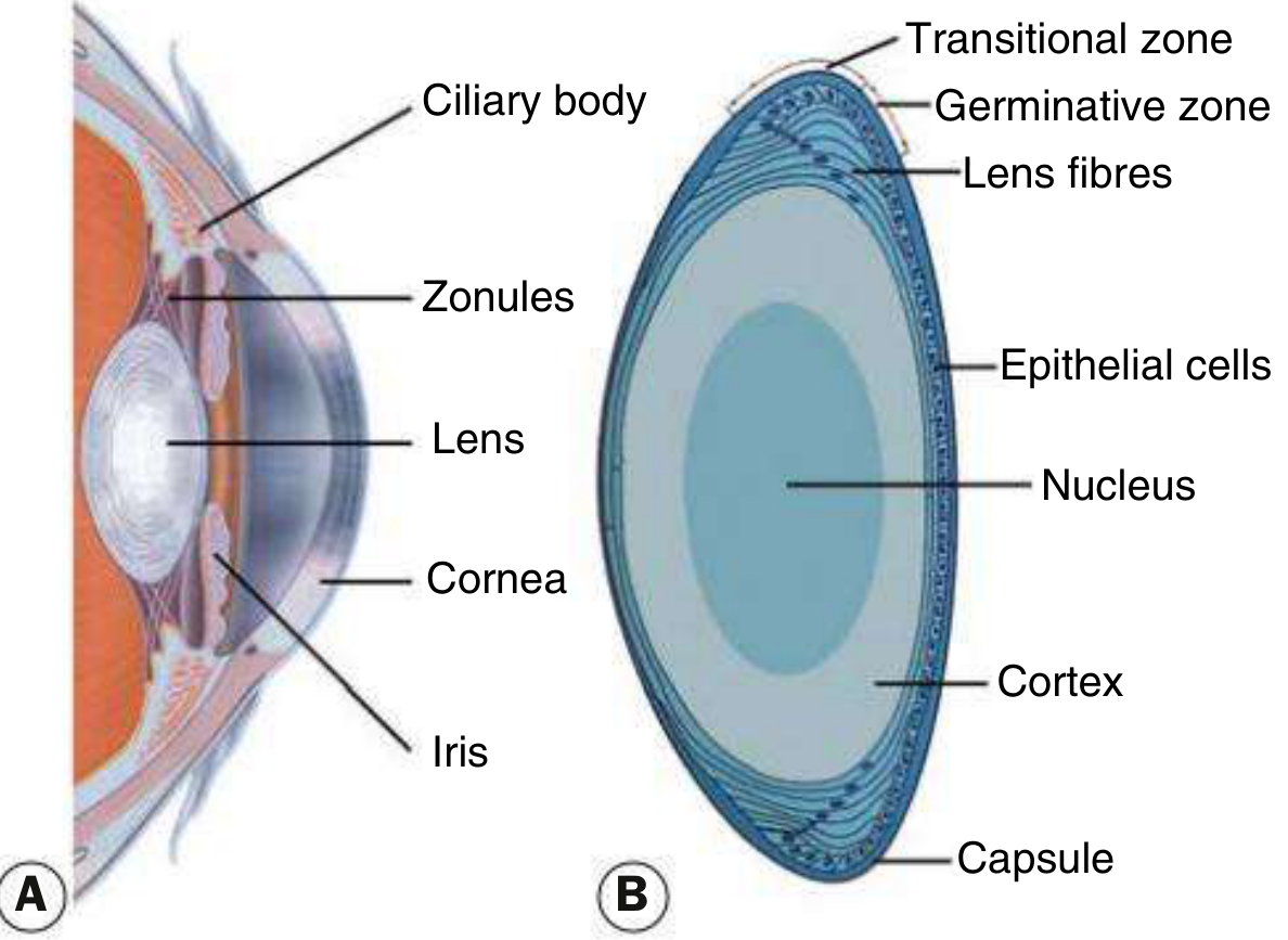
Kanski's Clinical Ophthalmology, Fig. 10.1 - Anterior segment anatomy and lens cross-section
Visual Acuity Assessment
In human ophthalmology, distance visual acuity (VA) is tested using the Snellen chart at standard distance. Categories for very poor vision include:
- Counting fingers (CF) - patient can count fingers at a specified distance (usually 1 metre)
- Hand movements (HM) - able to detect hand motion only
- Perception of light (PL) - can discern light only. In pure media opacity (dense cataract), the patient/animal can still determine direction of the light source
- No perception of light (NPL)
In veterinary patients, functional vision is assessed by the menace response, dazzle reflex, maze navigation in varying light conditions, and the pupillary light reflex (PLR) rather than chart-based testing.
Slit Lamp Biomicroscopy
The slit lamp is the cornerstone of anterior segment examination. It allows examination of:
- Eyelids and lid margins
- Conjunctiva (palpebral and bulbar)
- Cornea - surface, stroma, and endothelium
- Anterior chamber depth and presence of flare/cells (Tyndall effect)
- Iris and pupil
- Lens - capsule, cortex, and nucleus using retroillumination and oblique beam
The direct ophthalmoscope can also be used with a +15 D lens at 15-20 cm to detect lens and vitreous opacities via the red reflex (retroillumination). A cobalt blue filter after fluorescein instillation reveals corneal abrasions and ulcers. This technique is particularly practical for field veterinary examinations.
Fluorescein Staining (Corneal Examination)
Fluorescein stains epithelial defects and is the standard test for corneal ulceration in both human and veterinary practice. Rose Bengal stain is more sensitive for devitalized epithelial cells and mucous filaments (used to diagnose dry eye / keratoconjunctivitis sicca).
2. CONJUNCTIVITIS
Anatomy of the Conjunctiva
The conjunctiva is a transparent mucous membrane lining the inner eyelid surface and anterior globe, terminating at the corneoscleral limbus. It is divided into:
- Palpebral conjunctiva - firmly attached to the tarsal plates
- Fornical conjunctiva - loose and redundant
- Bulbar conjunctiva - covers the anterior sclera; continuous with corneal epithelium at the limbus. The palisades of Vogt at the limbus are the likely reservoir of corneal stem cells.
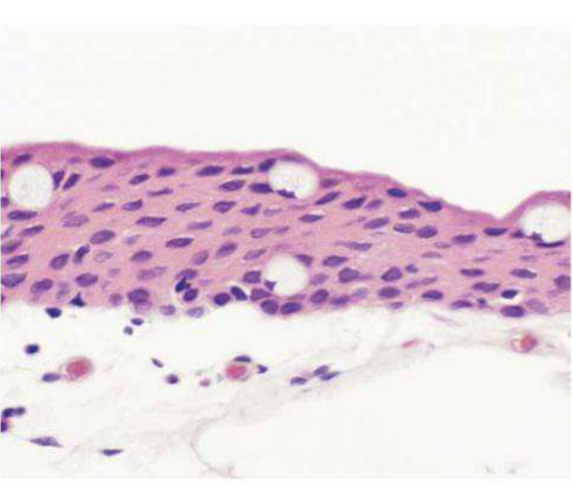
Kanski's Clinical Ophthalmology, Fig. 6.1 - Conjunctival histology showing non-keratinizing epithelium, goblet cells (clear oval cells), and stromal vasculature
The epithelium is non-keratinizing, approximately five cell layers deep. Goblet cells within the epithelium are densest inferonasally and in the fornices - they secrete mucus essential to the tear film. The stroma contains accessory lacrimal glands of Krause and Wolfring. Conjunctiva-associated lymphoid tissue (CALT) mediates ocular surface immune responses.
Symptoms and Signs of Conjunctival Inflammation
Symptoms:
- Lacrimation, grittiness, stinging, burning
- Itching is the hallmark of allergic disease
- Visual acuity is usually unaffected
- Significant pain, photophobia, or foreign body sensation suggest corneal involvement
Discharge types (diagnostically important):
| Discharge Type | Cause |
|---|---|
| Watery / serous | Viral or acute allergic conjunctivitis |
| Mucoid | Chronic allergic conjunctivitis or dry eye |
| Mucopurulent | Bacterial conjunctivitis |
| Purulent / hyperacute | Gonococcal or meningococcal infection |
Signs on examination:
- Conjunctival injection - superficial vessels (bright red, movable), vs. ciliary injection (deep purple-red, fixed - indicates intraocular inflammation)
- Chemosis - conjunctival oedema
- Papillae - raised, vascularized folds with a central fibrovascular core; seen in bacterial and allergic conjunctivitis
- Follicles - avascular lymphoid aggregates without a central core; typical of viral and chlamydial conjunctivitis
- Membranes/pseudomembranes - in severe bacterial (especially gonococcal) or adenoviral conjunctivitis
- Subconjunctival haemorrhage - adenoviral, gonococcal, mechanical
Types of Conjunctivitis
Bacterial Conjunctivitis
- Onset: Acute; usually bilateral (one eye 1-2 days before the other)
- Signs: Eyelids stuck together on waking, mucopurulent discharge
- Organisms: Staphylococcus aureus, Streptococcus pneumoniae, Haemophilus influenzae (in children), Moraxella catarrhalis
- Hyperacute purulent discharge is a red flag for Neisseria gonorrhoeae or N. meningitidis - requires urgent Gram stain (Gram-negative intracellular kidney-shaped diplococci) and culture on chocolate/Thayer-Martin agar
- Superficial corneal punctate epithelial erosions are common
- Peripheral corneal ulceration may occur in gonococcal/meningococcal infection and can rapidly perforate
- Treatment: Topical antibiotics (chloramphenicol, fusidic acid, fluoroquinolones); systemic for gonococcal/chlamydial
Viral Conjunctivitis
- Usually caused by adenovirus (epidemic keratoconjunctivitis - EKC)
- Features: Watery discharge, preauricular lymphadenopathy, follicular reaction
- Often self-limiting; highly contagious
Allergic Conjunctivitis
- Itching is the dominant symptom
- Types range from acute allergic (seasonal/perennial) to vernal keratoconjunctivitis (VKC) and atopic keratoconjunctivitis (AKC)
- VKC: giant cobblestone papillae on upper tarsal plate; Trantas dots at the limbus
- Treatment: Antihistamines, mast cell stabilizers (sodium cromoglicate, lodoxamide), topical steroids for severe cases
Chlamydial Conjunctivitis
- Adult: sexually transmitted Chlamydia trachomatis (serotypes D-K)
- Trachoma (serotypes A-C): leading infectious cause of blindness worldwide
- Chronic follicular conjunctivitis with pannus (fibrovascular invasion) and corneal scarring
Veterinary Note
In animals, the most common causes of conjunctivitis include:
- Dogs/cats: Bacterial (Staphylococcus, Streptococcus), viral (feline herpesvirus-1 in cats is a major cause), Chlamydophila felis (cats), Mycoplasma
- Horses: Bacterial, viral (Equine Herpesvirus), traumatic
- Cattle/sheep: Infectious bovine keratoconjunctivitis (Moraxella bovis - "pink eye")
3. CORNEAL EXAMINATION AND CONDITIONS
Normal Cornea
The cornea is the transparent avascular structure forming the anterior surface of the eye. It has five layers:
- Epithelium (with Bowman's layer beneath)
- Stroma (90% of corneal thickness)
- Descemet's membrane
- Endothelium
Central corneal thickness (CCT) - normal ~555 µm; measured by pachymetry; relevant for glaucoma evaluation (IOP correction) and before refractive surgery.
Examination Techniques
Slit lamp examination of the cornea:
- Diffuse illumination: Overall survey
- Focal/direct illumination: Detect opacities, vascularization
- Retroillumination: Detect early corneal haze, subtle opacities, endothelial changes
- Specular reflection: Examine endothelial cell morphology
- Fluorescein staining + cobalt blue light: Epithelial defects (ulcers, abrasions) stain bright green
- Rose Bengal staining: Devitalized epithelial cells and mucous filaments (dry eye)
Key Corneal Conditions (relevant to surgery)
Dry Eye / Keratoconjunctivitis Sicca (KCS)
- Punctate epithelial erosions that stain with fluorescein
- Filaments: mucus/cell strands attached at one end to the corneal surface (stain with rose Bengal)
- Mucous plaques
- Complications: epithelial breakdown, corneal melting, perforation, bacterial keratitis
- In dogs, KCS is a common and important surgical/medical condition (Schirmer tear test used for diagnosis)
Corneal Ulceration
- Superficial ulcers: Epithelial defects - visible with fluorescein; managed medically
- Stromal ulcers: Deeper; risk of perforation
- Descemetocele: Only Descemet's membrane remains - surgical emergency (corneoconjunctival transposition, conjunctival flap, or keratoplasty)
- Corneal perforation: Iris prolapse; treated with corneal suture + conjunctival flap or graft
Band Keratopathy
- Calcium deposition in the superficial cornea (Bowman's layer)
- Associated with chronic uveitis, hypercalcemia, or silicone oil tamponade in the eye
4. CATARACT
Anatomy
The lens is composed of:
- Capsule (outermost - elastic basement membrane)
- Epithelium (germinative zone anteriorly)
- Cortex
- Nucleus (oldest, most central fibres)
Classification by Location and Morphology
| Type | Features |
|---|---|
| Anterior subcapsular | Under the anterior capsule; fibrous metaplasia of lens epithelium |
| Posterior subcapsular (PSC) | Just in front of the posterior capsule; granular/plaque appearance on slit lamp; vacuolated on retroillumination (bladder/Wedl cells); profound effect on vision due to nodal point location; worsened by miosis and bright light |
| Nuclear sclerotic | Exaggerated aging change; yellowish-brown (urochrome pigment); associated with myopic shift ("second sight of the aged"); good red reflex but subtle cortex-nucleus distinction |
| Cortical | Clefts and vacuoles between lens fibres due to cortical hydration; cuneiform (wedge-shaped) or spoke-like opacities; starts infero-nasally |
| Immature | Partial opacification; red reflex present |
| Mature | Completely opaque; white lens; no red reflex |
| Hypermature | Shrunken wrinkled capsule from water leakage; risk of phacolytic uveitis |
| Morgagnian | Liquefied cortex with sunken brown nucleus; most advanced stage |
Causes of Cataract
- Age-related (senile): Most common in humans and dogs
- Diabetic: Sorbitol accumulation from glucose via aldose reductase pathway; cortical fluid vacuoles; "snowflake" pattern in young; accelerated nuclear sclerosis in older patients
- Traumatic: Penetrating injury (capsule disruption), blunt trauma (flower-shaped opacity), electrical/radiation injury; intralenticular foreign body
- Secondary to systemic disease: Myotonic dystrophy, hypocalcemia (fine lamellar cataract)
- Steroid-induced: Posterior subcapsular cataract from topical/systemic corticosteroids; significant glare symptoms
- Uveitic: Anterior plaques from fibrin, posterior synechiae causing iris-lens adhesion with sector opacification; glaukomflecken (focal lens epithelial infarcts) from acute angle-closure glaucoma
- Inherited/Congenital: Common in certain dog breeds (Labrador Retriever, Golden Retriever, Boston Terrier, etc.)
- High myopia: Associated with PSC and early nuclear sclerosis
Indications for Cataract Surgery
- Visual improvement - opacity sufficient to impair daily activities (or functional vision in animals)
- Medical indication - phacolytic glaucoma (lens proteins leak through hypermature capsule causing macrophage-mediated outflow obstruction), phacomorphic glaucoma (intumescent lens causing angle-closure), lens-induced uveitis
Surgical Management - Phacoemulsification
Preoperative assessment:
- Biometry (A-scan ultrasonography) to determine intraocular lens (IOL) power
- Systemic medications reviewed (alpha-blockers associated with intraoperative floppy iris syndrome - IFIS)
- Anticoagulant management per local protocol
- Diabetic patients: assess/photograph the retina before surgery (macular oedema may worsen postoperatively)
- Contact lens wearers: discontinue soft lenses 1 week, rigid gas-permeable (RGP) lenses 6 weeks before biometry to achieve corneal stability
- Previous refractive surgery (LASIK, PRK): standard IOL formulas are inaccurate; use modified formulas (Haigis-L, Masket, contact lens method)
Surgical technique - Phacoemulsification:
- Clear corneal incision (2.2-2.8 mm self-sealing, temporal or superior approach)
- Continuous curvilinear capsulorhexis (CCC) - circular opening in the anterior lens capsule (key step; prevents capsule radial tears)
- Hydrodissection - fluid wave under the capsule to free the lens nucleus
- Phacoemulsification - ultrasonic fragmentation and aspiration of the lens nucleus and cortex
- Cortex aspiration (irrigation/aspiration - I/A)
- IOL implantation into the capsular bag (standard foldable acrylic IOL)
- Wound closure (usually self-sealing; suture if necessary)
IOL considerations:
- Target refraction: usually emmetropia (±0.25 D myopia as safety margin)
- Monovision option: non-dominant eye targeted at ~1-2 D myopia for near
- Multifocal IOLs: provide near/intermediate/distance but may cause glare and reduced contrast sensitivity
- Postoperative refraction within 1.0 D of target in ~90% of cases
Postoperative complications:
- Posterior capsular opacification (PCO) - commonest late complication; "after-cataract"; Elschnig pearl formation from residual lens epithelial cells; treated by Nd:YAG laser capsulotomy
- Cystoid macular oedema (CME)
- Endophthalmitis - rare but devastating; prophylaxis with intracameral cefuroxime
- Posterior capsule rupture intraoperatively - risk of vitreous loss and dropped nucleus
- IFIS (from tamsulosin/alpha-blocker use) - floppy, billowing iris; managed with iris hooks or Malyugin ring
Veterinary note - Phacoemulsification in dogs/cats:
- Dogs are the most common veterinary patients for cataract surgery
- Preoperative electroretinography (ERG) is performed to confirm retinal function before surgery (dogs cannot communicate visual symptoms)
- Intravitreal gentamicin is NOT used (retinal toxicity); topical/systemic antibiotics used
- Diabetic dogs develop cataracts extremely rapidly (days to weeks) due to high aldose reductase activity
- Feline cataracts are less common and often secondary to uveitis
- Horses: cataract surgery is more technically challenging due to globe size and vitreal liquefaction
Sources: Kanski's Clinical Ophthalmology: A Systematic Approach, 10th Edition; Goldman-Cecil Medicine, 26th Edition; Robbins & Kumar Basic Pathology
Epidural anaesthesia detail
Reading File
Epidural anaesthesia is a regional anaesthetic technique involving injection of local anaesthetic (±opioids) into the epidural space to block nerve roots as they exit the spinal canal. It is widely used for surgical anaesthesia, obstetric analgesia, and postoperative pain management. The key anatomy involves the epidural space between the ligamentum flavum and the dura mater, typically accessed at the lumbar or thoracic level. Loss-of-resistance technique is the standard method of confirmation.
Searching sources...
Finding Sources
Finding Sources
Excellent - I have multiple high-quality sources. Let me read the main sections from Miller's Anesthesia (the gold standard reference), Morgan & Mikhail's Clinical Anesthesiology, and Bailey & Love's Surgery simultaneously.
Reading File
Reading File
Reading File
Excellent content from Morgan & Mikhail and Miller's. Let me now read more detailed technique, drugs, and complications sections:
Reading File
Reading File
Good - I have rich complication data from Morgan & Mikhail. Now let me read the technique, drugs, and more physiology sections from Morgan & Mikhail block 7:
Reading File
Reading File
Excellent - I now have very rich content. Let me fetch key diagrams and get the pharmacology/drug doses section:
Loading Image
Loading Image
Reading File
Excellent. Now let me get the local anaesthetic drug doses for epidural, the physiology of epidural block (sympathetic/sensory/motor effects), and the PDPH/specific complications section:
Reading File
Finding Sources
Let me get the drug doses table and the loss-of-resistance technique description:
Reading File
Finding Sources
Reading File
Finding Sources
I now have comprehensive, authoritative content from multiple sources. Let me compile the full detailed answer.
Epidural Anaesthesia - Comprehensive Reference
Sources: Morgan & Mikhail's Clinical Anesthesiology, 7th Edition | Miller's Anesthesia, 10th Edition | Bailey & Love's Short Practice of Surgery, 28th Edition
1. DEFINITION AND OVERVIEW
Epidural anaesthesia is a neuraxial regional technique in which a local anaesthetic (with or without opioids or adjuvants) is injected into the epidural space to block nerve roots as they exit the spinal canal. It can be performed as:
- Single-shot technique (limited duration)
- Catheter technique - allows continuous infusion, intermittent boluses, or patient-controlled epidural analgesia (PCEA)
Compared to spinal anaesthesia, epidural block has:
- Slower onset (10-20 minutes vs. 2-5 minutes for spinal)
- Larger drug volumes required
- More gradual sympathectomy - better haemodynamic control
- Ability to extend or prolong the block via catheter
- Segmental block possible - anaesthesia can be confined to specific nerve root levels
"Continuous epidural anesthesia is a neuraxial technique offering a range of applications wider than single-dose spinal anesthesia."
- Morgan & Mikhail's Clinical Anesthesiology, 7e
2. ANATOMY
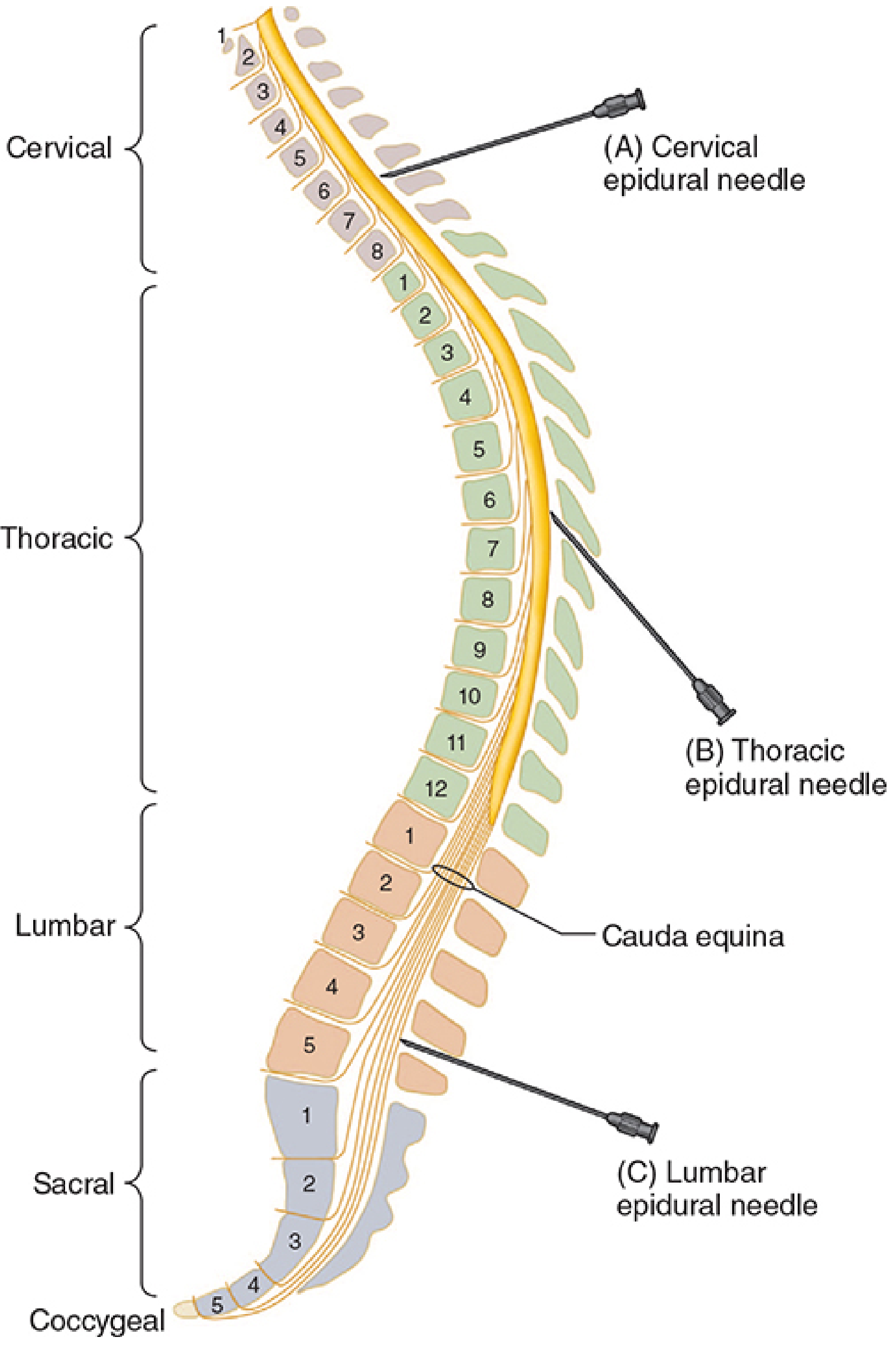
Morgan & Mikhail's Clinical Anesthesiology, Fig. 45-21 - Epidural needle angulation at different spinal levels
The Epidural Space
- Surrounds the dura mater posteriorly, laterally, and anteriorly
- Extends from the foramen magnum to the sacral hiatus
- Boundaries:
- Posterior: vertebral laminae + ligamentum flavum (key landmark)
- Anterior: posterior longitudinal ligament and vertebral body periosteum
- Lateral: pedicles and intervertebral foramina
Contents of the Epidural Space
- Fatty connective tissue (loose fat - more in infants and children up to 6-8 years)
- Batson's venous plexus (rich epidural veins - risk of intravascular injection)
- Lymphatics
- Nerve roots travelling to exit via intervertebral foramina
- Connective tissue septa/bands (may explain occasional unilateral block)
Spinal Levels and Tuffier's Line
- Tuffier's (intercristal) line - connects the two iliac crests, crosses the spinous processes at L4-L5 in adults and older children (crosses L5-S1 in infants up to 1 year)
- Spinal cord ends at L1 (conus medullaris) in adults - lower lumbar interspaces are safest
- Thoracic epidurals are technically more difficult due to acute downward angulation (30-50°) of spinous processes
Layers Traversed (Midline Approach, Posterior to Anterior)
- Skin and subcutaneous fat
- Supraspinous ligament
- Interspinous ligament
- Ligamentum flavum (fibrous, yellow, 3-5 mm thick in lumbar region) → entry into epidural space
- Epidural space
- Dura mater (avoid piercing)
- Subdural space
- Arachnoid mater
- Subarachnoid (intrathecal) space
3. INDICATIONS
| Category | Examples |
|---|---|
| Surgical anaesthesia | Lower limb, pelvic, abdominal, thoracic surgery |
| Obstetric analgesia | Labour analgesia, caesarean section |
| Postoperative pain | Major abdominal, thoracic, orthopaedic surgery; reduces respiratory complications; enables early mobilisation |
| Chronic pain management | Low back pain, cancer pain, CRPS |
| Paediatric surgery | Major abdominal, retroperitoneal, pelvic, thoracic procedures; pectus excavatum repair; scoliosis surgery |
| Trauma/acute pain | Rib fractures, burns |
4. CONTRAINDICATIONS
Absolute
- Patient refusal
- Coagulopathy / anticoagulation (risk of epidural haematoma)
- Infection at the insertion site
- Raised intracranial pressure (risk of brainstem herniation if dura accidentally punctured)
- Severe hypovolaemia (profound hypotension from sympathectomy)
Relative
- Sepsis / bacteraemia (risk of epidural abscess)
- Anatomical abnormalities of spine (scoliosis, previous spinal surgery, trauma)
- Pre-existing neurological disease (medicolegal risk)
- Demyelinating conditions
- Severe aortic stenosis or fixed cardiac output states
- Uncooperative patient
5. EQUIPMENT
Epidural Needles
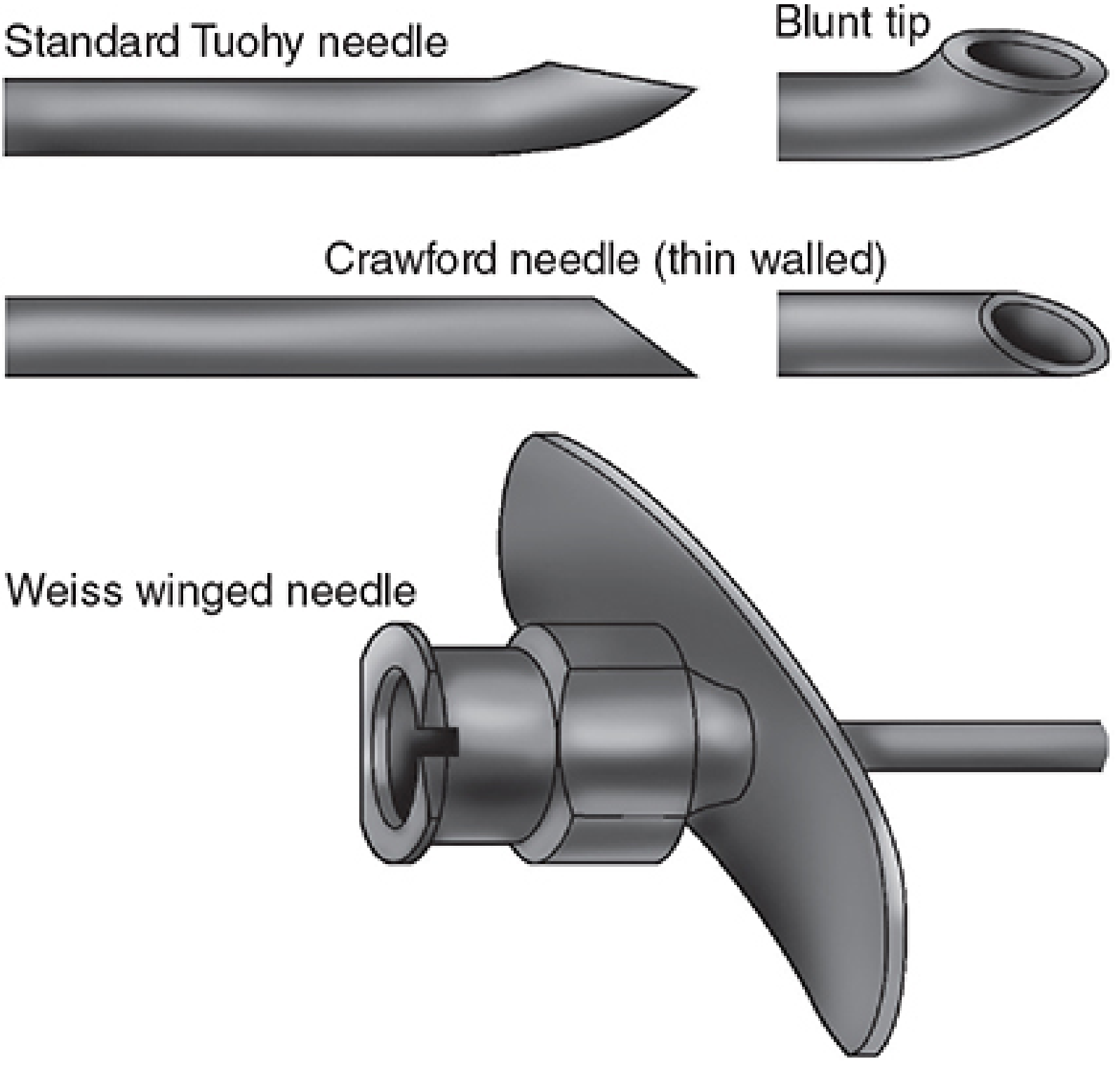
Morgan & Mikhail's Clinical Anesthesiology, Fig. 45-22 - Epidural needles
| Needle | Features |
|---|---|
| Tuohy (most common) | 17-18 gauge, 3-3.5 inches; blunt curved tip (Huber point) at 15-30°; theoretically deflects dura rather than penetrating it |
| Crawford | Thin-walled, straight; higher dural puncture incidence |
| Weiss winged | Tuohy-type with wings at hub to aid controlled advancement |
Epidural Catheters
- 19-20 gauge catheter passed through 17-18 gauge needle
- Advanced 2-6 cm into the epidural space
- Too short (<2 cm): risk of dislodgement
- Too long (>6 cm): risk of unilateral block, knotting, venous penetration, or exit through foramen
- Multi-orifice (closed tip + side ports) preferred - lower intravascular penetration rate, fewer paresthesias
- Spiral/spring-wire reinforced: kink-resistant
- Tunnelled subcutaneously for prolonged placement (>1 week)
6. PATIENT POSITIONING
| Level | Position | Approach |
|---|---|---|
| Lumbar (most common) | Lateral decubitus (foetal position) - preferred; or sitting | Midline or paramedian |
| Thoracic | Sitting or lateral decubitus | Midline or paramedian; acute angle (30-50°) required |
| Cervical | Sitting, neck flexed | Midline |
| Caudal | Lateral decubitus, prone jackknife (adults) | Via sacral hiatus |
Lateral decubitus is the preferred position for catheter placement in children - bends the spine forward, moves the spinal cord away from the ligamentum flavum, and expands the epidural space.
7. TECHNIQUE - STEP BY STEP
A. Preparation
- IV access established, resuscitation equipment and drugs (vasopressors, atropine, intralipid) at hand
- Standard monitoring: ECG, NIBP, SpO2
- Pre-loading with IV crystalloid (500-1000 mL) to attenuate sympathectomy-induced hypotension
B. Needle Placement (Midline Approach)
- Position patient (lateral or sitting), identify interspace (L2-L3, L3-L4, or L4-L5 for lumbar)
- Sterile preparation; infiltrate skin and subcutaneous tissue with local anaesthetic
- Insert epidural needle (stylet in place) through skin → supraspinous ligament → interspinous ligament (increased resistance felt)
- Remove stylet; attach syringe (glass syringe with saline or air for loss-of-resistance)
- Advance needle with continuous or repeating pressure on plunger
C. Confirming Epidural Space Entry
Loss of Resistance (LOR) Technique - preferred
- Needle within ligament: resistance prevents injection
- As tip enters epidural space: sudden loss of resistance - injection becomes easy
- Can use saline (avoids air embolism; LOR more reliable) or air (helps identify nerve injection but risks air embolism and pneumocephalus)
Hanging Drop Technique
- Hub of needle filled with saline so a drop hangs at the opening
- As needle enters epidural space, negative pressure sucks the drop in
- Less reliable if needle becomes plugged
D. Catheter Insertion (Catheter Technique)
- Confirm epidural space
- Thread catheter through needle, advance 2-6 cm beyond needle tip
- Hold catheter, withdraw needle over catheter
- Aspirate through catheter (check for blood = intravascular; CSF = intrathecal)
- Secure catheter to skin
8. TEST DOSE AND INCREMENTAL DOSING
Test Dose
- Standard: 3 mL of 1.5% lidocaine + epinephrine 1:200,000 (15 mcg)
- If intrathecal injection: 45 mg lidocaine produces rapid dense spinal block
- If intravascular injection: 15 mcg epinephrine produces ≥20% heart rate increase within 1 minute
- Limitations: false positives/negatives possible (uterine contractions, beta-blockers)
- Aspiration alone is insufficient - false-negative aspirations are common
Incremental Dosing
- Inject in 5 mL increments (allows early detection of inadvertent intravascular injection before seizure dose reached)
- Observe for: tinnitus, metallic taste, perioral numbness, slurred speech between doses
- Total dose typically given over 5-10 minutes
9. DRUGS USED IN EPIDURAL ANAESTHESIA
Local Anaesthetics
| Agent | Concentration | Onset | Duration | Use |
|---|---|---|---|---|
| Bupivacaine | 0.25-0.5% (surgical); 0.0625-0.125% (labour analgesia) | Slow (15-20 min) | Long (2-4 h) | Surgical anaesthesia; labour; postoperative |
| Ropivacaine | 0.2-0.75% | Slow-moderate | Long (2-4 h) | Labour, postoperative; less cardiotoxic than bupivacaine |
| Levobupivacaine | 0.25-0.5% | Slow | Long | Similar to ropivacaine |
| Lidocaine | 1.5-2% (surgical); 1% (postoperative) | Fast (5-10 min) | Short-medium (1-2 h) | Surgical; test dose |
| Chloroprocaine | 3% | Very fast (3-5 min) | Very short (<1 h) | Rapid onset situations; supplement inadequate block |
Large volumes needed - 10-20 mL for lumbar, up to 35 mL for caesarean section. Always in incremental doses.
Opioids (Adjuvants)
| Agent | Dose | Mechanism |
|---|---|---|
| Fentanyl | 50-100 mcg (bolus); 25-75 mcg/h infusion | Lipophilic; quick onset; segmental spinal action |
| Sufentanil | 10-20 mcg (bolus); 5-10 mcg/h | Most lipophilic; rapid onset |
| Morphine | 3-5 mg (bolus) | Hydrophilic; slow onset; prolonged analgesia 6-24 h; cephalad spread (risk of delayed respiratory depression) |
| Diamorphine | 2-4 mg | Intermediate lipophilicity |
Other Adjuvants
- Epinephrine 1:200,000 (5 mcg/mL) - prolongs block, reduces systemic absorption, acts as marker in test dose; use cautiously in thoracic epidurals (risk of spinal cord ischaemia)
- Clonidine (α2-agonist) - enhances analgesia; dose 1-2 mcg/kg
- Sodium bicarbonate - alkalinises LA solution → increases non-ionised free base → faster onset (1 mEq/10 mL lidocaine)
- Neostigmine - epidural use reduces opioid requirements (investigational)
10. PHYSIOLOGICAL EFFECTS
Sympathetic Blockade
- Preganglionic sympathetic fibres (smallest, B fibres) are blocked first
- Vasodilation in blocked segments → hypotension (most common significant side effect)
- Onset is more gradual than spinal → better haemodynamic control
- Blocked T1-T4 (cardiac accelerator fibres) → bradycardia
- T6-L2 blockade → bowel relaxation (facilitates abdominal surgery)
Sensory Blockade
- C and Aδ fibres blocked → pain and temperature loss
- Aβ fibres blocked → light touch, pressure
- Sensory block is the primary effect required
Motor Blockade
- Aα motor fibres - largest, most resistant to local anaesthetics
- Degree of motor block depends on concentration:
- Low concentration (0.0625-0.125% bupivacaine + opioid): sensory block only, minimal motor block - ideal for labour analgesia and postoperative pain
- High concentration (0.5% bupivacaine): dense motor block - for surgical anaesthesia
Block Height and Spread
Factors affecting cephalad spread:
- Volume of injectate (most important for epidural)
- Level of injection
- Speed of injection
- Patient positioning (gravity less important than with spinal)
- Age (reduced spread in elderly due to decreased epidural space compliance and narrowed foramina → reduce dose in elderly)
- Pregnancy (engorgement of epidural veins reduces space → reduce dose in pregnancy)
- Obesity (same effect)
11. SPECIAL APPLICATIONS
Labour Epidural Analgesia
- Dilute bupivacaine 0.0625-0.1% + fentanyl 2 mcg/mL (or sufentanil) as continuous infusion ± PCEA
- Provides excellent pain relief with minimal motor block (patient can push/walk)
- Maintenance: 10-15 mL/h continuous infusion, PCEA bolus 5-10 mL with 10-15 min lockout
Epidural for Caesarean Section
- Lidocaine 2% + epinephrine 1:200,000 (15-35 mL) most common in USA
- Bupivacaine 0.5% (15-25 mL) - widely used globally
- Add fentanyl 50-100 mcg or sufentanil 10-20 mcg for dense block
- Achieve T4 sensory level (nipple line) before proceeding
- Inject in 5 mL increments to avoid high block
- Epidural morphine 3-5 mg at end of surgery provides 6-24 h postoperative analgesia
Thoracic Epidural Analgesia (TEA)
- Inserted at T4-T8 for upper abdominal/thoracic surgery; T7-T10 for lower abdominal surgery
- Benefits: Excellent pain control; early extubation; reduced pulmonary complications; better bowel recovery; reduced cardiac ischaemia risk
- Uses: oesophagectomy, thoracotomy, nephrectomy, aortic surgery, major bowel surgery
- Infusion: dilute bupivacaine 0.125-0.25% + fentanyl at 5-10 mL/h
Combined Spinal-Epidural (CSE)
- "Needle-through-needle" technique: spinal needle through the epidural needle into the subarachnoid space; spinal dose given; needle withdrawn; epidural catheter placed
- Combines rapid dense onset of spinal with flexibility/prolongation of epidural
- Used for: labour (initial spinal injection for rapid relief, then epidural), caesarean section, major joint replacement surgery
- Caution: epidural drugs may pass through dural puncture → potentiated effect; titrate carefully
12. COMPLICATIONS
A. Cardiovascular
| Complication | Mechanism | Management |
|---|---|---|
| Hypotension (most common - 30%) | Sympathetic block → vasodilation and venodilation → reduced preload and SVR | IV fluids; ephedrine 5-10 mg IV (or phenylephrine 50-100 mcg IV if bradycardia also); left uterine displacement in pregnancy |
| Bradycardia | T1-T4 blockade (cardiac accelerators); Bezold-Jarisch reflex | Atropine 0.6 mg IV; ephedrine |
| Cardiac arrest | Severe hypotension + bradycardia → medullary hypoperfusion; incidence ~1:1500 | CPR; epinephrine; may need emergency delivery in obstetric cardiac arrest |
B. High / Total Block
- Cause: Excessive dose, inadvertent intrathecal injection of epidural dose, or exaggerated cephalad spread
- Signs: Dyspnoea → upper limb numbness → Horner syndrome → unconsciousness → apnea
- Apnoea more often from medullary hypoperfusion than phrenic nerve block (C3-C5)
- Treatment: Reassure; supplemental O2; intubate and ventilate; vasopressors + fluids; atropine for bradycardia
C. Post-Dural Puncture Headache (PDPH)
- Occurs after "wet tap" (accidental dural puncture with epidural needle) or unrecognised dural puncture
- Incidence: up to 20-50% following wet tap with large Tuohy needle in young pregnant women; <1% with 27G pencil-point spinal needle in elderly men
- Characteristics:
- Bilateral, frontal/occipital, extends into neck
- Postural: worse sitting/standing, relieved lying flat (hallmark)
- Onset 12-72 hours after procedure
- Associated with photophobia, nausea, tinnitus, diplopia (VI nerve traction)
- Mechanism: CSF leak from dural defect → intracranial hypotension → meningeal traction
- Risk factors: large needle gauge, cutting-point needle, female sex, young age, pregnancy, history of prior PDPH
- Conservative treatment:
- Recumbent positioning, oral/IV fluids, caffeine (vasoconsticts intracranial vessels + stimulates CSF production)
- NSAIDs, paracetamol, opioids
- Sphenopalatine ganglion block (intranasal LA application)
- Epidural blood patch (EBP): 15-20 mL autologous blood injected at the level of puncture; stops CSF leak by tamponade/clot formation; ~90% success rate with single patch; 90% of initial non-responders respond to second patch; generally not recommended prophylactically
D. Nerve Injury
| Injury | Cause | Feature |
|---|---|---|
| Radiculopathy | Direct needle/catheter trauma; neurotoxic drug | Dermatomal pain/paraesthesia |
| Epidural haematoma | Epidural vein laceration + coagulopathy | Severe back pain + progressive leg weakness → paralysis; MRI diagnosis; surgical decompression within 6-8 hours |
| Epidural abscess | Bacterial contamination (Staphylococcus most common) | Fever, back pain, leg weakness/paralysis; MRI diagnosis; surgical drainage + antibiotics |
| Cauda equina syndrome | Neurotoxicity from maldistributed drug (continuous spinal with high-concentration lignocaine); spinal haematoma/abscess | Saddle anaesthesia, bladder/bowel dysfunction, leg weakness |
| Anterior spinal artery syndrome | Prolonged severe hypotension + ↑intraspinal pressure | Bilateral leg paralysis with preserved position/vibration sense |
E. Other Complications
| Complication | Notes |
|---|---|
| Intravascular injection | Batson's plexus; LA systemic toxicity (LAST) - tinnitus → seizures → cardiovascular collapse; treat with 20% Intralipid emulsion |
| Inadvertent intrathecal injection | "High spinal" or total spinal; full epidural dose intrathecally causes cardiovascular collapse |
| Subdural injection | Rare; unpredictable extensive but patchy block with delayed onset |
| Inadequate/unilateral block | Catheter malposition, connective tissue septa, excessive catheter advancement into foramen |
| Urinary retention | Sacral parasympathetic blockade; catheterise |
| Backache | Local tissue trauma at insertion site; usually self-limiting |
| Catheter problems | Shearing, knotting, retention on removal |
13. EPIDURAL HAEMATOMA AND ABSCESS - KEY SAFETY POINTS
Epidural Haematoma
- Most feared complication of neuraxial anaesthesia
- Risk factors: anticoagulant use, platelet disorders, traumatic placement, indwelling catheter
- ASRA anticoagulation guidelines for timing of neuraxial block:
- Unfractionated heparin: hold 4-6 hours, check APTT
- LMWH (prophylactic): 12 hours before; 24 hours after removal
- LMWH (therapeutic): 24 hours before
- Warfarin: INR ≤1.5 before insertion/removal
- Aspirin/NSAIDs: not a contraindication alone
- Direct oral anticoagulants (DOACs): 48-72 hours (drug-specific)
Epidural Abscess
- Incidence: 1:1000-1:10,000 epidural catheter insertions
- Symptoms develop over hours to days: fever, back pain, meningism, then neurological deficit
- Surgical emergency if neurological deficits present
14. EPIDURAL vs. SPINAL ANAESTHESIA - COMPARISON
| Feature | Epidural | Spinal |
|---|---|---|
| Space entered | Epidural space | Subarachnoid space |
| Needle gauge | 17-18 G Tuohy | 25-27 G pencil-point |
| Drug volume | Large (10-30+ mL) | Small (1.5-4 mL) |
| Onset | Slow (10-20 min) | Fast (2-5 min) |
| Block density | Variable, may be patchy | Dense, reliable |
| Segmental block | Yes (valuable for analgesia without motor block) | Difficult |
| Catheter | Routinely used | Rarely used |
| Hypotension | More gradual | More rapid and severe |
| PDPH risk | Low (unless wet tap) | Higher (dependent on needle size/type) |
| Failure rate | Higher (~5-10%) | Lower (~1-2%) |
| Duration | Unlimited (via catheter) | Fixed (2-4 hours typical) |
15. SUMMARY OF CLINICAL PEARLS
- A segmental thoracic epidural provides excellent upper abdominal analgesia while sparing cervical and lumbar nerve roots - invaluable for major thoracoabdominal surgery
- Dilute LA + opioid = analgesia without motor block; concentrated LA = surgical block
- The Batson venous plexus in the epidural space means intravascular catheter placement is common - always aspirate and always test dose
- Pregnancy and obesity reduce the epidural space volume - reduce doses by 25-30%
- A blood patch cures ~90% of PDPHs; do not patch prophylactically
- Epidural haematoma presents as severe back pain with leg weakness - requires emergency MRI and decompressive laminectomy within 6-8 hours to avoid permanent paralysis
- Epidural morphine provides 6-24 hours of excellent post-surgical analgesia but carries risk of delayed respiratory depression (monitor for 24 hours)
Sources: Morgan & Mikhail's Clinical Anesthesiology, 7e | Miller's Anesthesia, 10e | Bailey & Love's Short Practice of Surgery, 28e | Goodman & Gilman's Pharmacological Basis of Therapeutics
This is a shared conversation. Sign in to Orris to start your own chat.