
Pt came to er with c/o While po uring water current line she got shocked nd entire left upper limb burned
Reading File
This is an electrical burn injury to the left upper limb - a clinical emergency. Let me search the medical library for comprehensive management guidance.
Searching sources...
Finding Sources
Finding Sources
Searching the Web
electrical burn injury management ER upper limb treatment guidelines 2024
Good results. Let me read the key sections on electrical burns from multiple textbooks simultaneously.
Reading File
Reading File
Finding Sources
Let me search for the dedicated electrical injury section from Rosen's and Tintinalli's directly.
Finding Sources
Finding Sources
Reading File
Finding Sources
The file is large. Let me read the electrical burn section directly - it starts around line 2221 per the DB.
Reading File
Finding Sources
Excellent content. Let me read the management sections.
Reading File
I now have comprehensive information. Let me compile the full clinical response.
Electrical Burn Injury - Left Upper Limb (ER Management)
Understanding the Mechanism
This patient sustained a low-voltage electrical contact injury (household AC current) while pouring water - water dramatically lowers skin resistance, increasing current flow and tissue damage. Key physics:
- AC household current (220-240V in most countries, 110V in North America) oscillates at 60 Hz
- AC causes tetanic muscle contraction - the "let-go" phenomenon - meaning the patient may have gripped the source involuntarily, prolonging contact
- Ventricular fibrillation occurs at 50-100 mA; asystole at >2A
- It is the amperage, not the voltage, that kills
- Water contact greatly reduces skin resistance (from ~100,000 ohms dry to ~1,000 ohms wet), dramatically increasing current through the body
The burn to the entire left upper limb indicates prolonged contact or high current flow through a large surface area.
Immediate ER Assessment - Primary Survey First
Airway, Breathing, Circulation (ABC)
- Airway: Secure if any facial/airway burns, altered consciousness, or stridor - anticipate early intubation before edema occludes airway
- Breathing: SpO2 continuous monitoring; supplemental O2
- Circulation: IV access (2 large-bore IVs), continuous cardiac monitoring (ECG) - this is mandatory. Ventricular fibrillation is the most common cause of death in electrical injury
- Check for pulse in the affected limb - note entry/exit points if visible
Disability (Neuro exam)
- GCS, level of consciousness
- Full spine assessment within 24 hours (electrical injury can cause vertebral fractures from tetanic contractions)
Critical Investigations to Order
| Investigation | Rationale |
|---|---|
| 12-lead ECG + continuous cardiac monitoring | Arrhythmia (VF, VT, AF, heart block) - mandatory |
| Serum CK (creatine kinase) | Rhabdomyolysis marker - often markedly elevated |
| Urine myoglobin / dipstick | Myoglobinuria = "tea/cola-colored" urine - renal risk |
| Serum BMP (electrolytes, creatinine, BUN) | Hyperkalemia (from cell lysis), AKI |
| CBC, coagulation | DIC risk |
| LFTs | Deep tissue injury effect |
| Troponin | Myocardial injury |
| Urine output (catheterize) | Continuous monitoring essential |
| X-rays of limb + spine | Fractures from tetanic contraction |
Fluid Resuscitation
Electrical burns cause deep tissue destruction far beyond the visible burn surface - internal "iceberg" injury. Standard Parkland formula based on body surface area (BSA) underestimates fluid needs.
Target urine output:
- 0.5 mL/kg/hr if low rhabdomyolysis risk
- 1.0-1.5 mL/kg/hr if myoglobinuria is present (to flush pigment and prevent acute tubular necrosis)
Use warmed isotonic crystalloid (Lactated Ringer's preferred). Monitor for hyperkalemia and hyperchloremia.
"Initiate fluid resuscitation with warmed, isotonic crystalloid IV fluids to a target urine output of 0.5 cc/kg/hour if low risk for rhabdomyolysis (greater than 1.0 cc/kg/hour if high risk for rhabdomyolysis). Monitor for the development of hyperkalemia and hyperchloremia."
- Current Surgical Therapy 14e
Wound Assessment - Left Upper Limb
Entry/Exit Points
- AC current creates contact points (not true entry/exit wounds like DC)
- Look for contact burns on the hand (where she held/touched the source) and potentially the feet (ground point)
Burn Depth Assessment
Electrical burns frequently have:
- Superficial skin appearance but severe deep tissue destruction (muscle, nerve, vascular)
- The true extent of injury may not be apparent for 24-72 hours as deep tissues demarcate
Compartment Syndrome - HIGH PRIORITY
The left upper limb is at high risk for compartment syndrome due to:
- Massive edema from fluid resuscitation and direct tissue injury
- Deep muscle destruction
- Circumferential burns
Monitor for the 5 P's (Pain with passive stretch, Pallor, Pulselessness, Paresthesia, Paralysis)
Compartment pressure measurement if clinically suspected. Fasciotomy if pressure >30 mmHg or within 30 mmHg of diastolic BP.
Cardiac Monitoring Protocol
- Low-voltage injuries (<1000V) with normal ECG on arrival and no arrhythmia: minimum 6-12 hours of monitoring
- Any abnormality on ECG, loss of consciousness, or continued symptoms: admit for 24-hour continuous cardiac monitoring
- Common arrhythmias: sinus tachycardia (most common), VF (most deadly), atrial fibrillation, BBB
Wound Care (Initial ER)
- Remove all jewelry and clothing from the affected limb immediately - these retain heat and may act as conductors
- Cover burns with clean, dry sterile dressings (not wet - avoid hypothermia)
- Do NOT apply ice
- Topical antimicrobials (silver sulfadiazine or bacitracin) after assessment
- Do NOT debride in the ER - extent of injury evolves over days
System-by-System Complications to Watch
| System | Complication |
|---|---|
| Cardiac | VF, VT, heart block, myocardial infarction |
| Renal | AKI from myoglobinuria (rhabdomyolysis) |
| Neurological | Peripheral nerve injury, spinal cord injury (delayed), seizures |
| Vascular | Thrombosis, aneurysm formation in limb vessels |
| Musculoskeletal | Compartment syndrome, deep muscle necrosis, fractures |
| Ophthalmic | Cataracts (delayed, weeks-months later) |
| Auditory | Tympanic membrane rupture, hearing loss |
Burn Center Transfer Criteria
Per the American Burn Association, this patient meets criteria for transfer to a burn center:
- Electrical burns (any voltage) qualify for burn center transfer
- Upper extremity involvement with possible deep tissue injury
- Need for multidisciplinary care (plastic surgery, hand surgery, vascular surgery)
"The American Burn Association referral criteria recommend that patients with high-voltage electrical trauma to the upper extremity should be referred to specialized burn centers experienced with these injuries."
- Current Surgical Therapy 14e
Summary Management Checklist
- Remove from source / confirm safe scene
- ABC + spine immobilization
- Cardiac monitor + 12-lead ECG
- 2 large-bore IV access + urinary catheter
- Labs: CK, BMP, CBC, troponin, coag, urine myoglobin
- IV fluid resuscitation (LR) - target UO 0.5-1.5 mL/kg/hr
- Remove jewelry/clothing from limb
- Wound assessment - document entry/exit contact points
- Neurovascular exam of limb - hourly (compartment syndrome vigilance)
- Pain management (IV opioids)
- Tetanus prophylaxis
- Consider early fasciotomy if compartment syndrome develops
- Refer to burn center
Sources: Current Surgical Therapy 14e, Rosen's Emergency Medicine, Campbell's Operative Orthopaedics 15e (2026), Miller's Anesthesia 10e
How to check compartment pressure
Compartment pressure can be measured using a needle manometer technique or a commercial device (Stryker monitor). The key threshold is >30 mmHg or within 30 mmHg of diastolic BP (delta P ≤30 mmHg) - either triggers fasciotomy.
Searching sources...
Finding Sources
Finding Sources
Reading File
Finding Sources
Reading File
Loading Image
Loading Image
Loading Image
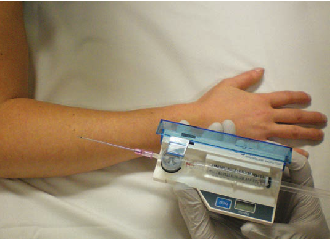
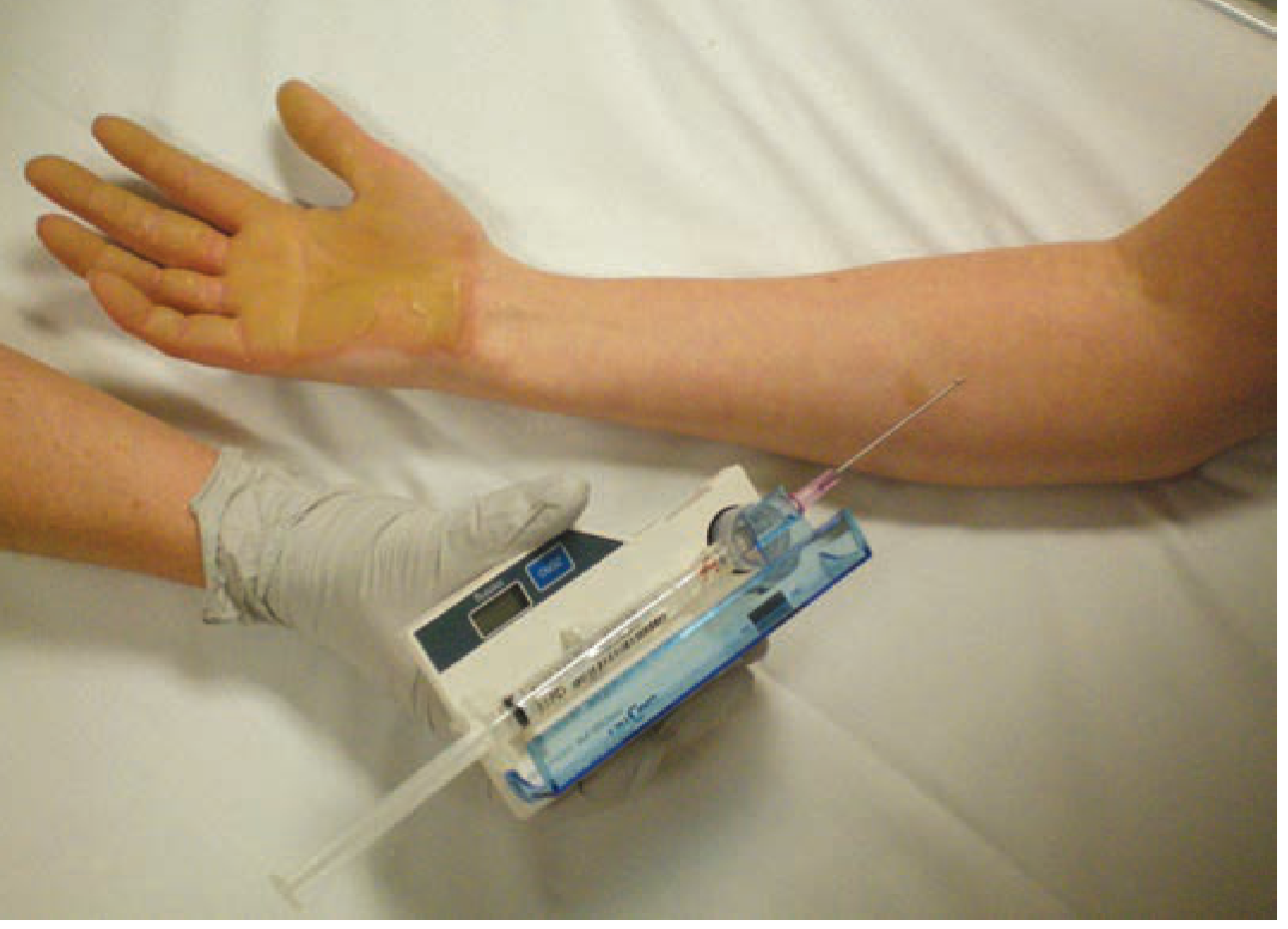
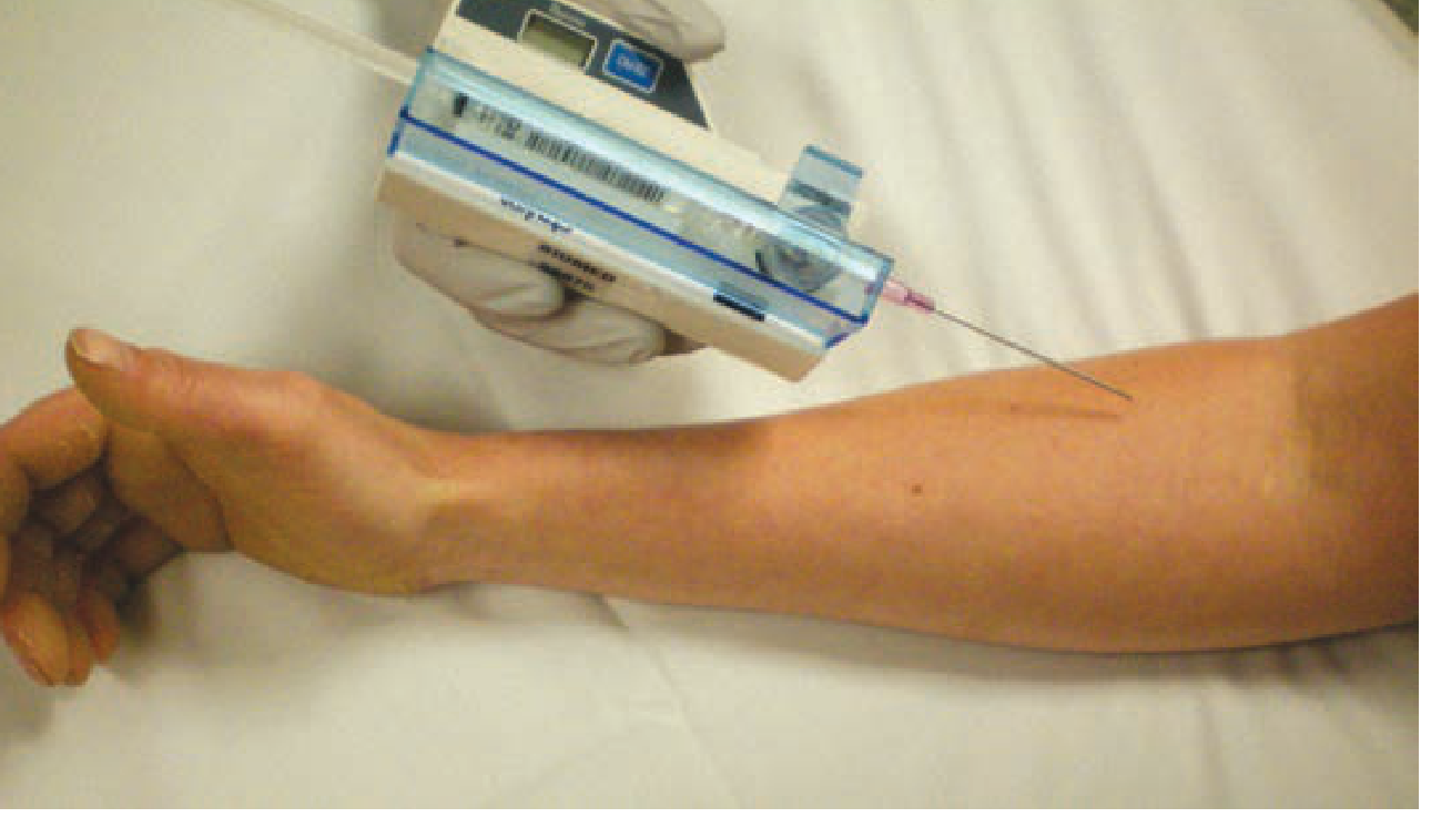
Compartment Pressure Measurement
Equipment (Stryker Intracompartmental Pressure Monitor)
The Stryker kit is the most commonly used commercial device. It contains:
- A saline-filled syringe
- A digital manometer
- A needle with a side port (18G needle typically)
Step-by-Step Technique
1. Setup
- Assemble: connect the manometer between the syringe and the needle
- Prime the system with saline - ensure no air bubbles
2. Position the limb
- Keep the limb at the level of the heart (not elevated, not dependent) - elevation falsely lowers readings
3. Needle insertion
- Clean the skin with antiseptic
- Insert the needle perpendicularly into the compartment to be measured
- Inject a few drops of saline to:
- Confirm the needle is not in a tendon
- Ensure no air pockets
- Open the tip against the compartment wall
4. Read the pressure
- The digital gauge gives the reading in mmHg
- Wait for the reading to stabilize
- Take 2 readings per compartment
- Also check adjacent compartments (pressure can spread)
5. Measurement site
- Obtain measurements within 5 cm of the injury/fracture site - this is where pressures are highest
Forearm Compartments (Relevant for This Patient)
For the left upper limb, you need to check 3 forearm compartments:
| Compartment | Needle Entry Site |
|---|---|
| Flexor (volar) | Volar (anterior) forearm, midline |
| Extensor (dorsal) | Dorsal forearm, midline |
| Mobile wad | Lateral forearm (brachioradialis muscle belly) |
The images below show the technique in the forearm:
Interpreting the Readings
| Pressure | Action |
|---|---|
| < 30 mmHg | Safe - no intervention needed |
| 30-45 mmHg | Borderline - use Delta P to decide |
| > 45 mmHg | Fasciotomy |
Delta P Rule (more reliable, especially in hypotensive patients)
Delta P = Diastolic BP - Compartment Pressure
- Delta P > 30-40 mmHg → can manage conservatively
- Delta P < 30 mmHg → perfusion is inadequate → fasciotomy
Example: If diastolic BP = 70 mmHg and compartment pressure = 45 mmHg → Delta P = 25 mmHg → fasciotomy needed
Important Notes
- Hands and feet have too many small compartments to measure reliably - if compartment syndrome is suspected there, many specialists proceed directly to surgical decompression without pressure measurement
- If no Stryker device is available, an arterial line setup or needle-manometer system can be improvised
- For obtunded or unresponsive patients (who cannot report pain), continuous intracompartmental monitoring has ~94% sensitivity and ~98% specificity
- If compartment syndrome is clinically obvious (tense limb, pain with passive stretch, absent pulses) - do not delay fasciotomy waiting for pressure readings
Time Window
| Tissue | Reversible injury | Irreversible damage |
|---|---|---|
| Muscle | < 3-4 hours | > 8 hours |
| Nerve | < 2 hours | > 8 hours |
Functional impairment is unlikely if compartment syndrome is diagnosed and treated within 6 hours of onset.
Source: Tintinalli's Emergency Medicine - A Comprehensive Study Guide
This is a shared conversation. Sign in to Orris to start your own chat.