
I’m preparing for derma quiz, test my knowledge and help me prepare accordingly
What's your level / exam context? · Which topics do you want to focus on? · How would you like to be tested?
🩺 MD/DNB Dermatology - MCQ Quiz Session
Q1 - Immunobullous Disorders
- A) BP180 (type XVII collagen)
- B) Desmoglein-3
- C) Desmoglein-1
- D) Type VII collagen
👉 Click to reveal answer & explanation
- BP180 → Bullous pemphigoid (subepidermal blistering)
- Desmoglein-1 only → Pemphigus foliaceus (superficial, no mucosal involvement, endemic form = fogo selvagem)
- Type VII collagen → Epidermolysis bullosa acquisita / Dystrophic EB
Q2 - Psoriasis Immunology
- A) IL-4 / IL-13 axis
- B) IL-5 axis
- C) IL-17A axis
- D) IL-23 / IL-12 axis
👉 Click to reveal answer & explanation
- IL-23 (produced by dendritic cells) → drives Th17 differentiation → IL-17A release
- IL-17A acts on keratinocytes → induces keratinocyte proliferation, chemokine production, neutrophil recruitment
| Target | Drug |
|---|---|
| IL-17A | Secukinumab, Ixekizumab |
| IL-17RA receptor | Brodalumab |
| IL-23 p19 subunit | Guselkumab, Risankizumab, Tildrakizumab |
| IL-12/23 p40 | Ustekinumab |
| TNF-α | Adalimumab, Etanercept |
- IL-4/IL-13 → Atopic dermatitis (dupilumab target)
- IL-5 → Eosinophilic conditions, urticaria
Q3 - Fungal Infections
- A) Trichophyton tonsurans
- B) Microsporum canis
- C) Trichophyton violaceum
- D) Trichophyton schoenleinii
👉 Click to reveal answer & explanation
- Microsporum canis (zoophilic, fluoresces green-yellow under Wood's lamp)
- Microsporum audouinii (anthropophilic, also ectothrix, Wood's lamp positive)
- T. verrucosum, T. mentagrophytes
- T. tonsurans (most common in USA - "black dot" tinea capitis)
- T. violaceum, T. soudanense
Q4 - Melanoma
- A) Stage IIA
- B) Stage IIB
- C) Stage IIC
- D) Stage IIIA
👉 Click to reveal answer & explanation
- T1: ≤1.0 mm
- T2: 1.01–2.0 mm
- T3: 2.01–4.0 mm
- T4: >4.0 mm
| Stage | T | Ulceration |
|---|---|---|
| IIA | T2b (ulcerated) or T3a (non-ulcerated) | +/- |
| IIB | T3b (ulcerated) or T4a (non-ulcerated) | +/- |
| IIC | T4b | Ulcerated, >4mm |
Correction: Answer is B) Stage IIB - T3b (3.2mm + ulcerated), N0, M0 = Stage IIB.
Q5 - Severe Cutaneous Adverse Reactions (SCARs)
- A) Age >40 years
- B) Body surface area detachment >10%
- C) Serum creatinine >140 μmol/L
- D) History of HIV infection
👉 Click to reveal answer & explanation
- Age >40 years
- Malignancy (active)
- Heart rate >120 bpm
- Initial epidermal detachment >10% BSA
- Serum urea >10 mmol/L (BUN >28 mg/dL)
- Serum glucose >14 mmol/L (>252 mg/dL)
- Serum bicarbonate <20 mmol/L
| Score | Mortality |
|---|---|
| 0–1 | ~3% |
| 2 | ~12% |
| 3 | ~35% |
| 4 | ~58% |
| ≥5 | >90% |
Q6 - Genodermatoses
- A) Netherton syndrome
- B) X-linked ichthyosis
- C) Lamellar ichthyosis
- D) Ichthyosis vulgaris
👉 Click to reveal answer & explanation
- AR inheritance; gene: TGM1 (transglutaminase-1, most common), ABCA12, CYP4F22, others
- Collodion baby at birth (tight, shiny membrane that cracks and peels)
- Ectropion and eclabium (everted eyelids and lips)
- Generalized large, dark, plate-like scales
- Minimal erythroderma in classic LI (vs. Nonbullous CIE which has erythroderma)
- Ichthyosis vulgaris: most common, filaggrin mutation (FLG), AD, "dirty neck," spares flexures, no collodion
- X-linked ichthyosis: males, steroid sulfatase deficiency, large dark scales, comma-shaped corneal opacities, cryptorchidism
- Netherton syndrome: AR, SPINK5 mutation, ichthyosis linearis circumflexa + trichorrhexis invaginata ("bamboo hair") + atopic diathesis
- EHK (Bullous CIE): KRT1/KRT10 mutations, blistering at birth then verrucous hyperkeratosis
Q7 - Contact Dermatitis
- A) Prick test (SPT)
- B) RAST/ImmunoCAP
- C) Repeat open application test (ROAT)
- D) Patch test
👉 Click to reveal answer & explanation
- Standard series (European Baseline Series / ICDRG standard): applied for 48 hours, readings at 48h and 96h (Day 4)
- Positive reaction: erythema, papules, vesicles at the patch site
- Grading (ICDRG): Negative (-), Doubtful (?+), Weak (+), Strong (++), Extreme (+++)
- Nickel sulfate (most common contact allergen worldwide)
- Fragrance mix, balsam of Peru
- Formaldehyde, parabens
- Rubber chemicals (thiuram mix, mercaptobenzothiazole)
- Chromate (cement workers)
- Neomycin, bacitracin
Q8 - Cutaneous Lymphoma
- A) Mycosis fungoides, erythrodermic variant
- B) Sézary syndrome
- C) Adult T-cell leukemia/lymphoma
- D) Pagetoid reticulosis
👉 Click to reveal answer & explanation
- Erythroderma (>80% BSA)
- Generalized lymphadenopathy
- Circulating Sézary cells >1000/μL (or >10% of lymphocytes) - large T-cells with hyperconvoluted cerebriform nuclei
- SS: erythroderma from the onset, blood involvement is defining; CD4:CD8 ratio >10:1 in blood
- Erythrodermic MF: progression from pre-existing patches/plaques of MF to erythroderma; less blood involvement
Q9 - Photodermatology
- A) Mees' lines
- B) Raindrop pigmentation
- C) Arsenical keratosis
- D) Hutchinson's sign
👉 Click to reveal answer & explanation
- Raindrop pigmentation: hyperpigmented macules on trunk (like raindrops)
- Arsenical keratoses: punctate hyperkeratotic lesions on palms and soles (classic finding)
- Leukomelanosis: mixture of hypo and hyperpigmentation
- Mees' lines: transverse white lines on nails (also seen with thallium, other heavy metals)
- Bowen's disease: multiple, in sun-protected areas (arsenical carcinogen effect)
- Peripheral vascular disease: "blackfoot disease" (Taiwan - arteriosclerosis obliterans)
Q10 - Acne & Rosacea
- A) Reduce isotretinoin dose
- B) Fundoscopy for papilledema
- C) CBC and LFTs
- D) MRI brain
👉 Click to reveal answer & explanation
- Immediate: Fundoscopy to confirm papilledema
- Stop isotretinoin immediately if pseudotumor cerebri is confirmed
- The condition is worsened by concurrent use of tetracycline antibiotics (both independently cause BIH - NEVER combine them)
| System | Side effect |
|---|---|
| Teratogenicity | Category X - strict pregnancy prevention (iPLEDGE in USA) |
| Mucocutaneous | Cheilitis (most common, >90%), dry skin, epistaxis |
| Lipids | Hypertriglyceridemia, elevated LDL |
| Liver | Elevated LFTs |
| Musculoskeletal | Myalgia, arthralgia, premature epiphyseal closure (in adolescents on high-dose) |
| Eyes | Dry eyes, night blindness (Vit A toxicity effect), impaired dark adaptation |
| Psychiatric | Depression (controversial; monitor) |
📊 How to Use This Session
- Work through the questions one by one
- Try to answer BEFORE revealing the explanation
- After you've gone through all 10, tell me your score and which topics you found tricky
- I'll generate a targeted second round focusing on your weak areas
| # | Topic | Difficulty |
|---|---|---|
| 1 | Immunobullous (PV) | ⭐⭐ |
| 2 | Psoriasis biologics | ⭐⭐ |
| 3 | Tinea capitis | ⭐⭐ |
| 4 | Melanoma staging | ⭐⭐⭐ |
| 5 | TEN/SCORTEN | ⭐⭐⭐ |
| 6 | Genodermatoses | ⭐⭐⭐ |
| 7 | Contact dermatitis | ⭐⭐ |
| 8 | Cutaneous lymphoma | ⭐⭐⭐ |
| 9 | Arsenical skin disease | ⭐⭐ |
| 10 | Isotretinoin toxicity | ⭐⭐ |
Create interactive visual and case based questions
psoriasis plaques silver scales erythematous
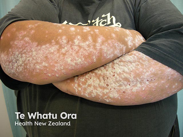
Clinical-wide dermatologic photography of the forearms in anterior view demonstrates extensive plaque psoriasis. Bilateral involvement of extensor surfaces presents as well‑demarcated erythematous plaques with adherent silvery scales, varying in thickness from moderate to dense. The plaques extend from near the elbows distally along the forearms, with areas of mild scaling and fissuring where plaques are thickest. There is no visible pustulation, ulceration, or atrophic change in this image, though surface texture ranges from smooth to raised. The morphological features—plaque morphology, silver-scale coating, and well‑defined borders—are characteristic of psoriasis vulgaris. The photograph captures subjective disease burden, distribution pattern, and surface level pathology, providing a visual record for assessment of extent and severity. Notably, the extensor-dominant distribution helps distinguish psoriasis from other inflammatory dermatoses such as eczema or tinea. This image complements clinical history and examination, enabling trajectory monitoring, treatment response evaluation, and patient education about prognosis and potential comorbidities (e.g., psoriatic arthritis, metabolic syndrome). It supports educational use for trainees and researchers studying plaque psoriasis morphology, distribution on the limbs, and therapeutic outcomes in chronic skin disease. Photographic documentation supports standardized severity scoring, helps track lesion evolution, and facilitates multi-site research collaborations evaluating topical and systemic therapeutic efficacy in diverse populations.
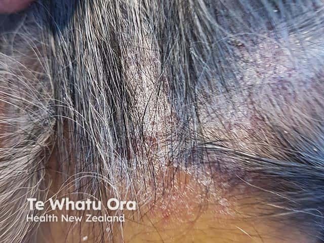
Clinical photography of the scalp depicting classic psoriasis plaque with silvery, adherent scale and erythematous background. Modality: External examination photography (dermatologic/clinical image) performed with standard bedside lighting, no dermoscopy or biopsy required for this description. Anatomical site: scalp skin, including crown and temporal regions, representing a hair-bearing area with regional alopecia potential from plaque coverage. Visual features: well-demarcated erythematous plaques, multilayered, lamellar silver scales, follicular plugging variations, mild edema at lesion margins, and diffuse desquamation. The plaques may extend to adjacent occipital scalp; involvement may be limited to seborrheic-like areas or generalized scalp involvement. The lesion morphology is plaque-type (psoriatic) with erythema, scaling, and mild but non-purulent irritation. Pathologic correlate (not shown) would be psoriatic epidermal hyperplasia with parakeratosis and neutrophilic microabscesses (Munro) on histology; clinically, this appearance correlates with psoriasis vulgaris, plaque type. Diagnostic significance: characteristic scalp psoriasis demonstrates a chronic relapsing course, responds to topical corticosteroids, vitamin D analogs, or combination therapies; helps differentiate from seborrheic dermatitis and tinea capitis. Clinical use: educational reference for dermatology trainees, triage for systemic evaluation, and documentation of typical scalp disease for monitoring treatment response and disease extent. The image serves as a representative example for scalp involvement in psoriasis and related inflammatory dermatoses.
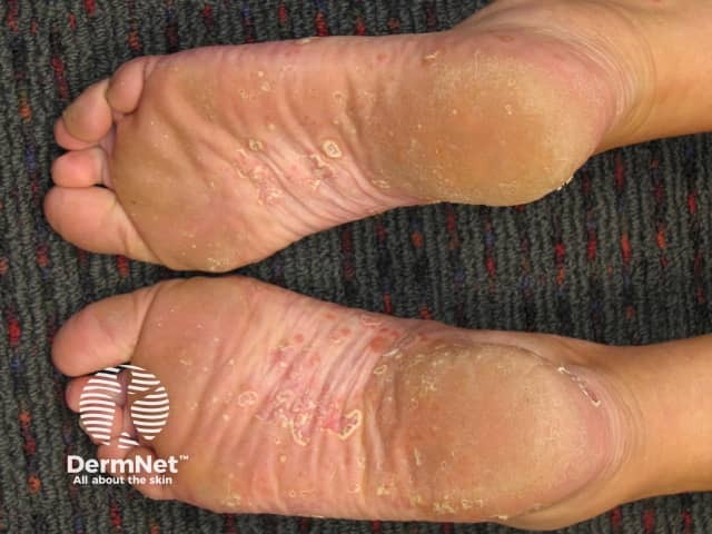
Clinical photography of bilateral plantar surfaces demonstrating palmoplantar psoriasis. The soles show thickened, erythematous plaques with well-defined borders and overlying white-to-silver scale, greatest at weight-bearing areas near the metatarsal heads and heels. Fissuring and cracking are present, producing painful splits in the stratum corneum. The surrounding skin is mildly desquamated with concentric layering patterns (lichenification) in some regions, and there is subtle hyperkeratosis along creases of the plantar surface. The palmar surfaces of the hands are not shown; focus is on the plantar aspect. The appearance is characteristic of chronic palmoplantar psoriasis: erythematous plaques with silvery scales, plaque morphology, and fissuring that may mimic eczema or fungal infection. The image lacks evidence of vesiculation or purulence. The texture is dry, cracked, and fissured with regional epidermal thickening. Clinically relevant features include involvement of both feet, symmetry, and the tendency for persistent, thickened scales with fissuring that can limit mobility and weight bearing. This image can support differential diagnosis against tinea pedis, contact dermatitis, or palmoplantar keratoderma, and may guide topical therapies (mid- to high-potency corticosteroids, vitamin D analogs) and multidisciplinary management. Correlation with patient history and joint symptoms is advised to assess psoriasis severity and psoriatic arthritis risk for treatment planning.
pemphigus vulgaris oral erosions flaccid blisters
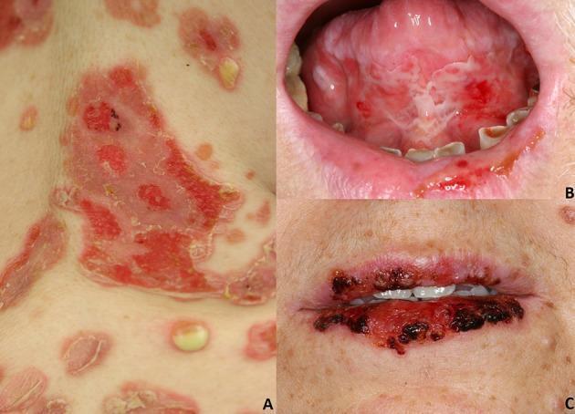
This composite of clinical photographs illustrates the mucocutaneous manifestations of pemphigus variants. Panel A (top left) displays Pemphigus vulgaris on the skin, showing large, irregular, erythematous erosions with peripheral peeling of the epidermis, consistent with ruptured flaccid blisters. A small, intact flaccid vesicle with yellowish fluid is visible in the lower quadrant. Panel B (top right) demonstrates oral involvement in Pemphigus vulgaris, featuring multiple painful-looking erosions and white macerated plaques on the ventral surface of the tongue and labial mucosa. Panel C (bottom right) showcases Paraneoplastic pemphigus, characterized by severe stomatitis with diffuse, hemorrhagic crusting and deep erosions across the entire vermillion border of the upper and lower lips. These images contrast the typical cutaneous and oral erosions of Pemphigus vulgaris with the more severe, hemorrhagic labial involvement often seen in paraneoplastic variants, serving as a diagnostic tool for identifying autoimmune blistering diseases and their clinical severity.
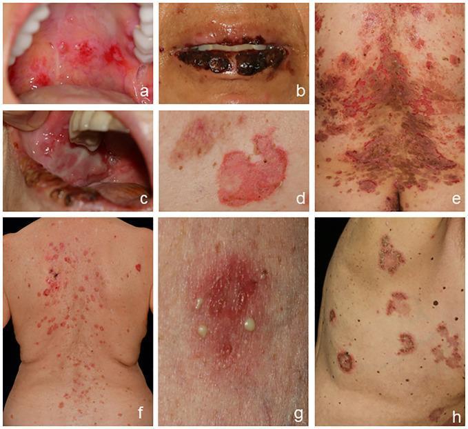
A composite of clinical photographs demonstrating the varied manifestations of autoimmune bullous diseases, specifically pemphigus subtypes. (A-C) Pemphigus vulgaris (PV) involving the oral cavity, showing bright red erosions on the soft palate and buccal mucosa, and hemorrhagic crusting of the lips. (D-E) Cutaneous PV exhibiting flaccid blisters that have ruptured into large, erythematous erosions and areas of postinflammatory hyperpigmentation on the trunk. (F) Pemphigus foliaceus (PF) presented as scaly, erythematous erosions localized to seborrheic areas on the back, appearing as crusts rather than intact blisters. (G-H) IgA pemphigus characterized by scattered pustules on an erythematous base and diagnostic annular (ring-shaped) or circinate patterns with peripheral vesicles and central crusting on the trunk. The collection illustrates key diagnostic features including mucous membrane involvement in PV versus the purely cutaneous, superficial exfoliation in PF, and the distinct pustular morphology of IgA pemphigus.
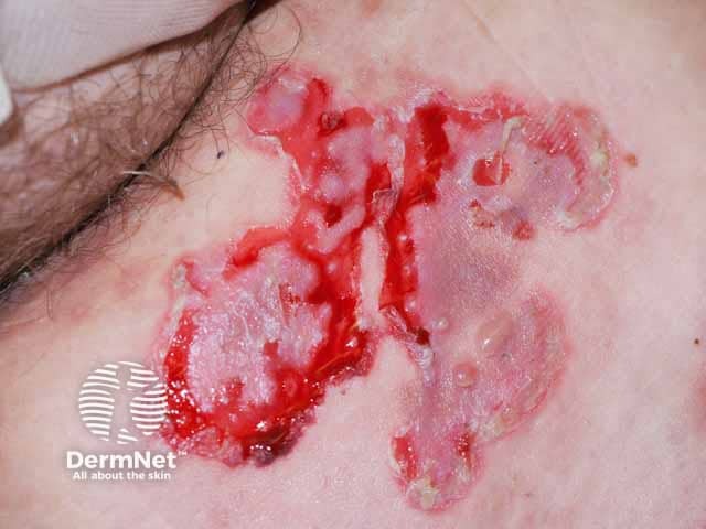
Clinical photography of a pemphigus vulgaris (PV) patient shows diffuse cutaneous erosions and ruptured, flaccid blisters with irregular edges exposing moist, erythematous bases. The close-up, high-magnification view captures superficial losses of epidermal integrity consistent with ruptured pemphigus bullae. Lesions appear tender, crusted at the periphery, and coalesce into larger erosive plaques with surrounding erythema. This presentation is classic for PV, in which intraepidermal acantholysis creates fragile blisters that rupture easily. Mucosal involvement is common in PV but is not depicted in this image. Clinically, a positive Nikolsky sign may accompany the erosions. Histologically, pemphigus vulgaris shows suprabasal acantholysis with tombstone basal cells; immunofluorescence reveals intercellular IgG. The image is relevant for dermatology, medical education, and clinical practice for recognizing active cutaneous PV and guiding management. Common treatment considerations include systemic corticosteroids, steroid-sparing immunosuppressants, and targeted biologics, with careful monitoring for flare progression. For research and training, annotate lesion age, localization, and wound characteristics, and correlate with serology (desmoglein antibodies) and biopsy findings when available. This image exemplifies PV skin involvement and supports differential diagnosis from bullous pemphigoid and other vesiculobullous diseases.
melanoma pigmented lesion skin ABCDE
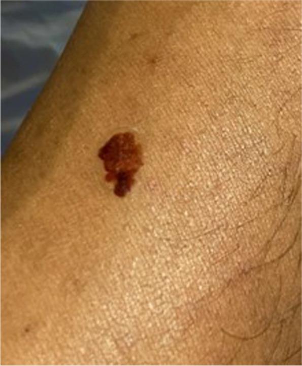
This clinical photograph shows a close-up view of a pigmented skin lesion, specifically a medical moulage representing a malignant melanoma in the form of a high-resolution transfer tattoo. The lesion exhibits the classic 'ABCDE' criteria for melanoma: it is highly asymmetric (Asymmetry), possesses irregular, ill-defined, and notched borders (Border), and displays significant color variegation (Color) with shades ranging from dark brown to tan and deep black-red. The surface appears textured and slightly elevated above the surrounding healthy skin. The image is used as an educational tool for medical training and Objective Structured Clinical Examinations (OSCEs) to teach students and healthcare professionals how to identify suspicious skin morphology on various skin tones. The surrounding skin appears healthy with no visible signs of acute inflammation, such as edema or surrounding erythema.
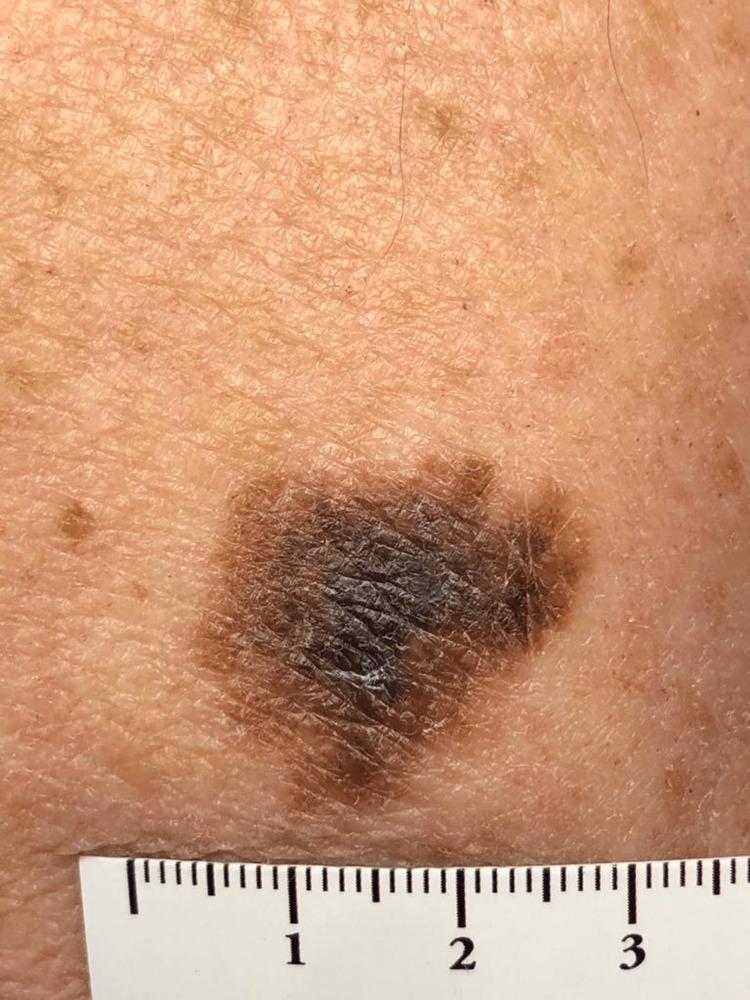
This clinical photograph shows a suspicious pigmented skin lesion on the left arm of an elderly patient, identified as melanoma in situ. The lesion is a large macule with a maximum diameter of approximately 2.5 to 3 cm as indicated by the adjacent metric ruler. It exhibits classic ABCDE features of melanoma, including significant asymmetry and highly irregular, notched borders. The color distribution is heterogeneous, displaying a variegated pattern of dark brown, tan, and focal areas of deep black pigment. The surface texture is uneven with subtle scaling and accentuation of skin markings, but without gross nodularity or ulceration. The surrounding skin shows signs of chronic actinic damage (dermatoheliosis), characterized by solar lentigines and textural changes. This image serves as a high-quality educational example of a primary cutaneous melanoma in its non-invasive stage, highlighting the importance of visual inspection and total skin examination in patients with high cumulative sun exposure.
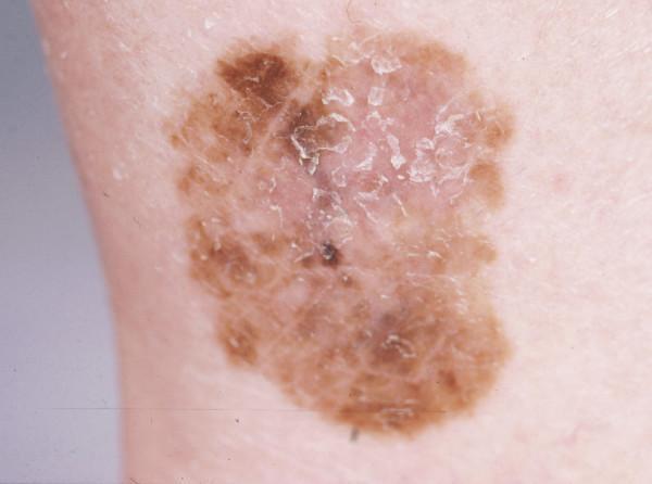
This clinical photograph shows a pigmented skin lesion on the ankle, characteristic of a superficial spreading melanoma. The lesion demonstrates the classic 'ABCDE' criteria: it is asymmetric in shape and shows significant border irregularity with poorly defined, notched margins. There is marked color variegation, with a primary palette of light to dark brown and focal areas of darker black-brown pigmentation. A central area of relative hypopigmentation suggests possible regression. The surface texture is uneven, with visible fine scaling and focal hyperkeratosis in the central portion. The lesion is relatively flat, consistent with the radial growth phase of superficial spreading melanoma. This image serves as a teaching example for dermatologic assessment of suspicious melanocytic lesions in a lower extremity location, highlighting the importance of recognizing irregular pigment distribution and evolving surface features in clinical oncology and dermatology.
toxic epidermal necrolysis skin detachment erosions
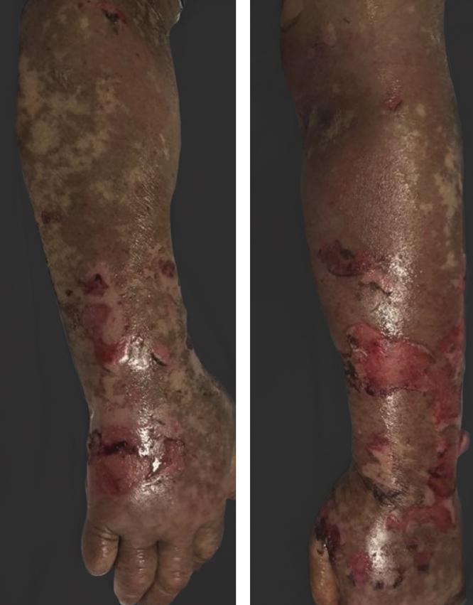
This clinical photograph displays a side-by-side view of an individual's upper extremity, demonstrating severe cutaneous pathology consistent with Toxic Epidermal Necrolysis (TEN). The image illustrates diffuse epidermal detachment and widespread erosions across the forearm, wrist, and dorsal hand. Visible features include large, irregular areas of denuded skin exposing the bright red, moist dermis, indicating significant loss of the epidermal layer. Surrounding the primary erosions, the skin shows evidence of confluent erythema, ruptured bullae (blisters), and mottled pigmentation changes with patches of dusky gray and tan discoloration. This presentation highlights a critical dermatological emergency often associated with drug-induced hypersensitivity reactions—in this specific context, potentially triggered by tyrosine kinase inhibitor therapy (vandetanib) following UV exposure. The visual findings serve as a classic representation of severe skin sloughing and full-thickness epidermal necrosis for educational use in dermatology and emergency medicine.
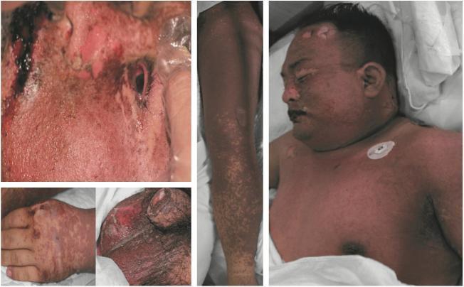
This composite clinical photograph illustrates the multisystem dermatological manifestations of Toxic Epidermal Necrolysis (TEN). The images demonstrate widespread epidermal detachment and mucocutaneous involvement across several anatomical regions. Facial views show significant erosions, hemorrhagic crusting, and denuded skin around the periorbital and perioral areas, characteristic of severe mucosal involvement. The trunk and upper extremities exhibit a confluent, dusky erythematous maculopapular rash with areas of epidermal sloughing and blistering, resulting in large patches of exposed, raw dermis. A detailed view of the genitalia reveals severe epidermal loss and ulceration, highlighting the involvement of non-oral mucosal surfaces. The hand and arm images show a mix of discrete and confluent erythematous lesions with positive Nikolsky-like signs of skin fragility. This visual presentation is indicative of a severe Cutaneous Adverse Reaction (SCAR), demonstrating the high morbidity associated with extensive integumentary failure and the critical need for intensive wound management in TEN cases.
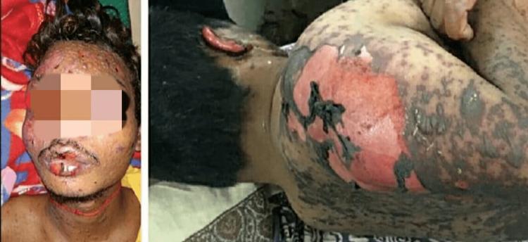
Clinical photograph of a patient exhibiting severe dermatological manifestations of Toxic Epidermal Necrolysis (TEN). The visual evidence includes extensive epidermal detachment and desquamation across the face, neck, and upper torso. On the face, there is significant mucosal involvement with hemorrhagic crusting and erosions of the lips, along with scattered vesiculobullous lesions on the cheeks and forehead. The lateral and posterior views of the upper body demonstrate large areas of denuded skin where the epidermis has sloughed off, revealing erythematous, raw dermis. Bordering these areas are gray-tinged, necrotic skin flaps and multiple scattered dusky macules and bullae. This presentation is characteristic of a severe cutaneous adverse reaction (SCAR), showing a positive Nikolsky sign where skin peels in large sheets. The distribution is generalized, involving both mucosal surfaces and a high percentage of total body surface area, illustrating the critical nature of the condition typically associated with drug-induced hypersensitivity.
tinea capitis black dot alopecia scalp child
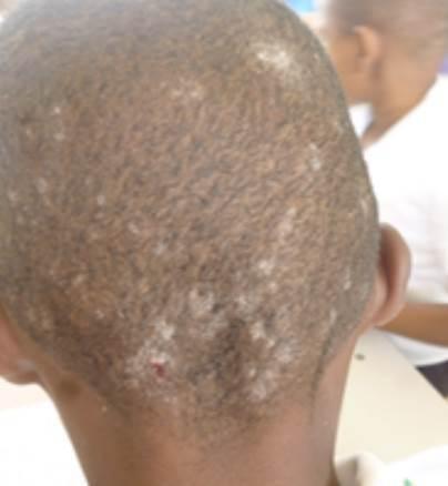
This clinical photograph displays a posterior view of a child's scalp exhibiting multiple discrete, circular patches of alopecia characteristic of Tinea capitis. The affected areas show visible scaling and a lighter, hypopigmented appearance compared to the adjacent healthy scalp. Within several of the alopecic patches, small dark specks are visible, suggesting 'black dot' alopecia caused by hairs breaking off at the skin surface. A focal area of erythema or a small excoriation is noted near the occipital hairline, potentially indicating irritation or secondary infection. The distribution is multifocal and concentrated across the vertex and occipital regions. These findings are consistent with an anthropophilic fungal infection, such as Trichophyton tonsurans, typically seen in pediatric populations and characterized by endothrix hair shaft invasion.
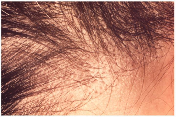
This clinical photograph shows the scalp of an adult male exhibiting signs of non-inflammatory tinea capitis, specifically a 'black dot' alopecia pattern typical of Trichophyton violaceum infection. The image demonstrates a localized area of significant hair thinning and patchy alopecia. High-resolution detail reveals numerous broken hair shafts that have fractured at the level of the scalp surface, appearing as small, dark, comedo-like plugs or 'black dots.' The underlying skin shows mild, diffuse fine scaling without evidence of severe inflammation, kerion formation, or scarring. The hair distribution is uneven, with short, brittle shafts interspersed between areas of complete hair loss. This visual illustrates the endothrix invasion pattern where fungal spores weaken the hair shaft internally, leading to easy breakage. Educational focus is on the diagnostic identification of dermatophytosis of the scalp in an adult and the differentiation between various alopecia morphologies.
dermatitis herpetiformis vesicles elbow pruritic
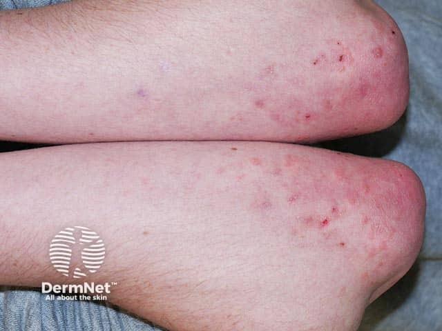
This high-resolution clinical photograph shows bilateral elbow skin with a pruritic vesiculopapular eruption typical of dermatitis herpetiformis. The extensor surfaces of both elbows display clustered vesicles and papules on a background of erythema, with adjacent crusting from scratching. Lesions are symmetrically distributed on the antecubital and olecranon regions, reflecting the classic DH predilection for extensor surfaces. The skin appears non-ulcerated, with variable edema and a mosaic of small vesicles that may be grouped in crops. In DermNet terms, the rash is intensely pruritic and may present as vesicles, papules, and urticarial plaques; this image captures the hallmark pattern without deep infection. Pathophysiology involves IgA immune complex deposition at dermal papillae, correlating with gluten sensitivity and potential celiac disease. Clinically, the image would prompt serologic assessments for celiac disease (tTG IgA) and consideration of skin biopsy to reveal granular IgA deposition. Treatment includes a gluten-free diet to address the underlying disease and dapsone for pruritus relief. This image is valuable for dermatology education and pattern-recognition training, aiding differential diagnosis with vesiculobullous conditions such as linear IgA disease, contact dermatitis, and eczema herpeticum.
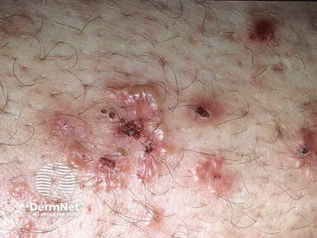
Imaging modality: Clinical photography of a skin eruption on the extensor surface of the elbow. The image shows clustered vesicles and vesiculopapular plaques on an erythematous base with surrounding excoriations and crusts. Lesions are pruritic and arranged in grouped clusters, predominantly on the extensor aspect of the elbow (antecubital region) with variable edema and superficial erosions. The appearance is highly suggestive of dermatitis herpetiformis in the appropriate clinical context, though differential diagnoses include dyshidrotic dermatitis and other vesiculobullous disorders. No biopsy or immunofluorescence data are provided; if obtained, direct immunofluorescence would be expected to reveal granular IgA deposits in dermal papillae, supporting DH. Clinically, DH is associated with gluten sensitivity and celiac disease; patients often present with intensely itchy vesicles that erupt in crops on elbows, knees, buttocks, and scalp. This image captures the papulovesicular eruption with vesicle grouping and crusting after rupture. Diagnostic significance lies in recognizing extensor surface involvement and pruritic grouped vesicles, guiding serologic testing for celiac disease and consideration of a gluten-free diet and dapsone therapy for symptomatic relief. Educationally, the image is useful for pattern recognition of DH, correlation with histopathology, and discussion of differential diagnoses, patient counseling, and clinical management in dermatology and gastroenterology practice.
acanthosis nigricans hyperpigmentation neck axilla insulin resistance
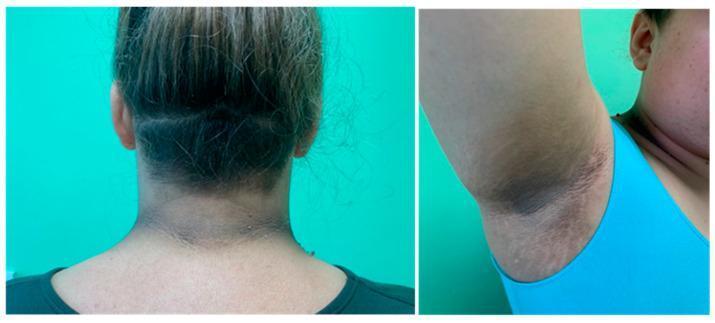
Clinical photograph showing classic dermatological manifestations of Acanthosis nigricans in two key anatomical locations: the posterior neck and the axilla (armpit). The left panel displays the back of the neck, revealing a broad, horizontal band of symmetric, grey-brown hyperpigmentation. The skin exhibits characteristic 'velvety' thickening (hyperkeratosis) and accentuated skin lines. The right panel shows a similar presentation in the axillary vault, where the hyperpigmentation is more pronounced and well-demarcated against the surrounding normal skin, following the folds of the intertriginous area. These visible changes are significant clinical markers often associated with systemic conditions such as insulin resistance, obesity, and metabolic syndrome. The image serves as an educational tool for identifying the typical morphology and distribution of this cutaneous sign, emphasizing the importance of recognizing texture and color changes in intertriginous zones for diagnostic assessment.
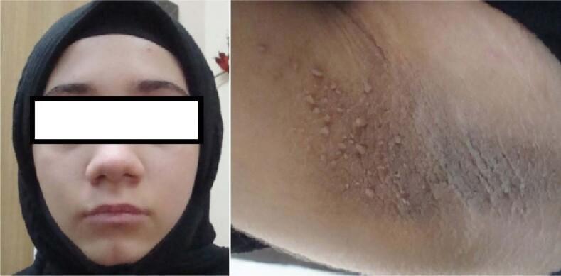
Two-panel clinical photograph displaying the phenotypic characteristics of a patient with Type A insulin resistance syndrome. The left panel shows a frontal view of the face, appearing largely unremarkable except for mild facial fullness. The right panel is a close-up image of the axillary region, demonstrating classic features of acanthosis nigricans. Key visual findings in the axilla include significant hyperpigmentation and skin thickening with a velvety, verrucous, or 'leathery' texture. Multiple small, flesh-colored to light-brown pedunculated papules consistent with skin tags (acrochordons) are visible, scattered across the hyperpigmented area. These skin changes are indicative of severe hyperinsulinemia and resultant hyperkeratosis and papillomatosis. The content is suitable for medical education regarding endocrine-related dermatological manifestations and clinical signs of systemic insulin resistance.
mkdir -p /tmp/workspace/derma-quiz
~/derma-quiz/derma_quiz.html
<!DOCTYPE html>
<html lang="en">
<head>
<meta charset="UTF-8">
<meta name="viewport" content="width=device-width, initial-scale=1.0">
<title>MD/DNB Dermatology - Visual & Case-Based Quiz</title>
<style>
  :root {
    --bg: #0f1117;
    --card: #1a1d2e;
    --card2: #22263a;
    --accent: #7c6af7;
    --accent2: #5b8af5;
    --green: #22c55e;
    --red: #ef4444;
    --yellow: #f59e0b;
    --text: #e8eaf0;
    --muted: #9095b0;
    --border: #2d3155;
    --radius: 14px;
  }

  * { box-sizing: border-box; margin: 0; padding: 0; }

  body {
    font-family: 'Segoe UI', system-ui, sans-serif;
    background: var(--bg);
    color: var(--text);
    min-height: 100vh;
    padding: 0;
  }

  /* ─── HEADER ─────────────────────────────── */
  .header {
    background: linear-gradient(135deg, #1a1d2e 0%, #12152a 100%);
    border-bottom: 1px solid var(--border);
    padding: 18px 32px;
    display: flex;
    align-items: center;
    justify-content: space-between;
    position: sticky;
    top: 0;
    z-index: 100;
  }
  .header-title { display: flex; align-items: center; gap: 12px; }
  .header-title .icon { font-size: 26px; }
  .header-title h1 { font-size: 18px; font-weight: 700; color: var(--text); }
  .header-title p { font-size: 12px; color: var(--muted); }
  .score-pill {
    background: var(--card2);
    border: 1px solid var(--border);
    border-radius: 50px;
    padding: 8px 20px;
    font-size: 14px;
    font-weight: 600;
    color: var(--accent);
  }

  /* ─── PROGRESS ───────────────────────────── */
  .progress-wrap {
    background: var(--card);
    border-bottom: 1px solid var(--border);
    padding: 12px 32px;
  }
  .progress-bar-outer {
    background: var(--border);
    border-radius: 50px;
    height: 6px;
    overflow: hidden;
  }
  .progress-bar-inner {
    height: 100%;
    background: linear-gradient(90deg, var(--accent), var(--accent2));
    border-radius: 50px;
    transition: width 0.6s ease;
  }
  .progress-labels {
    display: flex;
    justify-content: space-between;
    font-size: 11px;
    color: var(--muted);
    margin-top: 6px;
  }

  /* ─── MAIN LAYOUT ────────────────────────── */
  .main {
    max-width: 900px;
    margin: 0 auto;
    padding: 32px 20px 80px;
  }

  /* ─── QUIZ CARD ──────────────────────────── */
  .quiz-card {
    background: var(--card);
    border: 1px solid var(--border);
    border-radius: var(--radius);
    overflow: hidden;
    display: none;
    animation: fadeUp 0.4s ease;
  }
  .quiz-card.active { display: block; }

  @keyframes fadeUp {
    from { opacity: 0; transform: translateY(16px); }
    to   { opacity: 1; transform: translateY(0); }
  }

  .card-header {
    display: flex;
    align-items: center;
    gap: 12px;
    padding: 16px 24px;
    border-bottom: 1px solid var(--border);
    background: var(--card2);
  }
  .q-number {
    background: linear-gradient(135deg, var(--accent), var(--accent2));
    color: #fff;
    font-size: 12px;
    font-weight: 700;
    padding: 4px 12px;
    border-radius: 50px;
  }
  .q-tag {
    background: rgba(124,106,247,0.15);
    color: var(--accent);
    font-size: 11px;
    font-weight: 600;
    padding: 3px 10px;
    border-radius: 50px;
    border: 1px solid rgba(124,106,247,0.3);
  }
  .q-type-tag {
    margin-left: auto;
    background: rgba(91,138,245,0.12);
    color: var(--accent2);
    font-size: 11px;
    font-weight: 600;
    padding: 3px 10px;
    border-radius: 50px;
    border: 1px solid rgba(91,138,245,0.25);
  }

  .card-body { padding: 24px; }

  /* Clinical scenario box */
  .scenario-box {
    background: linear-gradient(135deg, rgba(91,138,245,0.08), rgba(124,106,247,0.05));
    border: 1px solid rgba(91,138,245,0.25);
    border-left: 4px solid var(--accent2);
    border-radius: 10px;
    padding: 18px 20px;
    margin-bottom: 20px;
    font-size: 14.5px;
    line-height: 1.7;
  }
  .scenario-box .case-label {
    font-size: 10px;
    font-weight: 700;
    text-transform: uppercase;
    letter-spacing: 1.5px;
    color: var(--accent2);
    margin-bottom: 8px;
  }
  .scenario-box strong { color: #fff; }

  /* Image area */
  .image-frame {
    background: #0a0c18;
    border: 1px solid var(--border);
    border-radius: 10px;
    overflow: hidden;
    margin-bottom: 20px;
    position: relative;
  }
  .image-frame img {
    width: 100%;
    max-height: 360px;
    object-fit: cover;
    display: block;
    cursor: zoom-in;
    transition: transform 0.3s;
  }
  .image-frame img:hover { transform: scale(1.02); }
  .image-caption {
    background: rgba(0,0,0,0.6);
    color: var(--muted);
    font-size: 11px;
    padding: 6px 12px;
    text-align: center;
  }
  .zoom-hint {
    position: absolute;
    top: 10px;
    right: 10px;
    background: rgba(0,0,0,0.7);
    color: #fff;
    font-size: 11px;
    padding: 4px 10px;
    border-radius: 50px;
    pointer-events: none;
  }

  /* Lightbox */
  .lightbox {
    display: none;
    position: fixed;
    inset: 0;
    background: rgba(0,0,0,0.92);
    z-index: 1000;
    align-items: center;
    justify-content: center;
    cursor: zoom-out;
  }
  .lightbox.open { display: flex; }
  .lightbox img { max-width: 92vw; max-height: 90vh; border-radius: 8px; }

  /* Question text */
  .question-text {
    font-size: 16px;
    font-weight: 600;
    color: #fff;
    margin-bottom: 20px;
    line-height: 1.6;
  }

  /* Options */
  .options-grid {
    display: grid;
    grid-template-columns: 1fr 1fr;
    gap: 10px;
    margin-bottom: 20px;
  }
  @media (max-width: 600px) { .options-grid { grid-template-columns: 1fr; } }

  .option-btn {
    background: var(--card2);
    border: 1.5px solid var(--border);
    border-radius: 10px;
    padding: 14px 16px;
    text-align: left;
    color: var(--text);
    font-size: 14px;
    cursor: pointer;
    transition: all 0.2s;
    display: flex;
    align-items: flex-start;
    gap: 12px;
  }
  .option-btn:hover:not(:disabled) {
    border-color: var(--accent);
    background: rgba(124,106,247,0.1);
    transform: translateY(-1px);
  }
  .option-btn:disabled { cursor: default; }
  .option-label {
    background: var(--border);
    color: var(--muted);
    border-radius: 6px;
    width: 26px;
    height: 26px;
    display: flex;
    align-items: center;
    justify-content: center;
    font-size: 12px;
    font-weight: 700;
    flex-shrink: 0;
    transition: all 0.2s;
  }
  .option-text { line-height: 1.5; padding-top: 2px; }

  /* States */
  .option-btn.correct {
    border-color: var(--green);
    background: rgba(34,197,94,0.12);
  }
  .option-btn.correct .option-label {
    background: var(--green);
    color: #fff;
  }
  .option-btn.wrong {
    border-color: var(--red);
    background: rgba(239,68,68,0.1);
  }
  .option-btn.wrong .option-label {
    background: var(--red);
    color: #fff;
  }
  .option-btn.selected-wrong {
    border-color: var(--red);
    background: rgba(239,68,68,0.15);
  }
  .option-btn.selected-wrong .option-label {
    background: var(--red);
    color: #fff;
  }

  /* Reveal button */
  .reveal-btn {
    background: linear-gradient(135deg, var(--accent), var(--accent2));
    border: none;
    border-radius: 10px;
    color: #fff;
    font-size: 14px;
    font-weight: 600;
    padding: 12px 24px;
    cursor: pointer;
    display: none;
    align-items: center;
    gap: 8px;
    margin-bottom: 16px;
    transition: opacity 0.2s, transform 0.2s;
  }
  .reveal-btn:hover { opacity: 0.9; transform: translateY(-1px); }
  .reveal-btn.show { display: flex; }

  /* Explanation */
  .explanation {
    display: none;
    background: var(--card2);
    border: 1px solid var(--border);
    border-radius: 10px;
    padding: 20px;
    margin-top: 4px;
    animation: fadeUp 0.3s ease;
  }
  .explanation.show { display: block; }
  .explanation-header {
    display: flex;
    align-items: center;
    gap: 8px;
    font-size: 13px;
    font-weight: 700;
    color: var(--accent);
    text-transform: uppercase;
    letter-spacing: 1px;
    margin-bottom: 12px;
  }
  .explanation p {
    font-size: 14px;
    line-height: 1.75;
    color: var(--text);
    margin-bottom: 10px;
  }
  .explanation p:last-child { margin-bottom: 0; }
  .explanation strong { color: #fff; }
  .explanation em { color: var(--accent2); font-style: normal; font-weight: 600; }

  /* Key points table */
  .key-table {
    width: 100%;
    border-collapse: collapse;
    margin-top: 12px;
    font-size: 13px;
  }
  .key-table th {
    background: rgba(124,106,247,0.15);
    color: var(--accent);
    padding: 8px 12px;
    text-align: left;
    font-size: 11px;
    text-transform: uppercase;
    letter-spacing: 0.8px;
  }
  .key-table td {
    padding: 8px 12px;
    border-bottom: 1px solid var(--border);
    color: var(--text);
  }
  .key-table tr:last-child td { border-bottom: none; }
  .key-table tr:nth-child(even) td { background: rgba(255,255,255,0.02); }

  /* Feedback badges */
  .feedback-badge {
    display: none;
    align-items: center;
    gap: 8px;
    padding: 10px 16px;
    border-radius: 10px;
    font-size: 14px;
    font-weight: 600;
    margin-bottom: 14px;
  }
  .feedback-badge.show { display: flex; }
  .feedback-badge.correct-fb { background: rgba(34,197,94,0.15); border: 1px solid rgba(34,197,94,0.3); color: var(--green); }
  .feedback-badge.wrong-fb { background: rgba(239,68,68,0.12); border: 1px solid rgba(239,68,68,0.25); color: var(--red); }

  /* Next button */
  .next-btn {
    background: var(--card2);
    border: 1.5px solid var(--border);
    border-radius: 10px;
    color: var(--text);
    font-size: 14px;
    font-weight: 600;
    padding: 12px 28px;
    cursor: pointer;
    display: none;
    align-items: center;
    gap: 8px;
    transition: all 0.2s;
    margin-top: 16px;
  }
  .next-btn:hover { border-color: var(--accent); color: var(--accent); }
  .next-btn.show { display: flex; }

  /* ─── RESULTS SCREEN ─────────────────────── */
  #results-screen {
    display: none;
    text-align: center;
    padding: 40px 20px;
  }
  .trophy { font-size: 72px; margin-bottom: 16px; }
  .results-title { font-size: 26px; font-weight: 700; color: #fff; margin-bottom: 8px; }
  .results-sub { font-size: 15px; color: var(--muted); margin-bottom: 32px; }
  .score-circle {
    width: 140px;
    height: 140px;
    border-radius: 50%;
    background: conic-gradient(var(--accent) var(--pct), var(--border) 0);
    display: flex;
    align-items: center;
    justify-content: center;
    margin: 0 auto 32px;
    position: relative;
  }
  .score-circle::before {
    content: '';
    position: absolute;
    inset: 12px;
    background: var(--card);
    border-radius: 50%;
  }
  .score-circle-text {
    position: relative;
    z-index: 1;
    font-size: 28px;
    font-weight: 800;
    color: var(--accent);
  }
  .results-grid {
    display: grid;
    grid-template-columns: repeat(3, 1fr);
    gap: 16px;
    max-width: 500px;
    margin: 0 auto 32px;
  }
  .results-stat {
    background: var(--card2);
    border: 1px solid var(--border);
    border-radius: 12px;
    padding: 16px;
  }
  .results-stat-num { font-size: 28px; font-weight: 800; }
  .results-stat-num.green { color: var(--green); }
  .results-stat-num.red { color: var(--red); }
  .results-stat-num.yellow { color: var(--yellow); }
  .results-stat-label { font-size: 12px; color: var(--muted); margin-top: 4px; }
  .restart-btn {
    background: linear-gradient(135deg, var(--accent), var(--accent2));
    border: none;
    border-radius: 10px;
    color: #fff;
    font-size: 15px;
    font-weight: 700;
    padding: 14px 36px;
    cursor: pointer;
    transition: opacity 0.2s, transform 0.2s;
  }
  .restart-btn:hover { opacity: 0.9; transform: translateY(-1px); }

  /* ─── INTRO SCREEN ───────────────────────── */
  #intro-screen { padding: 20px 0; }
  .intro-card {
    background: var(--card);
    border: 1px solid var(--border);
    border-radius: var(--radius);
    padding: 40px 32px;
    text-align: center;
    margin-bottom: 24px;
  }
  .intro-icon { font-size: 56px; margin-bottom: 16px; }
  .intro-title { font-size: 24px; font-weight: 800; color: #fff; margin-bottom: 8px; }
  .intro-sub { font-size: 14px; color: var(--muted); line-height: 1.7; margin-bottom: 28px; }
  .topic-pills { display: flex; flex-wrap: wrap; gap: 8px; justify-content: center; margin-bottom: 28px; }
  .topic-pill {
    background: rgba(124,106,247,0.12);
    border: 1px solid rgba(124,106,247,0.3);
    color: var(--accent);
    font-size: 12px;
    font-weight: 600;
    padding: 5px 14px;
    border-radius: 50px;
  }
  .start-btn {
    background: linear-gradient(135deg, var(--accent), var(--accent2));
    border: none;
    border-radius: 12px;
    color: #fff;
    font-size: 16px;
    font-weight: 700;
    padding: 16px 48px;
    cursor: pointer;
    transition: all 0.2s;
    display: inline-flex;
    align-items: center;
    gap: 10px;
  }
  .start-btn:hover { opacity: 0.9; transform: translateY(-2px); }

  /* Misc */
  .highlight-box {
    background: rgba(245,158,11,0.08);
    border: 1px solid rgba(245,158,11,0.25);
    border-radius: 8px;
    padding: 10px 14px;
    margin-top: 10px;
    font-size: 13px;
    color: var(--yellow);
  }
  .highlight-box strong { color: #fff; }

  .source-cite {
    font-size: 11px;
    color: var(--muted);
    margin-top: 10px;
    font-style: italic;
  }
</style>
</head>
<body>

<!-- Lightbox -->
<div class="lightbox" id="lightbox" onclick="closeLightbox()">
  <img id="lightbox-img" src="" alt="Zoomed image">
</div>

<!-- Header -->
<div class="header">
  <div class="header-title">
    <span class="icon">🩺</span>
    <div>
      <h1>Dermatology Visual Quiz</h1>
      <p>MD/DNB Level · Visual & Case-Based</p>
    </div>
  </div>
  <div class="score-pill" id="header-score">Score: 0 / 0</div>
</div>

<!-- Progress -->
<div class="progress-wrap" id="progress-wrap" style="display:none">
  <div class="progress-bar-outer">
    <div class="progress-bar-inner" id="progress-bar" style="width:0%"></div>
  </div>
  <div class="progress-labels">
    <span id="progress-label">Question 1 of 8</span>
    <span id="progress-pct">0%</span>
  </div>
</div>

<div class="main">

  <!-- ── INTRO ── -->
  <div id="intro-screen">
    <div class="intro-card">
      <div class="intro-icon">🔬</div>
      <div class="intro-title">Visual & Case-Based Dermatology Quiz</div>
      <div class="intro-sub">
        8 high-yield questions with real clinical photographs from the medical library.<br>
        Each case simulates a patient encounter — examine the image, read the history, select your answer.
      </div>
      <div class="topic-pills">
        <span class="topic-pill">📸 Image-Based</span>
        <span class="topic-pill">🏥 Case Scenarios</span>
        <span class="topic-pill">🧬 Immunobullous</span>
        <span class="topic-pill">🍄 Fungal Infections</span>
        <span class="topic-pill">⚡ SCARs / TEN</span>
        <span class="topic-pill">🔵 Psoriasis</span>
        <span class="topic-pill">🎯 Melanoma</span>
        <span class="topic-pill">🧪 Paraneoplastic</span>
      </div>
      <button class="start-btn" onclick="startQuiz()">
        <span>Start Quiz</span> <span>→</span>
      </button>
    </div>
  </div>

  <!-- ── QUIZ ── -->
  <div id="quiz-screen" style="display:none">
    <!-- Questions injected here by JS -->
  </div>

  <!-- ── RESULTS ── -->
  <div id="results-screen">
    <div class="trophy" id="trophy-emoji">🏆</div>
    <div class="results-title" id="results-title">Quiz Complete!</div>
    <div class="results-sub" id="results-sub">Here's how you did:</div>
    <div class="score-circle" id="score-circle">
      <span class="score-circle-text" id="score-circle-text">0%</span>
    </div>
    <div class="results-grid">
      <div class="results-stat">
        <div class="results-stat-num green" id="r-correct">0</div>
        <div class="results-stat-label">Correct</div>
      </div>
      <div class="results-stat">
        <div class="results-stat-num red" id="r-wrong">0</div>
        <div class="results-stat-label">Wrong</div>
      </div>
      <div class="results-stat">
        <div class="results-stat-num yellow" id="r-total">0</div>
        <div class="results-stat-label">Total</div>
      </div>
    </div>
    <button class="restart-btn" onclick="restartQuiz()">🔄 Restart Quiz</button>
  </div>

</div>

<script>
const questions = [
  {
    id: 1,
    type: "IMAGE-BASED",
    tag: "Psoriasis",
    imageUrl: "https://cdn.orris.care/cdss_images/DermNetNZ_1760030315647_975f5a92-978b-44ce-8bb2-91f9904a9835.jpg",
    imageCaption: "Clinical photograph — extensor surfaces of forearms",
    scenario: `<div class="case-label">🏥 Clinical Scenario</div>A 35-year-old male presents with a 5-year history of recurring itchy skin lesions. He notes that they worsen in winter and improve in summer. Examination reveals the findings shown in the image above. Family history is positive for a similar skin condition.`,
    question: "Looking at the image and clinical history, which histopathological feature is MOST characteristic of this condition?",
    options: [
      "Spongiosis with eosinophils in the dermis",
      "Parakeratosis, Munro microabscesses, and suprapapillary thinning",
      "Subepidermal blister with eosinophils",
      "Lichenoid infiltrate with colloid bodies"
    ],
    correct: 1,
    explanation: `<p>The image shows classic <strong>plaque psoriasis</strong> — well-demarcated erythematous plaques with silvery-white scales on extensor surfaces (elbows/forearms), which is pathognomonic.</p>
    <p><strong>Histopathology of psoriasis:</strong></p>
    <table class="key-table">
      <tr><th>Feature</th><th>Significance</th></tr>
      <tr><td>Parakeratosis</td><td>Retained nuclei in stratum corneum — rapid keratinocyte turnover</td></tr>
      <tr><td>Munro microabscesses</td><td>Neutrophil collections in stratum corneum (pathognomonic)</td></tr>
      <tr><td>Kogoj spongiform pustules</td><td>Neutrophils in upper spinous layer (pustular psoriasis)</td></tr>
      <tr><td>Suprapapillary thinning</td><td>Thinned epidermis over dermal papillae (Auspitz sign basis)</td></tr>
      <tr><td>Dilated tortuous capillaries</td><td>In dermal papillae — explains Auspitz sign (bleeding on scale removal)</td></tr>
      <tr><td>Regular acanthosis</td><td>Elongated rete ridges, "test tube" shape</td></tr>
    </table>
    <div class="highlight-box">💡 <strong>Exam pearl:</strong> Auspitz sign = pinpoint bleeding on removal of psoriatic scale (due to dilated papillary capillaries). Positive in psoriasis but NOT specific.</div>
    <p class="source-cite">Source: Fitzpatrick's Dermatology; Dermatology 2-Volume Set 5e</p>`
  },

  {
    id: 2,
    type: "IMAGE-BASED",
    tag: "Immunobullous",
    imageUrl: "https://cdn.orris.care/cdss_images/pmc_clinical_VQA_5211d73fe6d78c2398fdd4070ffff6e07aa469c9611d8235e8e6349965846d03.jpg",
    imageCaption: "Composite: Panel A = cutaneous PV, Panel B = oral PV, Panel C = paraneoplastic pemphigus",
    scenario: `<div class="case-label">🏥 Clinical Scenario</div>A 50-year-old woman presents with a 3-month history of painful oral ulcers followed by skin blisters. The blisters rupture easily, leaving large raw erosions. <strong>Nikolsky sign is positive.</strong> Her ELISA shows elevated anti-desmoglein-3 (Dsg-3) antibodies with negative anti-Dsg-1. Direct immunofluorescence (DIF) of perilesional skin shows <em>intercellular IgG deposition in a fishnet/chicken-wire pattern.</em>`,
    question: "Based on the clinical image and findings, what is the PRIMARY site of blister formation on histology?",
    options: [
      "Subepidermal (below the basement membrane)",
      "Suprabasal intraepidermal (above basal cell layer)",
      "Subcorneal intraepidermal (below stratum corneum)",
      "Within the stratum granulosum"
    ],
    correct: 1,
    explanation: `<p>This is <strong>Pemphigus Vulgaris (PV)</strong>. The anti-Dsg-3 only pattern confirms mucosal-dominant disease.</p>
    <p><strong>Level of blister formation — key differentiator:</strong></p>
    <table class="key-table">
      <tr><th>Disease</th><th>Level</th><th>Target Antigen</th></tr>
      <tr><td>Pemphigus Vulgaris</td><td>Suprabasal (intraepidermal)</td><td>Dsg-3 (± Dsg-1)</td></tr>
      <tr><td>Pemphigus Foliaceus</td><td>Subcorneal (intraepidermal)</td><td>Dsg-1 only</td></tr>
      <tr><td>Bullous Pemphigoid</td><td>Subepidermal</td><td>BP180, BP230</td></tr>
      <tr><td>EBA / Bullous SLE</td><td>Subepidermal</td><td>Type VII collagen</td></tr>
      <tr><td>Dermatitis Herpetiformis</td><td>Subepidermal</td><td>Epidermal transglutaminase (IgA)</td></tr>
    </table>
    <p><strong>"Tombstone" pattern:</strong> In PV, basal cells remain attached to the basement membrane but lose connections to each other (acantholysis), creating a row of tombstone-shaped basal cells.</p>
    <div class="highlight-box">💡 <strong>DIF patterns:</strong><br>• PV: Intercellular IgG + C3 (fishnet/chicken-wire)<br>• BP: Linear IgG + C3 at BMZ<br>• DH: Granular IgA at dermal papillae<br>• Linear IgA: Linear IgA at BMZ</div>
    <p class="source-cite">Source: Robbins Basic Pathology; Fitzpatrick's Dermatology</p>`
  },

  {
    id: 3,
    type: "IMAGE-BASED",
    tag: "Tinea Capitis",
    imageUrl: "https://cdn.orris.care/cdss_images/pmc_clinical_VQA_89a45ba8dad785ed248efe63664ae53f69d912d180090ab9cfd974a4e6633dfb.jpg",
    imageCaption: "Posterior scalp — child with patchy alopecia",
    scenario: `<div class="case-label">🏥 Clinical Scenario</div>A 9-year-old boy is brought with patchy hair loss for 6 weeks. As shown in the image, there are multiple discrete areas of alopecia with broken hair shafts appearing as dark dots on the scalp surface. KOH preparation shows arthroconidia <em>inside</em> the hair shaft. <strong>Wood's lamp examination is NEGATIVE.</strong>`,
    question: "Which organism is MOST likely responsible AND what is the correct first-line oral treatment?",
    options: [
      "Microsporum canis — Fluconazole",
      "Trichophyton tonsurans — Griseofulvin or Terbinafine",
      "Trichophyton violaceum — Itraconazole",
      "Microsporum audouinii — Terbinafine"
    ],
    correct: 1,
    explanation: `<p>The image shows <strong>black dot tinea capitis</strong> — an endothrix pattern where fungal hyphae grow <em>within</em> the hair shaft, causing it to break at scalp level, leaving dark stubs ("black dots").</p>
    <p><strong>Wood's lamp negative + endothrix = Trichophyton species</strong> (most commonly <em>T. tonsurans</em> in North America/UK)</p>
    <table class="key-table">
      <tr><th>Feature</th><th>Ectothrix</th><th>Endothrix</th></tr>
      <tr><td>Spore location</td><td>Outside hair shaft</td><td>Inside hair shaft</td></tr>
      <tr><td>Wood's lamp</td><td>✅ Positive (green-yellow for Microsporum)</td><td>❌ Negative</td></tr>
      <tr><td>Organisms</td><td>M. canis, M. audouinii</td><td>T. tonsurans, T. violaceum</td></tr>
      <tr><td>Appearance</td><td>Diffuse scaling, broken hairs</td><td>"Black dot" alopecia</td></tr>
    </table>
    <p><strong>Treatment:</strong> Systemic antifungals are mandatory (topicals do not penetrate the hair shaft):<br>
    • <em>Griseofulvin</em> — traditional first-line (micro: 20–25 mg/kg/day for 6–8 weeks)<br>
    • <em>Terbinafine</em> — preferred for <em>Trichophyton</em> spp. (more effective, shorter course)<br>
    • <em>Itraconazole</em> — alternative for Microsporum (as terbinafine less effective for Microsporum)</p>
    <div class="highlight-box">💡 <strong>Key exam point:</strong> Terbinafine > Griseofulvin for T. tonsurans; Griseofulvin or Itraconazole preferred for Microsporum species.</div>
    <p class="source-cite">Source: Andrews' Diseases of the Skin; Dermatology 5e</p>`
  },

  {
    id: 4,
    type: "IMAGE-BASED",
    tag: "Melanoma",
    imageUrl: "https://cdn.orris.care/cdss_images/pmc_clinical_VQA_1b644877a0cf4fd7755e01ce60f3313c581587fa3773b3ec5bdbe4a65457ea8c.jpg",
    imageCaption: "Pigmented lesion on arm — ruler visible (approx. 2.5–3 cm)",
    scenario: `<div class="case-label">🏥 Clinical Scenario</div>A 68-year-old fair-skinned man presents with a pigmented lesion on his left arm (shown above) that has been slowly enlarging over 2 years. It is now approximately 2.5 cm. He has chronic sun damage. The lesion is flat with no palpable nodule. <strong>Dermoscopy shows regression structures, atypical pigment network, and irregular streaks.</strong>`,
    question: "What is the MOST appropriate initial management step for this lesion?",
    options: [
      "Shave biopsy to remove the lesion",
      "Punch biopsy from the most suspicious area",
      "Excisional biopsy with 2mm margins down to subcutaneous fat",
      "Wide local excision with 1cm margins as definitive treatment"
    ],
    correct: 2,
    explanation: `<p>This lesion is highly suspicious for <strong>melanoma in situ (lentigo maligna type)</strong> given: large size, irregular borders, color variegation, chronic sun-damaged skin, and dermoscopic features.</p>
    <p><strong>Biopsy of choice = Complete EXCISIONAL biopsy with 2mm margins to subcutaneous fat</strong></p>
    <p>This allows:</p>
    <ul style="padding-left:18px; line-height:2; font-size:14px; color:var(--text)">
      <li>Accurate Breslow thickness measurement</li>
      <li>Assessment of ulceration, mitotic rate, lymphovascular invasion</li>
      <li>Complete staging</li>
    </ul>
    <table class="key-table">
      <tr><th>Biopsy Type</th><th>Status for Melanoma</th><th>Reason</th></tr>
      <tr><td>Excisional biopsy (2mm margin)</td><td>✅ Gold standard</td><td>Full thickness, complete lesion</td></tr>
      <tr><td>Punch biopsy (partial)</td><td>⚠️ Only if excision not feasible</td><td>May miss thickest area</td></tr>
      <tr><td>Shave biopsy</td><td>❌ Contraindicated</td><td>Transects lesion, inaccurate Breslow</td></tr>
      <tr><td>Wide excision first</td><td>❌ Not before diagnosis</td><td>Must confirm dx + stage first</td></tr>
    </table>
    <p><strong>Breslow thickness</strong> is the single most important prognostic factor and determines subsequent wide excision margins and sentinel lymph node biopsy (SLNB) indication.</p>
    <div class="highlight-box">💡 SLNB is indicated for melanomas ≥0.8mm Breslow thickness (or any T1b: ulcerated or mitosis ≥1/mm²)</div>
    <p class="source-cite">Source: Harrison's Principles 22E; Fitzpatrick's Dermatology</p>`
  },

  {
    id: 5,
    type: "IMAGE-BASED",
    tag: "SCARs / TEN",
    imageUrl: "https://cdn.orris.care/cdss_images/pmc_clinical_VQA_cb15f9d58c8fa61368f888926e3b803ce59f74832323882cd1ca004a91497183.jpg",
    imageCaption: "Multisystem mucocutaneous involvement — face, trunk, genitalia, extremities",
    scenario: `<div class="case-label">🏥 Clinical Scenario</div>A 42-year-old HIV-positive man started <strong>cotrimoxazole</strong> for PCP prophylaxis 3 weeks ago. He now presents with fever (39.2°C), painful widespread erosions as shown, mucosal involvement of eyes/mouth/genitalia, and >35% BSA epidermal detachment. SCORTEN calculated = 3.`,
    question: "Regarding management of this patient, which statement is MOST accurate?",
    options: [
      "IV immunoglobulin (IVIg) has proven mortality benefit in randomised trials",
      "Systemic corticosteroids are the mainstay and should be started immediately",
      "Stop the causative drug, admit to burns unit, supportive care; SCORTEN guides prognosis",
      "Cyclosporine is contraindicated due to HIV status"
    ],
    correct: 2,
    explanation: `<p>This is <strong>Toxic Epidermal Necrolysis (TEN)</strong> — >30% BSA detachment, drug-induced (cotrimoxazole is a top culprit, especially in HIV+ patients). SCORTEN = 3 predicts ~35% mortality.</p>
    <p><strong>Management priorities:</strong></p>
    <table class="key-table">
      <tr><th>Step</th><th>Action</th></tr>
      <tr><td>1. Stop causative drug</td><td>Immediately — halving the drug half-life improves survival</td></tr>
      <tr><td>2. Transfer to burns unit / ICU</td><td>Specialized wound care, temperature regulation, infection control</td></tr>
      <tr><td>3. Supportive care</td><td>Fluid replacement (similar to burns), nutritional support, eye care (ophthalmology), analgesia</td></tr>
      <tr><td>4. Wound care</td><td>Non-adherent dressings; debridement of necrotic skin</td></tr>
    </table>
    <p><strong>Immunomodulatory treatments (controversial):</strong></p>
    <table class="key-table">
      <tr><th>Drug</th><th>Evidence</th></tr>
      <tr><td>Cyclosporine</td><td>Best current evidence (retrospective/case series) — may reduce mortality; NOT contraindicated in HIV per se</td></tr>
      <tr><td>IVIg</td><td>No proven mortality benefit in RCTs (EuroSCAR data); some use in refractory cases</td></tr>
      <tr><td>Systemic corticosteroids</td><td>Controversial — increased risk of infection and sepsis; generally NOT recommended</td></tr>
      <tr><td>Etanercept / TNF-α blockers</td><td>Emerging evidence; some benefit shown in case series</td></tr>
    </table>
    <div class="highlight-box">💡 <strong>Top TEN-causing drugs:</strong> Allopurinol (most common globally), Cotrimoxazole, Anticonvulsants (carbamazepine, phenytoin, lamotrigine), NSAIDs (oxicam class), Nevirapine</div>
    <p class="source-cite">Source: Dermatology 2-Volume Set 5e; Fitzpatrick's Dermatology</p>`
  },

  {
    id: 6,
    type: "IMAGE-BASED",
    tag: "Dermatitis Herpetiformis",
    imageUrl: "https://cdn.orris.care/cdss_images/DermNetNZ_1760030710023_8292c368-690c-4d02-8ac7-21df7a9d409a.jpg",
    imageCaption: "Pruritic vesiculopapular eruption — bilateral elbows (extensor surfaces)",
    scenario: `<div class="case-label">🏥 Clinical Scenario</div>A 28-year-old woman presents with an intensely itchy rash on both elbows for 4 months. She scratches constantly and most vesicles are already excoriated. She also reports loose stools and mild bloating. Skin biopsy with direct immunofluorescence shows <strong>granular IgA deposits in dermal papillae.</strong> Anti-tissue transglutaminase (tTG) IgA antibodies are markedly elevated.`,
    question: "What is the correct diagnosis AND which investigation would confirm the associated systemic disease?",
    options: [
      "Bullous pemphigoid — anti-BP180 ELISA",
      "Dermatitis herpetiformis — small bowel biopsy (duodenal)",
      "Linear IgA bullous dermatosis — anti-LAD-1 antibodies",
      "Scabies — skin scraping for mites"
    ],
    correct: 1,
    explanation: `<p>This is classic <strong>Dermatitis Herpetiformis (DH)</strong> — the cutaneous manifestation of <strong>celiac disease (gluten-sensitive enteropathy)</strong>.</p>
    <p><strong>Diagnostic triad of DH:</strong></p>
    <table class="key-table">
      <tr><th>Feature</th><th>Finding</th></tr>
      <tr><td>Skin biopsy (DIF)</td><td>Granular IgA in dermal papillae (pathognomonic)</td></tr>
      <tr><td>Serology</td><td>Anti-tTG IgA, anti-EMA IgA, anti-epidermal TG (anti-eTG) IgA</td></tr>
      <tr><td>GI confirmation</td><td>Small bowel (duodenal) biopsy — villous atrophy, crypt hyperplasia</td></tr>
    </table>
    <p><strong>Management:</strong><br>
    • <em>Dapsone</em> — rapid relief of pruritus/blistering (within days); does NOT treat GI disease<br>
    • <em>Gluten-free diet</em> — treats both skin and GI disease; may allow dapsone to be discontinued<br>
    • Monitor for dapsone side effects: hemolysis (G6PD screen before starting), methemoglobinemia, agranulocytosis</p>
    <div class="highlight-box">💡 <strong>Key DH facts:</strong><br>
    • Most patients have subclinical/silent celiac disease (only ~30% have GI symptoms)<br>
    • Associated with HLA-DQ2 and HLA-DQ8<br>
    • Nikolsky sign is NEGATIVE (unlike pemphigus)<br>
    • Intensely pruritic — patients scratch so much, intact vesicles are rarely seen</div>
    <p class="source-cite">Source: Textbook of Family Medicine 9e; Dermatology 5e</p>`
  },

  {
    id: 7,
    type: "CASE-BASED",
    tag: "Acanthosis Nigricans",
    imageUrl: "https://cdn.orris.care/cdss_images/pmc_clinical_VQA_a09dbe93e53b8354a3fd527eebaa53f873393f155eb7740b1fc2121190790701.jpg",
    imageCaption: "Hyperpigmented velvety skin — posterior neck and axilla",
    scenario: `<div class="case-label">🏥 Clinical Scenario</div>A 55-year-old man presents with rapidly progressive darkening of the skin over his neck and armpits (as shown) over just 3–4 months. He has unintentionally lost 8 kg in 3 months. He denies diabetes. <strong>BMI is 21 kg/m².</strong> He is not on any medications. The hyperpigmentation is also noted on his palms (tripe palms) and oral mucosa.`,
    question: "What is the MOST important next investigation in this patient?",
    options: [
      "Fasting blood glucose and HbA1c",
      "Thyroid function tests",
      "CT chest/abdomen/pelvis with upper GI endoscopy to screen for malignancy",
      "Skin biopsy for histopathology"
    ],
    correct: 2,
    explanation: `<p>This is <strong>Malignant Acanthosis Nigricans</strong> — a paraneoplastic dermatosis. The key differentiating features from benign (insulin-resistance related) AN are:</p>
    <table class="key-table">
      <tr><th>Feature</th><th>Benign AN</th><th>Malignant AN</th></tr>
      <tr><td>Onset</td><td>Gradual</td><td>Rapid (weeks–months)</td></tr>
      <tr><td>BMI</td><td>Usually obese/overweight</td><td>Normal or underweight</td></tr>
      <tr><td>Associated findings</td><td>DM, PCOS, drugs</td><td>Weight loss, tripe palms, mucosal involvement</td></tr>
      <tr><td>Extent</td><td>Neck, axillae, groin</td><td>Extensive, includes palms, oral mucosa</td></tr>
      <tr><td>Associated cancer</td><td>None</td><td>Gastric adenocarcinoma (60%), other GI, lung</td></tr>
    </table>
    <p><strong>Tripe palms</strong> (hyperkeratotic, rugose palms resembling tripe) + AN = strongly associated with gastric or lung cancer.</p>
    <p><strong>Most common associated malignancy:</strong> <em>Gastric adenocarcinoma</em> (~60%), followed by other GI adenocarcinomas, lung, uterine.</p>
    <div class="highlight-box">💡 <strong>Paraneoplastic clues in this case:</strong><br>
    • Rapid onset + low BMI = red flag against benign AN<br>
    • Mucosal + palmar involvement = malignancy until proven otherwise<br>
    • Weight loss + AN = urgent oncologic workup mandatory</div>
    <p class="source-cite">Source: Fitzpatrick's Dermatology; Dermatology 5e; Sleisenger & Fordtran</p>`
  },

  {
    id: 8,
    type: "CASE-BASED",
    tag: "Pemphigus Foliaceus vs PV",
    imageUrl: "https://cdn.orris.care/cdss_images/pmc_clinical_VQA_7584f047d5b13f1200ffed6e56259597bbf53f91c7db1bc143f2af53625b47cd.jpg",
    imageCaption: "Composite: PV oral + cutaneous, Pemphigus Foliaceus (back), IgA pemphigus (trunk)",
    scenario: `<div class="case-label">🏥 Clinical Scenario</div>Look carefully at the composite image. Panel F shows a patient from rural Brazil with scaly, erythematous erosions localized to <em>seborrheic areas</em> (face, scalp, chest, back) with <strong>NO oral involvement</strong>. Nikolsky sign is positive. ELISA: anti-Dsg-1 strongly positive, anti-Dsg-3 negative. The patient's neighbour has the same condition.`,
    question: "What is the diagnosis and what unique epidemiological feature is associated with this variant?",
    options: [
      "Pemphigus vulgaris — associated with HLA-DR4",
      "Bullous pemphigoid — associated with elderly population",
      "Pemphigus foliaceus (fogo selvagem) — endemic in certain rural areas of Brazil, possibly triggered by Simulium black fly bites",
      "IgA pemphigus — associated with IgA monoclonal gammopathy"
    ],
    correct: 2,
    explanation: `<p>This is <strong>Pemphigus Foliaceus (PF)</strong>, specifically its endemic form <strong>"Fogo Selvagem"</strong> (Portuguese for "wildfire") — endemic in rural Brazil and parts of South America.</p>
    <table class="key-table">
      <tr><th>Feature</th><th>Pemphigus Vulgaris</th><th>Pemphigus Foliaceus</th></tr>
      <tr><td>Blister level</td><td>Suprabasal</td><td>Subcorneal / granular layer</td></tr>
      <tr><td>Target antigen</td><td>Dsg-3 (± Dsg-1)</td><td>Dsg-1 only</td></tr>
      <tr><td>Oral involvement</td><td>Yes (often first)</td><td>NO</td></tr>
      <tr><td>Distribution</td><td>Trunk + mucosae</td><td>Seborrheic areas (face, scalp, chest, back)</td></tr>
      <tr><td>Nikolsky sign</td><td>Positive</td><td>Positive</td></tr>
      <tr><td>DIF</td><td>Intercellular IgG (full epidermis)</td><td>Intercellular IgG (superficial)</td></tr>
    </table>
    <p><strong>Fogo Selvagem (endemic PF):</strong></p>
    <ul style="padding-left:18px; line-height:2; font-size:14px; color:var(--text)">
      <li>Endemic in Limão Verde and rural areas of Brazil</li>
      <li>Clusters in families (not contagious — shared environmental trigger)</li>
      <li>Associated with <em>Simulium pruinosum</em> (black fly) bites as possible environmental trigger</li>
      <li>Salivary antigens from black fly may trigger cross-reactive immune response to Dsg-1</li>
      <li>HLA-DRB1*0404 strongly associated</li>
    </ul>
    <div class="highlight-box">💡 <strong>Desmoglein compensation theory:</strong><br>
    • Dsg-3 is expressed in mucosa AND deep epidermis<br>
    • Dsg-1 is expressed in SUPERFICIAL epidermis only<br>
    • In PF (anti-Dsg-1 only): superficial blistering, no mucosal disease (Dsg-3 compensates in mucosa and deep skin)</div>
    <p class="source-cite">Source: Fitzpatrick's Dermatology; Dermatology 2-Volume Set 5e</p>`
  }
];

let currentQ = 0;
let score = 0;
let answered = 0;

function startQuiz() {
  document.getElementById('intro-screen').style.display = 'none';
  document.getElementById('quiz-screen').style.display = 'block';
  document.getElementById('progress-wrap').style.display = 'block';
  renderQuestion(0);
  updateProgress();
}

function renderQuestion(index) {
  const q = questions[index];
  const qs = document.getElementById('quiz-screen');

  const optionsHTML = q.options.map((opt, i) => {
    const labels = ['A','B','C','D'];
    return `<button class="option-btn" id="opt-${index}-${i}" onclick="selectAnswer(${index}, ${i})">\
      <span class="option-label">${labels[i]}</span>\
      <span class="option-text">${opt}</span>\
    </button>`;
  }).join('');

  const imageHTML = q.imageUrl ? `
    <div class="image-frame" onclick="openLightbox('${q.imageUrl}')">
      <span class="zoom-hint">🔍 Click to zoom</span>
      <img src="${q.imageUrl}" alt="${q.imageCaption}" loading="lazy">
      <div class="image-caption">📸 ${q.imageCaption}</div>
    </div>` : '';

  qs.innerHTML = `
    <div class="quiz-card active" id="qcard-${index}">
      <div class="card-header">
        <span class="q-number">Q${index + 1} of ${questions.length}</span>
        <span class="q-tag">${q.tag}</span>
        <span class="q-type-tag">${q.type}</span>
      </div>
      <div class="card-body">
        ${imageHTML}
        <div class="scenario-box">${q.scenario}</div>
        <div class="question-text">❓ ${q.question}</div>
        <div class="options-grid" id="options-${index}">${optionsHTML}</div>
        <div class="feedback-badge" id="feedback-${index}"></div>
        <button class="reveal-btn" id="reveal-btn-${index}" onclick="revealExplanation(${index})">
          📖 Show Explanation
        </button>
        <div class="explanation" id="explanation-${index}">
          <div class="explanation-header">🎓 Explanation</div>
          ${q.explanation}
        </div>
        <button class="next-btn" id="next-btn-${index}" onclick="nextQuestion(${index})">
          ${index < questions.length - 1 ? 'Next Question →' : '📊 View Results'}
        </button>
      </div>
    </div>`;
}

function selectAnswer(qIndex, optIndex) {
  const q = questions[qIndex];
  const allOpts = document.querySelectorAll(`#options-${qIndex} .option-btn`);

  // disable all
  allOpts.forEach(btn => btn.disabled = true);

  const fb = document.getElementById(`feedback-${qIndex}`);
  const revealBtn = document.getElementById(`reveal-btn-${qIndex}`);
  const nextBtn = document.getElementById(`next-btn-${qIndex}`);

  if (optIndex === q.correct) {
    allOpts[optIndex].classList.add('correct');
    score++;
    answered++;
    fb.className = 'feedback-badge correct-fb show';
    fb.innerHTML = '✅ Correct! Well done.';
  } else {
    allOpts[optIndex].classList.add('selected-wrong');
    allOpts[q.correct].classList.add('correct');
    answered++;
    fb.className = 'feedback-badge wrong-fb show';
    fb.innerHTML = `❌ Incorrect. The correct answer is <strong>${['A','B','C','D'][q.correct]}</strong>.`;
  }

  revealBtn.classList.add('show');
  nextBtn.classList.add('show');
  updateScore();
  updateProgress();
}

function revealExplanation(qIndex) {
  document.getElementById(`explanation-${qIndex}`).classList.add('show');
  document.getElementById(`reveal-btn-${qIndex}`).style.display = 'none';
}

function nextQuestion(qIndex) {
  currentQ = qIndex + 1;
  if (currentQ >= questions.length) {
    showResults();
  } else {
    renderQuestion(currentQ);
    updateProgress();
    window.scrollTo({ top: 0, behavior: 'smooth' });
  }
}

function updateScore() {
  document.getElementById('header-score').textContent = `Score: ${score} / ${answered}`;
}

function updateProgress() {
  const pct = Math.round((currentQ / questions.length) * 100);
  document.getElementById('progress-bar').style.width = pct + '%';
  document.getElementById('progress-label').textContent = `Question ${currentQ + 1} of ${questions.length}`;
  document.getElementById('progress-pct').textContent = pct + '%';
}

function showResults() {
  document.getElementById('quiz-screen').style.display = 'none';
  document.getElementById('progress-wrap').style.display = 'none';
  const rs = document.getElementById('results-screen');
  rs.style.display = 'block';

  const pct = Math.round((score / questions.length) * 100);
  const wrong = questions.length - score;

  document.getElementById('r-correct').textContent = score;
  document.getElementById('r-wrong').textContent = wrong;
  document.getElementById('r-total').textContent = questions.length;
  document.getElementById('score-circle-text').textContent = pct + '%';
  document.getElementById('score-circle').style.setProperty('--pct', pct + '%');
  document.getElementById('header-score').textContent = `Final: ${score} / ${questions.length}`;

  if (pct >= 80) {
    document.getElementById('trophy-emoji').textContent = '🏆';
    document.getElementById('results-title').textContent = 'Outstanding Performance!';
    document.getElementById('results-sub').textContent = 'You\'re well prepared for your derma quiz!';
  } else if (pct >= 60) {
    document.getElementById('trophy-emoji').textContent = '🥈';
    document.getElementById('results-title').textContent = 'Good Effort!';
    document.getElementById('results-sub').textContent = 'Review the explanations for missed questions and try again.';
  } else {
    document.getElementById('trophy-emoji').textContent = '📚';
    document.getElementById('results-title').textContent = 'Keep Studying!';
    document.getElementById('results-sub').textContent = 'Review the explanations carefully — these are high-yield topics!';
  }
}

function restartQuiz() {
  currentQ = 0;
  score = 0;
  answered = 0;
  document.getElementById('results-screen').style.display = 'none';
  document.getElementById('header-score').textContent = 'Score: 0 / 0';
  document.getElementById('quiz-screen').style.display = 'block';
  document.getElementById('progress-wrap').style.display = 'block';
  renderQuestion(0);
  updateProgress();
  window.scrollTo({ top: 0, behavior: 'smooth' });
}

function openLightbox(url) {
  document.getElementById('lightbox-img').src = url;
  document.getElementById('lightbox').classList.add('open');
}

function closeLightbox() {
  document.getElementById('lightbox').classList.remove('open');
}

document.addEventListener('keydown', e => {
  if (e.key === 'Escape') closeLightbox();
});
</script>
</body>
</html>
wc -l /tmp/workspace/derma-quiz/derma_quiz.html && echo "File size: $(du -sh /tmp/workspace/derma-quiz/derma_quiz.html | cut -f1)"
derma quiz
HTML File · HTML
What's inside the quiz
| # | Topic | Type | Image |
|---|---|---|---|
| 1 | Plaque psoriasis histopathology | Image-based | Bilateral forearm plaques |
| 2 | Pemphigus vulgaris — blister level | Image-based | PV oral + cutaneous composite |
| 3 | Tinea capitis — black dot pattern | Image-based | Scalp alopecia in a child |
| 4 | Melanoma — biopsy approach | Image-based | Pigmented lesion with ruler |
| 5 | TEN management (SCORTEN 3) | Image-based | Multi-site epidermal detachment |
| 6 | Dermatitis herpetiformis + celiac | Image-based | Elbow vesicles |
| 7 | Malignant acanthosis nigricans | Case-based | Neck + axilla hyperpigmentation |
| 8 | PF vs PV (Fogo Selvagem) | Image-based | Pemphigus composite panel |
Features
- Click images to zoom (full-screen lightbox)
- Instant color-coded feedback (correct = green, wrong = red + reveals right answer)
- Expandable explanations with tables, differentials, and exam pearls after each answer
- Live score tracker in the header
- Animated progress bar
- Results screen with score circle and performance message
- Restart to attempt again