


Differential diagnosis?
multiple exophytic fungating nodules scalp neck nevus sebaceous carcinoma
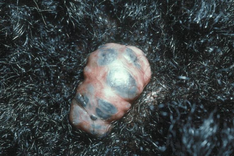
This clinical photograph displays a 2.5 x 1.5 cm pedunculated, exophytic nodule located on the scalp, surrounded by dense, dark, curly hair. The lesion exhibits a multi-lobulated, irregular morphology with a complex pigmentary pattern. The base of the nodule is predominantly flesh-toned to erythematous, interspersed with distinct, irregular macules of dark brown, slate-gray, and bluish pigmentation. The surface appears smooth but uneven due to the internal lobulations and variable pigment distribution. Clinically, this presentation is characteristic of a pigmented basal cell carcinoma, which may arise within a pre-existing lesion such as a nevus sebaceous. The image serves as an educational example of dermatologic oncology, highlighting the morphological diversity of basal cell carcinoma in patients with darker skin phototypes and the importance of evaluating scalp nodules that exhibit changes in size, texture, or pigmentation.
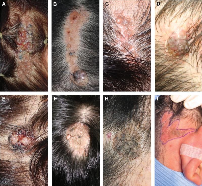
This composite clinical photograph displays a series of scalp lesions illustrating the malignant transformation of Nevus Sebaceous (NS) into Basal Cell Carcinoma (BCC). The images demonstrate varied morphological presentations: (A) and (B) show linear, brownish, and yellowish alopecia plaques with superimposed blackish pigmented nodules and focal ulcerations. (C) and (F) illustrate classic NS features including verrucous, yellowish-tan plaques with a characteristic cobblestone or mammillated surface. (D) and (E) highlight suspicious morphological changes such as rapid size expansion, protruding masses, and hemorrhagic crusting. (H) shows a densely textured, dark pigmented plaque. (I) provides a surgical perspective with preoperative markings for wide excision of a large, irregular plaque on the temporal scalp. These images emphasize the clinical indicators of malignant degeneration within a pre-existing congenital lesion, specifically targeting the transition from benign epidermal/sebaceous hyperplasia to invasive basaloid cell nests. The collection serves as an educational reference for dermatologic oncology and surgical planning for scalp adnexal tumors.
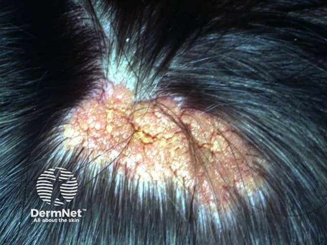
Sebaceous nevus on the scalp is a congenital cutaneous hamartoma characterized by an orange-yellow plaque with thickened, velvety surface and coarse, smooth to pebbly texture. In this close‑up clinical photography, the lesion is localized to the hair-bearing scalp, extending across several square centimeters. The overlying epidermis appears hyperkeratotic with a mosaic of pedunculated, papillomatous projections and mottled coloration ranging from pale yellow to tan to orange. Hair density around the lesion is variably reduced, and surrounding skin shows mild erythema consistent with chronic irritation. The lesion corresponds anatomically to ectopic sebaceous glands with diminished follicular development, commonly arising on the scalp, face, or neck. Histologically, nevus sebaceus features sebaceous gland hyperplasia with immature hair follicles and sebaceous differentiation embedded in a fibrous stroma. Clinically, this entity is part of the spectrum of adnexal hamartomas and is usually present at birth or early childhood, enlarging in early life and stabilizing thereafter. The significance lies in recognition of potential secondary neoplasms within nevus sebaceus, notably syringocystadenoma papilliferum or basal cell carcinoma, though malignant transformation is uncommon. Management often involves observation for changes or excision if cosmetically disfiguring or for risk of neoplasm, especially in adulthood. The image supports educational discussion of clinical appearance, differential diagnoses, and excision indications.
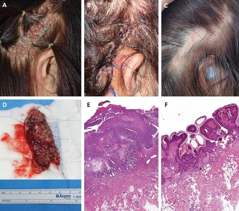
A multi-panel medical figure documenting the clinical presentation, surgical management, and histopathology of basal cell carcinoma (BCC) arising from nevus sebaceous (NS). (A) Clinical photograph of the right temporal scalp showing a linear, brownish, verrucous plaque (NS) with focal blackish nodules and superficial ulceration, suggestive of malignant transformation. (B) Immediate postoperative view showing a local flap reconstruction with blue nylon sutures covering the excision site. (C) Long-term follow-up showing a stable, well-healed surgical scar with appropriate hair coverage. (D) Gross surgical specimen on gauze, measuring approximately 8 cm in length, showing the excised cutaneous lesion. (E) Hematoxylin and eosin (H&E) stained micrograph at low magnification demonstrating BCC characterized by basaloid epithelial cell nests with distinct peripheral palisading and stromal retraction. (F) H&E stained micrograph of the background NS lesion, highlighting epidermal papillomatosis, verrucous hyperplasia, and prominent sebaceous glands. This sequence illustrates the clinical progression and diagnostic features of secondary malignancy within a congenital sebaceous nevus.
multiple fungating tumor masses scalp ulcerated verrucous squamous cell carcinoma
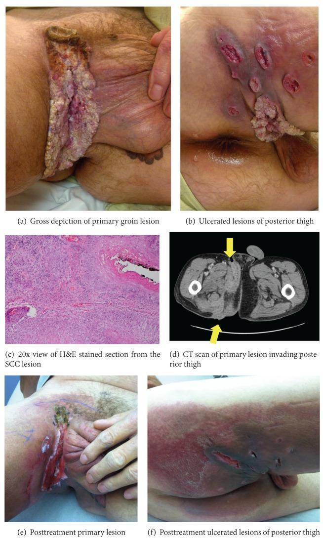
This composite educational graphic illustrates the clinical presentation, histopathology, and imaging of Squamous Cell Carcinoma (SCC) with verrucous features in the groin and thigh. Clinical photographs (a, b) show a large, fungating, exophytic growth in the right inguinal region extending to the posterior thigh, characterized by irregular borders, verrucous texture, and multiple satellite ulcerated lesions. A 20x magnification H&E stained photomicrograph (c) demonstrates superficially invasive malignant keratinocytes consistent with SCC. An axial CT scan of the pelvis (d) highlights an enhancing soft tissue density (arrows) invading the right quadriceps muscle and gluteal folds. Post-treatment photographs (e, f) document the disease progression following chemoradiotherapy, showing a reduction in the primary tumor mass, residual ulceration, and dark necrotic changes at the edges of the posterior thigh lesions. This comparison serves as a clinical timeline for monitoring therapeutic response in advanced cutaneous malignancy.
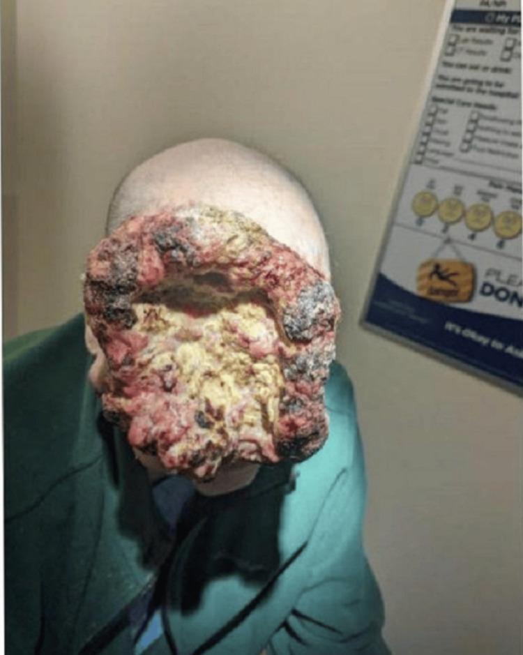
This clinical photograph displays a massive, locally advanced malignant tumor, identified contextually as squamous cell carcinoma, involving the right facial and frontal scalp regions. The lesion is characterized by prominently raised, exophytic, 'cauliflower-like' margins with a highly irregular and verrucous texture. The center of the mass is deeply depressed and exhibits extensive yellow-white slough and purulent-appearing material consistent with significant tissue necrosis. Throughout the lesion, there are variegated colors including erythematous pink areas of friable tissue and dark, blackened crusting or scabbing indicative of chronic capillary bleeding and hemorrhage. The mass is large enough to cause complete mechanical obstruction of the right ocular region and severe periorbital edema. This image serves as a significant example of dermatologic oncology, illustrating disease progression in a patient with limited access to care and the clinical presentation of a fungating facial malignancy with intracranial extension.
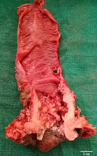
Macroscopic photograph of an anal canal/perianal surgical specimen illustrating an ulcerating, fungating squamous cell carcinoma. The lesion is distal to the dentate line, with a friable, red-tan mass protruding from the anal verge and invading surrounding perianal skin. The gross appearance is polypoid to verrucous in architecture, with irregular, necrotic surfaces and visible areas of tissue destruction at the margin. Early stages may be small, mobile, and exophytic; with progression, the tumor can extend proximally into the rectum, where it may resemble primary rectal adenocarcinoma on a gross exam. In distal growth, the tumor breaches the anal skin, producing ulceration and secondary inflammatory change that can mimic benign inflammatory processes. The current specimen demonstrates advanced disease with a conspicuous, ulcerated surface and anastomotic or transmural extension toward nearby tissues. Basaloid variants, if present, characteristically arise proximal to the dentate line and may be difficult to detect clinically on external examination. Case history provided by pathology and surgical teams contextualizes the image within a typical clinical presentation of anorectal malignancy. Correlation with histology and immunohistochemistry remains essential for definitive classification and therapeutic planning. This image supports education on gross pathology, differential diagnosis, and surgical oncology considerations. Educational utility for clinical trainees.
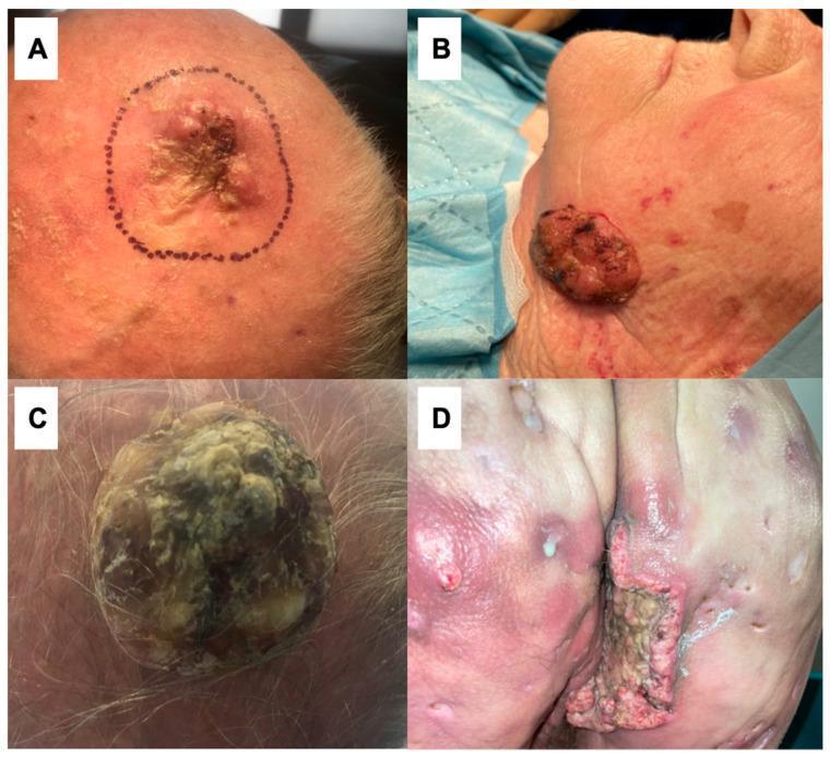
This composite of four clinical photographs (A-D) demonstrates various morphological presentations of giant cutaneous squamous cell carcinoma (cSCC), a malignant keratinocytic neoplasm. (A) Shows a large, irregular, ulcerated plaque on the scalp, marked with a surgical margin, featuring central crusting and perilesional erythema. (B) Displays a prominent, exophytic, reddish-brown nodular mass on the left cheek with a friable, glistening surface and associated actinic damage on surrounding skin. (C) Illustrates a massive, dome-shaped verrucous tumor on the scalp with a hyperkeratotic, multi-lobulated surface containing yellowish-tan and gray keratinous debris. (D) Presents an extensive, deep ulcerated cSCC on the buttock occurring in the setting of chronic hidradenitis suppurativa (HS). This lesion shows irregular, everted borders and a necrotic base, surrounded by inflamed tissue with scarring, sinus tracts, and pustules characteristic of Hurley stage III HS. These cases highlight the clinical diversity of cSCC, its association with chronic inflammatory conditions, and the potential for significant local tissue destruction in advanced stages.
Differential Diagnosis
Clinical Summary of Findings
- Dozens of exophytic, lobulated, dome-shaped nodules across the entire scalp (vertex, parietal, occipital)
- Massive confluent fungating/cauliflower-like tumor on the posterior neck and right shoulder (>15 cm)
- Erythematous, pink-red to violaceous color with ulceration and hemorrhagic crusting
- Background diffuse hyperpigmentation and lichenification of adjacent skin
- Alopecia over and around lesions
Differential Diagnoses (in order of probability)
1. Malignant Transformation in Nevus Sebaceous (Most Likely)
- The multifocal scalp distribution with discrete smaller nodules over a background of hyperpigmented/lichenified skin, combined with one dominant massively growing tumor, fits this pattern well
- The background skin changes (hyperpigmentation, scaling) may represent the underlying sebaceous nevus
2. Advanced Cutaneous Squamous Cell Carcinoma (SCC) - Multiple Primary or Metastatic
- SCC can present as multiple exophytic, fungating, verrucous, cauliflower-like masses on the scalp, especially in photodamaged or immunocompromised individuals
- The massive ulcerated neck mass with satellite scalp nodules would be consistent with locally advanced or metastatic SCC
- In the Indian subcontinent, SCC arising from chronic wounds, burn scars, or chronic inflammatory conditions (Marjolin's ulcer) can present this aggressively
3. Scalp Angiosarcoma
- Angiosarcoma of the scalp classically presents in elderly patients as multifocal violaceous, hemorrhagic, ulcerated nodules and plaques on the scalp/forehead
- The violaceous-red color, multifocal nature, bleeding, and scalp distribution are consistent
- Can grow to massive sizes rapidly
- More common in elderly males, but can rarely occur in younger patients
- Key distinguishing feature: lesions often start as flat bruise-like patches that later become nodular
4. Cutaneous Metastases to the Scalp (Alopecia Neoplastica)
- Scalp is a common site for metastatic deposits from internal malignancies (lung, renal cell, thyroid, colon)
- Can present as multiple firm, skin-colored to erythematous nodules with alopecia
- The firmness, multifocality, and alopecia fit
- However, the degree of exophytic, fungating growth would be atypical for pure metastases without a very large primary
5. Pilomatrixoma / Trichilemmal Carcinoma (Multiple / Malignant)
- Multiple pilomatrixomas occur in rare conditions (myotonic dystrophy, Gardner syndrome, Rubinstein-Taybi syndrome)
- Malignant trichilemmal carcinoma can present as large, ulcerated scalp masses
- Less likely given the sheer number and size of lesions
6. Lymphoma - Primary Cutaneous B-cell or T-cell Lymphoma
- Scalp is a favored site for primary cutaneous follicle center lymphoma (PCFCL) and other cutaneous lymphomas
- Can present as multiple pink-red nodules/tumors
- Less likely to be this fungating, but should be excluded with biopsy
7. Proliferating Trichilemmal Cyst (Trichilemmal Carcinoma)
- Arises from the outer root sheath, almost exclusively on the scalp
- Can start as a cyst and undergo malignant transformation into a large, ulcerated, exophytic tumor
- The solitary large neck/shoulder mass could represent malignant transformation, but the multifocality is unusual
8. Epidermodysplasia Verruciformis (EV) with Malignant Transformation
- A rare genodermatosis with abnormal susceptibility to HPV, causing widespread flat warts and SCC
- Less common to present as this degree of fungating tumor, but if there is immunodeficiency (HIV, transplant), EV-associated SCC can be aggressive and multifocal
Key Recommended Next Steps
| Investigation | Purpose |
|---|---|
| Punch/incisional biopsy (from largest mass + a smaller scalp nodule) | Histopathology - gold standard |
| Immunohistochemistry | Differentiate SCC, BCC, angiosarcoma (CD31, ERG), lymphoma (CD20, CD3) |
| CT head, neck, chest, abdomen | Assess depth of invasion, lymphadenopathy, distant metastases |
| HIV serology | Immunosuppression predisposes to aggressive cutaneous malignancy |
| Dermoscopy | If any lesions accessible without biopsy |