

3d086dfb-3f0a-49b1-86bb-831bc1d433aa.jpg
Answer acc to pathology
analyse_media
Reading File
Finding Sources
Finding Sources
Finding Sources
Finding Sources
Finding Sources
Reading File
Reading File
Reading File
Finding Sources
Finding Sources
Finding Sources
Finding Sources
Reading File
Reading File
Reading File
Reading File
Reading File
Reading File
Reading File
Reading File
Reading File
I now have all the content needed. Let me compile a comprehensive answer from Robbins & Kumar Basic Pathology.
Pathology Answers
Q1. Difference between Leiomyoma & Leiomyosarcoma
(Robbins & Kumar Basic Pathology)
| Feature | Leiomyoma (Benign) | Leiomyosarcoma (Malignant) |
|---|---|---|
| Nature | Benign smooth muscle tumor | Malignant smooth muscle tumor |
| Common site | Uterus (fibroids), skin (pilar leiomyoma), GI tract | Deep soft tissues of extremities, retroperitoneum, uterus, great vessels (IVC) |
| Age/Sex | Common in reproductive age women | Primarily older adults; more common in women |
| Size | Usually 1-2 cm | Can grow very large (retroperitoneal) |
| Gross | Well-circumscribed, firm mass | Painless, firm mass; retroperitoneal tumors may be huge |
| Microscopy | Fascicles of densely eosinophilic spindle cells with minimal atypia and extremely rare mitotic figures | Interweaving fascicles of eosinophilic spindle cells to sheets of pleomorphic cells; mitotic activity and necrosis are common |
| Nuclear atypia | Absent / minimal | Present (pleomorphic, hyperchromatic nuclei) |
| Mitoses | Extremely rare | Frequent (key criterion) |
| Necrosis | Absent | Common |
| Karyotype | Simple / normal | Complex karyotype with multiple chromosomal gains and losses (genomic instability) |
| Genetics | Germline FH (fumarate hydratase) mutations in hereditary multiple leiomyoma syndrome | Complex karyotypic abnormalities; underlying genomic instability |
| IHC | Smooth muscle actin (+) | Smooth muscle proteins (+) on IHC (aids diagnosis) |
| Prognosis | Cured surgically (solitary lesions) | Superficial: good prognosis. Retroperitoneal: difficult to control; metastasizes to lungs; often fatal |
| Metastasis | Never | Yes - especially to lungs |
Key distinguishing criteria (pathological): number of mitoses per HPF, degree of nuclear atypia, and presence of necrosis.
Q2. Short Note on Retinoblastoma
(Robbins & Kumar Basic Pathology)
Definition: The most common primary intraocular malignancy of children. A "small round blue cell" tumor of the retina.
Genetics (Two-hit hypothesis - Knudson)
- Hereditary (40%): Germline mutation in the RB tumor suppressor gene (chromosome 13q14). Typically bilateral and multifocal. Autosomal dominant pattern of inheritance. Also increased risk of osteosarcoma and other soft tissue tumors.
- Sporadic (60%): Somatic mutations in RB gene. Typically unilateral and unifocal.
- Both alleles of RB must be inactivated ("two hits") for tumor to develop.
Morphology
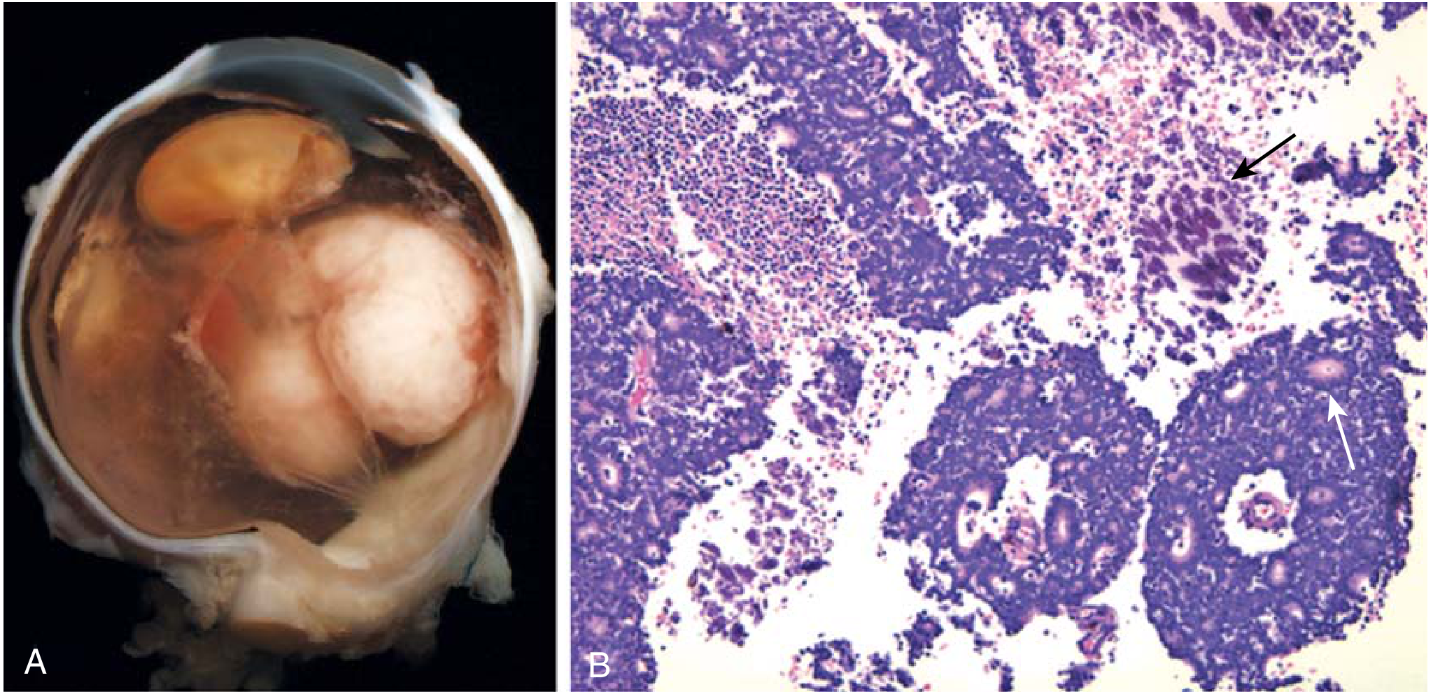
- Gross: Nodular masses, usually in the posterior retina, sometimes with satellite seedlings.
- Microscopy:
- Undifferentiated elements: small round cells with hyperchromatic nuclei (resembling retinoblasts) - "small round blue cells"
- Differentiated elements: Flexner-Wintersteiner rosettes (single layer of cuboidal/columnar cells around a central lumen - reflecting photoreceptor differentiation) and fleurettes
- Viable tumor cells cluster around blood vessels; necrosis in avascular areas
- Focal dystrophic calcification is characteristic
Spread
- To brain, skull, bone marrow
- Seldom to lungs (unlike most sarcomas)
- Prognosis worsened by: extraocular extension, invasion along optic nerve, choroidal invasion
Treatment
- Chemoreduction (including selective intra-ophthalmic artery delivery) followed by laser/cryopexy
- Goal: preserve vision + eradicate tumor
Q3. Short Note on Rhabdomyosarcoma (RMS)
(Robbins & Kumar Basic Pathology)
Definition: A malignant mesenchymal tumor with skeletal muscle differentiation. The most common soft tissue sarcoma of childhood and adolescence.
Subtypes (4)
| Subtype | Frequency | Age | Site | Morphology | Genetics | Prognosis |
|---|---|---|---|---|---|---|
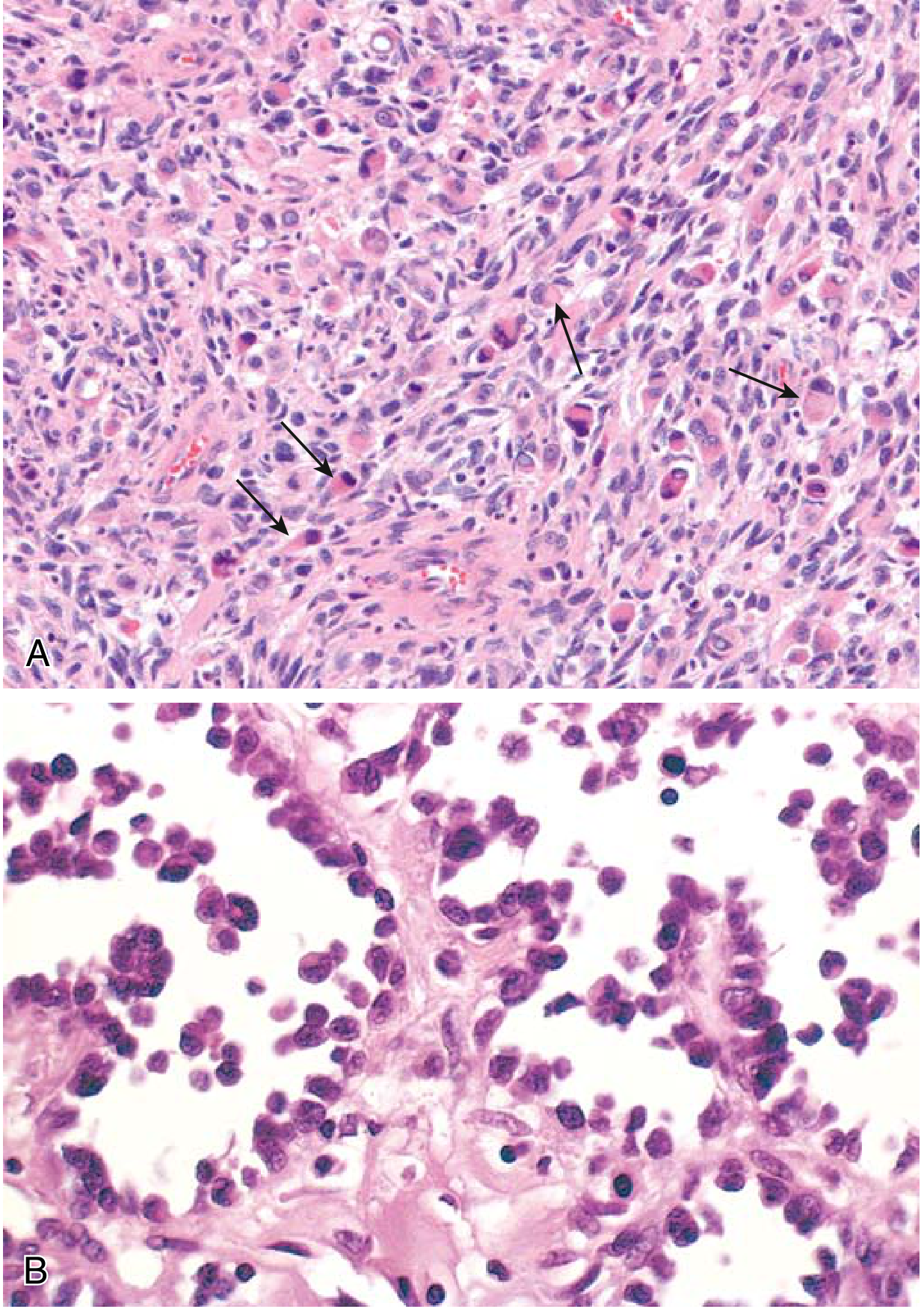
| Embryonal | 50% | 1-5 years | Head/neck, genitourinary | Primitive round and spindle cells in myxoid stroma; strap cells with cross-striations ("rhabdomyoblasts") | LOH 11p15 | Better |
| Alveolar | 20% | 5-15 years | Extremities | Round cells divided by fibrous septa into alveolar-like spaces | t(2;13) → PAX3-FOXO1 fusion; t(1;13) → PAX7-FOXO1 | Worse |
| Pleomorphic | 20% | Adults | Deep extremities | Large, bizarre, multinucleate eosinophilic cells | Complex karyotype | Often fatal |
| Spindle cell/Sclerosing | 10% | All ages | Variable | Fusiform cells in fascicles/storiform pattern; dense sclerotic stroma | - | Variable |
Key Points
- Sarcoma botryoides: grape-like variant of embryonal RMS; develops in hollow viscera (urinary bladder, vagina) - best prognosis
- Arises from undifferentiated mesenchymal stem cells (not from pre-existing skeletal muscle)
- IHC: positive for myogenin, MyoD1, desmin, muscle-specific actin
- Head and neck is the second most common site (orbit is most common within H&N)
- Treatment: surgery + chemotherapy ± radiation; COG protocols
Q4. Classification of Soft Tissue Tumours
(Robbins & Kumar Basic Pathology, Table 19.3)
By convention, soft tissue = nonepithelial tissue exclusive of skeleton, joints, CNS, and hematopoietic/lymphoid tissues. Benign soft tissue tumors are 100 times more frequent than sarcomas.
Classification by Tissue of Origin
| Category | Benign | Malignant |
|---|---|---|
| Adipose tissue | Lipoma | Liposarcoma (well-differentiated, myxoid, pleomorphic) |
| Fibrous / myofibroblastic | Nodular fasciitis, Deep fibromatosis (desmoid) | Fibrosarcoma |
| Skeletal muscle | Rhabdomyoma | Rhabdomyosarcoma (embryonal, alveolar, pleomorphic, spindle cell/sclerosing) |
| Smooth muscle | Leiomyoma | Leiomyosarcoma |
| Vascular | Hemangioma | Angiosarcoma, Kaposi sarcoma |
| Peripheral nerve | Schwannoma, Neurofibroma | Malignant peripheral nerve sheath tumor (MPNST) |
| Cartilage | - | Extraskeletal chondrosarcoma |
| Bone | - | Extraskeletal osteosarcoma |
| Uncertain origin | - | Synovial sarcoma, Undifferentiated pleomorphic sarcoma (UPS), Ewing sarcoma |
| Uncertain/other | Tenosynovial giant cell tumor | - |
Classification by Karyotypic Pattern
- Simple karyotype (15-20%): Single or few chromosomal changes (e.g., Ewing sarcoma, synovial sarcoma). Young patients, monomorphic appearance.
- Complex karyotype (80-85%): Aneuploid/polyploid with multiple gains and losses (e.g., leiomyosarcoma, undifferentiated pleomorphic sarcoma). Older adults, pleomorphic cells.
Classification by Clinical Behavior
- Benign - self-limited, minimal treatment needed
- Intermediate grade - locally aggressive, minimal metastatic risk
- Malignant (sarcomas) - high risk of metastasis and mortality; cause 2% of cancer deaths despite being <1% of all cancers
Sources: Robbins & Kumar Basic Pathology, Cummings Otolaryngology
This is a shared conversation. Sign in to Orris to start your own chat.