Shaft of bone
Reading File
Finding Sources
Finding Sources
Reading File
Finding Sources
Reading File
Reading File
Loading Image
Shaft of a Bone (Diaphysis)
The shaft of a bone is anatomically called the diaphysis (Greek: "growing between"). It is the elongated, cylindrical middle portion of a long bone, situated between the two expanded ends.
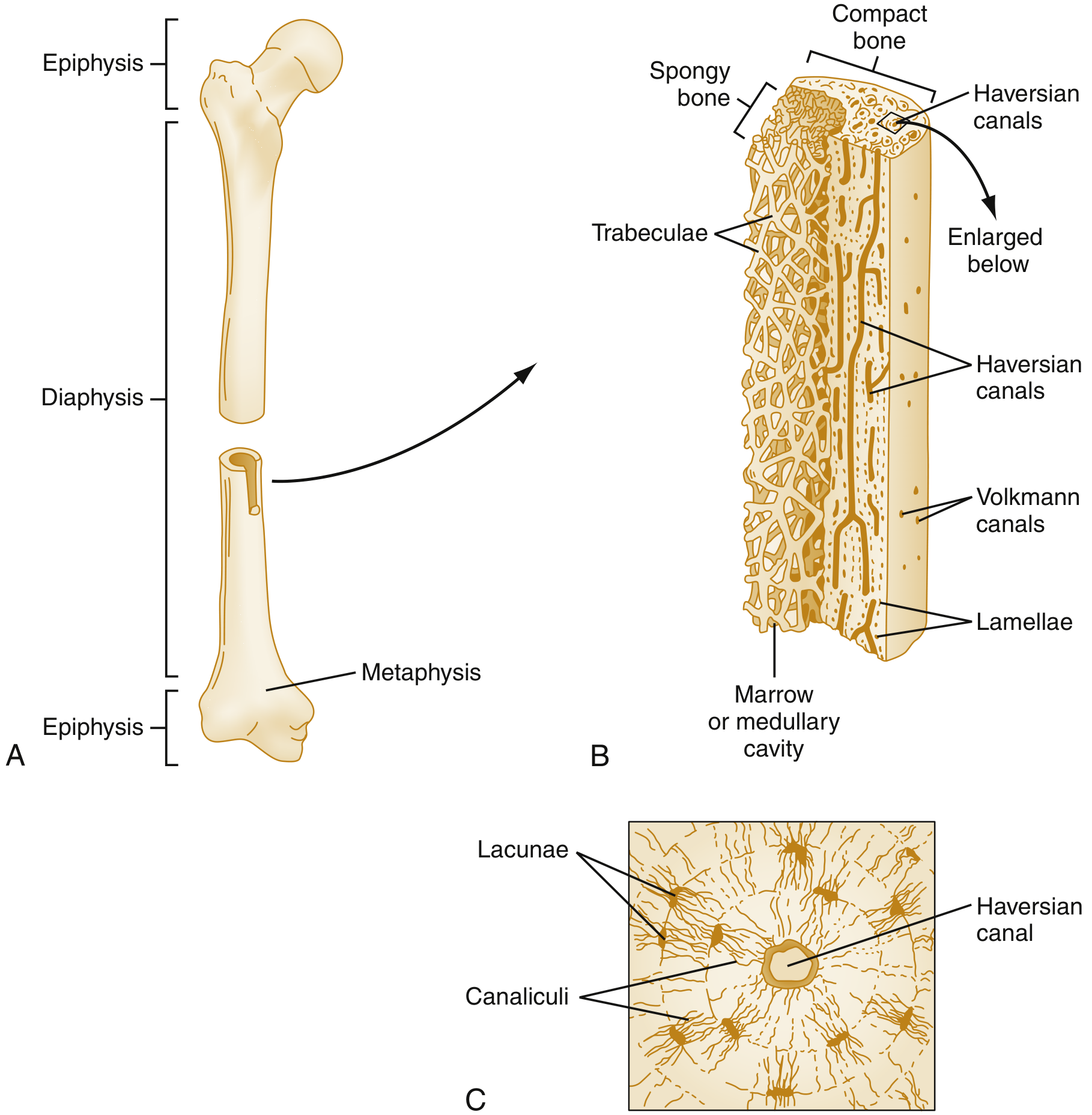
Gross Structure
The diaphysis is composed of three concentric layers:
| Layer | Description |
|---|---|
| Periosteum | Fibrous outer covering with two layers: outer fibrous layer and inner osteogenic (cambium) layer containing osteoblasts. Attached to bone by Sharpey's fibers. |
| Compact (cortical) bone | Dense, solid bone forming the wall of the shaft - provides strength and rigidity. Thicker here than anywhere else in the bone. |
| Medullary (marrow) cavity | Central hollow space lined by the endosteum; contains yellow (fatty) marrow in adults. |
"The diaphysis is the shaft of the bone and contains the compact cortical bone with an overlying periosteum and a medullary canal containing marrow." - Rosen's Emergency Medicine
Microscopic Structure
Compact bone of the diaphysis is organized as lamellar bone with:
- Osteons (Haversian systems) - long cylinders running parallel to the long axis of the diaphysis; each consists of 5-20 concentric lamellae surrounding a central Haversian canal that houses blood vessels and nerves.
- Haversian canals - longitudinal channels containing microvascular supply.
- Volkmann's (perforating) canals - transverse canals connecting adjacent Haversian canals, the marrow cavity, and the periosteum. They cross lamellae and lack concentric organization.
- Interstitial lamellae - remnants of old, partially resorbed osteons; wedged between intact osteons.
- Circumferential lamellae - outer lamellae immediately beneath the periosteum, and inner lamellae around the marrow cavity; these enclose and strengthen the region between them.
- Lacunae and canaliculi - small spaces housing osteocytes, with thin tunnels (canaliculi) connecting them for nutrient/waste exchange.
Relations of the Diaphysis
- Proximally and distally it transitions into the metaphysis - the junctional, flared region with abundant trabecular bone and a thinning cortex.
- Beyond that lies the epiphysis at each end - a cap of trabecular bone covered by articular cartilage (in the mature skeleton).
Clinical Relevance
- The periosteum of the diaphysis is well-vascularized and contains osteoprogenitor cells critical for fracture healing. In adults it is firmly attached to underlying bone, which limits subperiosteal abscess formation in osteomyelitis.
- In hematogenous osteomyelitis, infection most commonly seeds the metaphysis (not the diaphysis itself) due to slow sinusoidal blood flow there; it can then spread laterally through Volkmann canals to lift the periosteum.
- Compact bone is 5-10% turned over annually via continuous remodeling - osteoclasts tunnel through old osteons, and osteoblasts deposit new concentric lamellae to form new osteons.
Sources: Rosen's Emergency Medicine, p. 2720 | Junqueira's Basic Histology, 17e, p. 371-372
This is a shared conversation. Sign in to Orris to start your own chat.