I now have enough information to provide a thorough clinical analysis. Let me compile the complete response.
Clinical Considerations: Right-Sided Back Pain Above the Hip in a 50-Year-Old Woman
This is a clinically important case that needs careful evaluation. The combination of right-sided pain above the hip (loin/flank region), chronicity, aggravation by activity, and local warmth points to several distinct diagnostic categories.
Anatomical Location - What's in That Region?
The area above the hip on the right side, in the back, corresponds to the right loin / right costovertebral angle (CVA) / right lumbar region. Structures here include:
- Right kidney and ureter
- Lumbar spine (L1-L5), facet joints, paraspinal muscles
- Sacroiliac (SI) joint (if slightly lower)
- Right psoas muscle
- Retroperitoneal structures (aorta, lymph nodes)
- Ascending colon / hepatic flexure (referred pain)
- Thoracolumbar fascia and erector spinae
Priority Differential Diagnosis
1. Renal/Urological Causes (Must Rule Out First)
| Condition | Key Features in This Case |
|---|
| Chronic Pyelonephritis | Chronic flank pain, fever episodes, CVA tenderness, local warmth due to ongoing infection/inflammation |
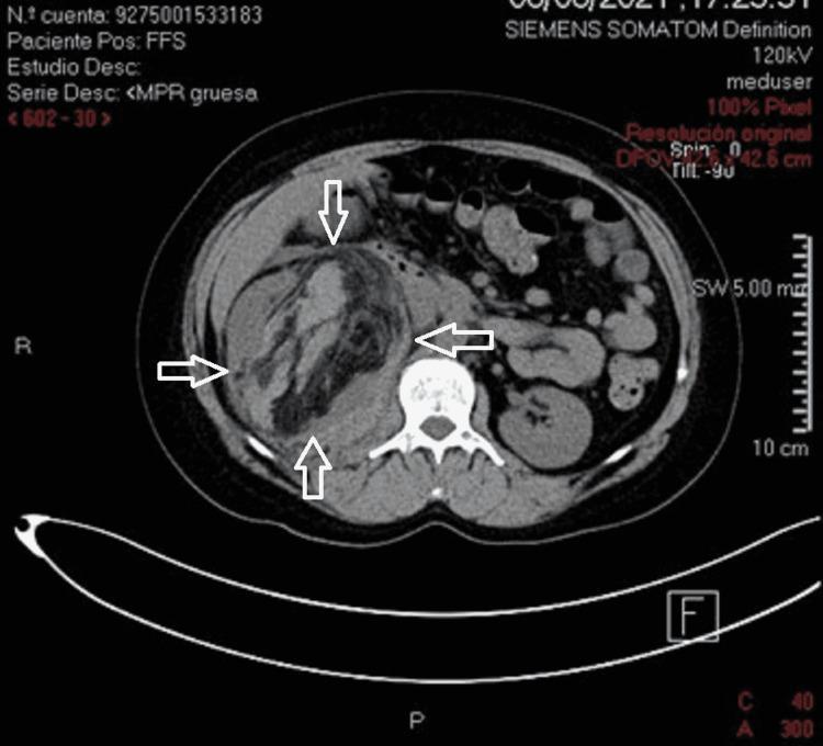
| Xanthogranulomatous Pyelonephritis (XGP) | Classic presentation - middle-aged females, chronic flank pain, fever, malaise, palpable mass; associated with obstruction and infection |
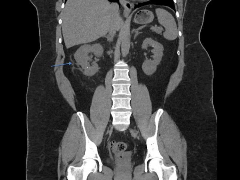
| Nephrolithiasis (Staghorn calculi) | Can cause chronic right loin pain, especially if partial obstruction; pain worse with activity |
| Hydronephrosis / UPJ obstruction | Chronic dull flank pain aggravated by increased fluid intake or activity |
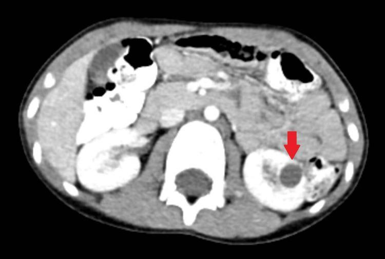
| Renal cyst / Polycystic kidney disease | Can cause chronic dull aching flank pain |
| Perinephric abscess | Flank pain + local heat - important to exclude in chronic cases |
From Comprehensive Clinical Nephrology, 7th Ed: "Patients with xanthogranulomatous pyelonephritis are characteristically middle-aged females and have chronic symptoms such as flank pain, fever, chills, and malaise. Flank tenderness, a palpable mass, and irritative voiding symptoms are common."
The local temperature increase is a very significant clue here - it suggests active inflammation or infection in the local tissue, raising the suspicion for renal/perirenal infection or inflammatory musculoskeletal pathology.
2. Musculoskeletal Causes
| Condition | Key Features |
|---|
| Lumbar facet joint arthropathy / OA | Very common at 50 years; right-sided, worse with activity/standing; tenderness at paraspinal area |
| Lumbar muscle strain (chronic) | Chronic paraspinal muscle pain, worsens with exertion |
| Sacroiliitis | Pain at/above the sacroiliac area; can be unilateral |
| Lumbar disc disease / Radiculopathy | Focal back pain with possible radiation to leg |
| Vertebral osteomyelitis / Discitis | Chronic back pain + local warmth + tenderness = do NOT miss; may be insidious |
| Myofascial pain syndrome | Trigger points in right quadratus lumborum, iliocostalis |
From Symptom to Diagnosis, 4th Ed: "The primary task when evaluating patients with low back pain is to identify those who have a serious cause of back pain that requires specific, and sometimes rapid, diagnosis."
3. Inflammatory / Systemic Causes
| Condition | Key Features |
|---|
| Axial spondyloarthritis / Ankylosing spondylitis | Though typically younger onset (<45 yrs) and bilateral, unilateral presentations occur; morning stiffness, improvement with exercise |
| Psoriatic arthropathy | Unilateral sacroiliitis possible |
| Reactive arthritis | Preceding infection history |
| Spinal metastasis / Myeloma | Age >50, pain not relieved by rest, nocturnal pain - RED FLAG |
4. Visceral Referred Pain
| Condition | Key Features |
|---|
| Cholecystitis / Gallstones | Right-sided, referred to back, postprandial aggravation |
| Hepatic pathology (abscess, cyst) | Right upper quadrant/back pain |
| Ascending colon pathology | Right-sided colonic mass, diverticulitis |
| Retroperitoneal lymphadenopathy | Chronic dull back pain |
Red Flags to Screen for Urgently (per Symptom to Diagnosis, 4th Ed, Table 7-1)
Given her age is over 50, ALL of the following must be actively sought:
- Unexplained weight loss - malignancy
- Nocturnal pain waking from sleep - malignancy, infection
- Fever - infection (osteomyelitis, pyelonephritis, abscess)
- Urinary symptoms (dysuria, frequency, hematuria) - renal/urological cause
- Bowel/bladder dysfunction - cauda equina syndrome
- History of cancer - spinal metastasis
- Prolonged corticosteroid use - compression fracture
Significance of LOCAL TEMPERATURE INCREASE
This is a key clinical clue that narrows the diagnosis significantly:
- Superficial warmth over the right loin/back suggests active local inflammation or infection
- Most important considerations when warmth is present:
- Perinephric/renal abscess - urgent
- Xanthogranulomatous pyelonephritis - chronic infection
- Vertebral osteomyelitis or paraspinal abscess
- Inflammatory arthritis of the right SI joint / facet joint
- Deep muscle inflammation/myositis
- Soft tissue cellulitis overlying the area (less likely if longstanding)
Recommended Evaluation
History (Detailed)
- Duration, character (dull/sharp/colicky), radiation
- Urinary symptoms: hematuria, dysuria, frequency, foul-smelling urine
- Bowel habits
- Episodes of fever, chills, sweating
- Menstrual history (menopause status) - osteoporosis risk
- Weight loss
- Morning stiffness
- Family history of kidney disease, cancer, inflammatory arthritis
- Activity-related worsening vs. rest - if worsens with work, suggests mechanical component
Physical Examination
- CVA (Costovertebral angle) tenderness - percuss the right flank firmly; positive in renal pathology
- Assess local warmth - is it superficial (skin) or deep?
- Spinal palpation - point tenderness on spinous processes suggests bone pathology
- Paraspinal muscle tenderness
- SLR (Straight Leg Raise) - for nerve root compression
- Abdominal exam - organomegaly, mass
- Gait and posture assessment
Investigations - First Line
| Test | Purpose |
|---|
| Urine routine & microscopy | Hematuria, pyuria, casts - renal/urological cause |
| Urine culture & sensitivity | Identify infecting organism |
| CBC | Leukocytosis (infection), anemia (malignancy, chronic disease) |
| CRP / ESR | Inflammatory/infective marker - important given local warmth |
| Renal function (creatinine, BUN) | Assess kidney function |
| Serum calcium, phosphate | Nephrolithiasis, malignancy |
| X-ray lumbar spine (AP & lateral) | Disc space loss, vertebral fractures, osteophytes, sacroiliitis |
| Ultrasound abdomen & kidneys | Hydronephrosis, calculi, cysts, masses, abscess |
Investigations - Second Line (based on findings)
| Test | When |
|---|
| CT abdomen/pelvis (with contrast) | Suspected renal mass, abscess, XGP, retroperitoneal pathology |
| NCCT (non-contrast CT) | Gold standard for renal calculi |
| MRI lumbar spine | Suspected disc disease, osteomyelitis, malignancy, nerve compression |
| Bone scan / PET scan | Suspected metastasis |
| HLA-B27 | Suspected ankylosing spondylitis |
| ANA, RF, Anti-CCP | Inflammatory arthritis workup |
| PSA / tumor markers | If malignancy suspected |
Summary of Most Likely Diagnoses (Ranked for This Patient)
| Priority | Diagnosis | Why |
|---|
| Most likely | Chronic musculoskeletal pain (lumbar facet/paraspinal) | Chronic, activity-related, common at 50 |
| Must not miss | Renal/perirenal infection (XGP, abscess, chronic pyelonephritis) | Local warmth + chronic + female + middle-age |
| Must not miss | Vertebral osteomyelitis / spinal infection | Local warmth + chronicity |
| Must not miss | Spinal malignancy / metastasis | Age >50 = red flag |
| Consider | Nephrolithiasis with partial obstruction | Chronic flank pain |
| Consider | Sacroiliitis / inflammatory arthritis | Unilateral pain |
| Consider | Visceral referred pain (gallbladder, colon) | Right-sided location |
Important Note on Perimenopause at Age 50
At 50 years, she is likely perimenopausal or recently postmenopausal. This adds additional considerations:
- Osteoporosis - increased fracture risk
- Osteoporotic vertebral compression fracture - can present as chronic low back pain
- Baseline DEXA scan should be considered
- Hormonal changes can also increase susceptibility to urinary tract infections
The combination of chronicity + activity aggravation + local warmth in a 50-year-old woman makes this a case where both musculoskeletal AND renal/infective etiologies must be actively investigated simultaneously. Do not assume it is purely mechanical until infection and serious pathology have been excluded with basic investigations. Starting with urine analysis, CBC, CRP/ESR, and a renal ultrasound alongside a lumbar X-ray would be the most practical initial workup.
Sources: Symptom to Diagnosis: An Evidence Based Guide, 4th Ed; Comprehensive Clinical Nephrology, 7th Ed; Goldman-Cecil Medicine, International Edition; Bradley and Daroff's Neurology in Clinical Practice