
Blood (formation, RBC indices) explanation in simple way
Reading File
Now let me search the library for blood formation and RBC indices content in parallel.
Finding Sources
Finding Sources
Finding Sources
Reading File
Reading File
Finding Sources
Reading File
Reading File
Reading File
Reading File
Now let me get the hemopoiesis (blood cell formation) content from the histology textbook:
Reading File
Reading File
Reading File
I now have all the information I need to give a thorough, simple explanation. Let me also fetch the hemopoiesis diagram image:
Loading Image
I have everything I need. Here is a clear, simple, well-organized explanation:
Blood: Formation & RBC Indices - Simple Explanation
Part 1 - What Is Blood?
Blood is a fluid connective tissue (yes, it counts as connective tissue!) that flows through the cardiovascular system. An average adult has about 6 litres of it - roughly 7-8% of body weight.
Blood has two main parts:
- Plasma (the liquid - ~55%) - mostly water (91-92%), proteins (albumin, globulins, fibrinogen), electrolytes, glucose, hormones
- Formed elements (the cells - ~45%) - red blood cells (RBCs), white blood cells (WBCs), and platelets
Part 2 - Blood Formation (Hemopoiesis/Hematopoiesis)
Hemopoiesis = the continuous process of making ALL blood cells. Think of it as a factory that never stops, because blood cells have a limited lifespan and must be replaced constantly.
RBCs live ~120 days | Platelets live ~10 days | WBCs live variable, short periods
Where does it happen?
Blood formation starts in the embryo and shifts location over time:
| Phase | Location | When |
|---|---|---|
| 1. Yolk-sac phase | Yolk sac "blood islands" | Week 3 of gestation |
| 2. Hepatic phase | Liver (+ spleen) | 2nd month to birth |
| 3. Bone marrow phase | Red bone marrow | 2nd trimester onward - continues for LIFE |
After birth, the red bone marrow is the sole factory. In adults it's found in flat bones (skull, sternum, ribs, pelvis) and the ends of long bones.
The Family Tree of Blood Cells
All blood cells come from one common ancestor: the Hemopoietic Stem Cell (HSC).
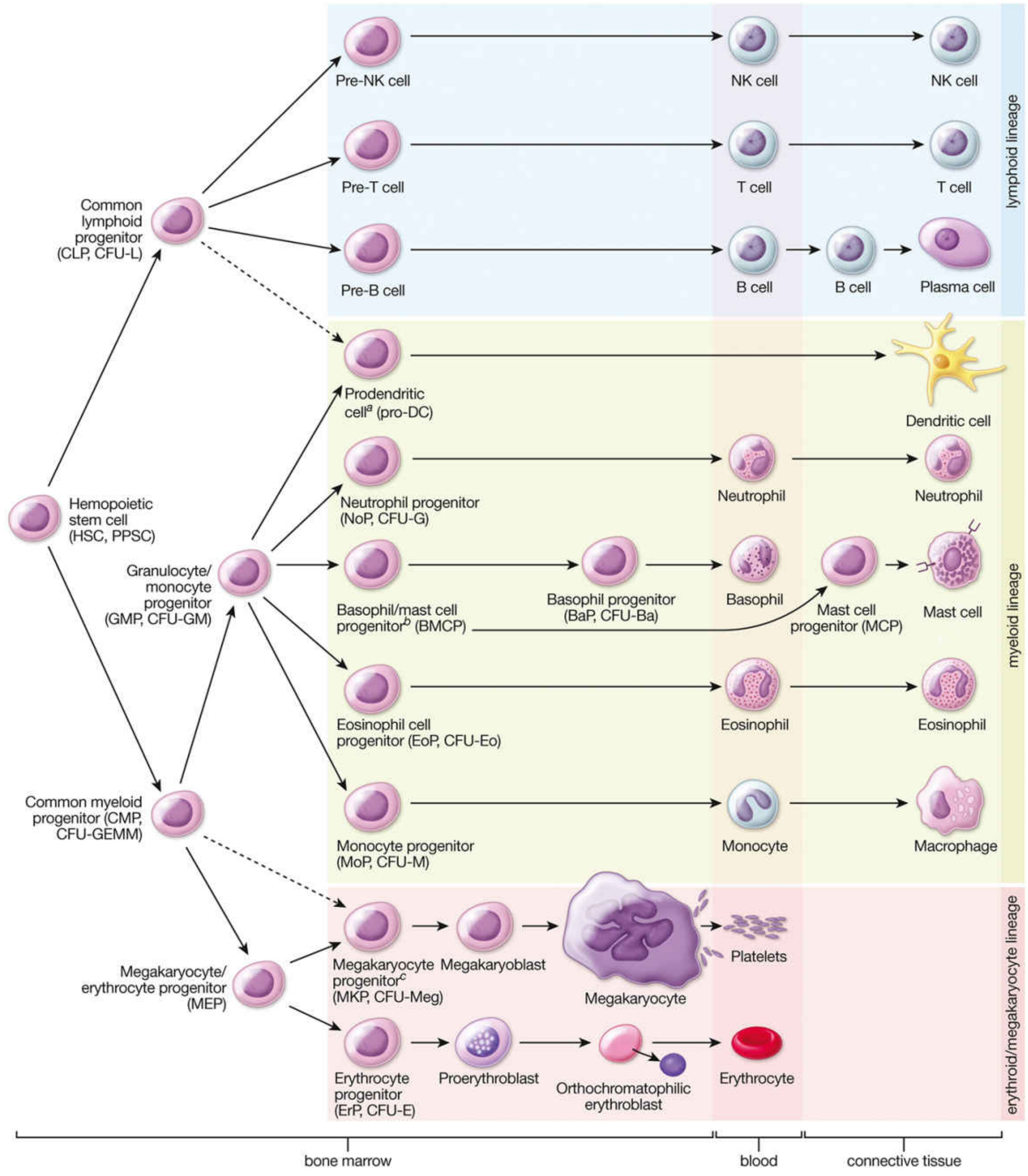
The HSC splits into two major "families":
1. Common Myeloid Progenitor (CMP) → gives rise to:
- RBCs (erythrocytes) via the erythroid line
- Platelets (thrombocytes) via megakaryocytes
- Neutrophils, eosinophils, basophils (granulocytes)
- Monocytes → become macrophages in tissues
2. Common Lymphoid Progenitor (CLP) → gives rise to:
- T cells, B cells (→ plasma cells), NK cells
Part 3 - RBC Formation Specifically (Erythropoiesis)
This is the most clinically important lineage. Here is the maturation sequence:
Hemopoietic Stem Cell
       ↓
Erythrocyte Progenitor (ErP)
       ↓
Proerythroblast  ← large cell, basophilic (blue) cytoplasm, big nucleus
       ↓
Basophilic erythroblast  ← starts making hemoglobin
       ↓
Polychromatophilic erythroblast  ← mixed pink/blue; actively dividing
       ↓
Orthochromatophilic erythroblast (Normoblast)
    ← cytoplasm now pink (full of Hgb), nucleus becomes dense & small
    ← NUCLEUS IS EJECTED here (Howell-Jolly bodies = leftover fragments)
       ↓
Reticulocyte (Polychromatophilic erythrocyte)
    ← no nucleus, still has ribosomes making Hgb
    ← stays in marrow ~24 hrs, then blood ~24 hrs
    ← normally 1-2% of circulating RBCs
       ↓
Mature Erythrocyte (RBC)
    ← biconcave disc, no nucleus, no organelles, full of hemoglobin
Key driver: The hormone erythropoietin (EPO) - made by the kidneys when they sense low oxygen. EPO signals the marrow to produce more RBCs.
Part 4 - RBC Indices (Wintrobe Indices)
These were developed by Maxwell Wintrobe in 1932 and are still used in every CBC (Complete Blood Count) today. They tell you the size and hemoglobin content of your RBCs, which is key to classifying anaemia.
You only need three raw values to calculate them: RBC count, Haemoglobin (Hb), and Haematocrit (HCT).
The Four Main Indices
1. MCV - Mean Corpuscular Volume
"How big is an average RBC?"
Formula: MCV (fL) = 10 × HCT (%) ÷ RBC (millions/µL)
Normal range: ~80-96 fL
| Result | Name | What it means |
|---|---|---|
| Low MCV (<80 fL) | Microcytic | Small RBCs - think iron deficiency, thalassaemia |
| Normal MCV | Normocytic | Normal-sized - e.g. acute blood loss, renal anaemia |
| High MCV (>96 fL) | Macrocytic | Large RBCs - think B12 or folate deficiency |
2. MCH - Mean Corpuscular Haemoglobin
"How much haemoglobin is in an average RBC?"
Formula: MCH (pg) = Hb (g/dL) × 10 ÷ RBC (millions/µL)
Normal range: ~27-33 pg
- Low = Hypochromic (pale RBCs, not enough Hgb)
- Normal = Normochromic
- High = occurs with macrocytic states
3. MCHC - Mean Corpuscular Haemoglobin Concentration
"What is the CONCENTRATION of haemoglobin inside the RBC?"
Formula: MCHC (g/dL) = Hb (g/dL) × 100 ÷ HCT (%)
Normal range: ~32-36 g/dL
- Low = Hypochromic (diluted, pale RBCs)
- Normal = Normochromic
- High = Seen in hereditary spherocytosis (densely packed cells)
4. RDW - Red Cell Distribution Width
"Are all my RBCs a uniform size, or are they different sizes?"
- Normal RDW = uniform sizes (isocytosis)
- High RDW = mix of sizes (anisocytosis) - can help distinguish, e.g., iron deficiency anaemia (high RDW) vs. thalassaemia trait (normal-low RDW), even if both are microcytic
Quick Summary Table
| Index | Measures | Normal | Low means | High means |
|---|---|---|---|---|
| MCV | RBC size (fL) | 80-96 | Microcytic | Macrocytic |
| MCH | Hgb per RBC (pg) | 27-33 | Hypochromic | Macrocytic |
| MCHC | Hgb concentration (g/dL) | 32-36 | Hypochromic | Spherocytosis |
| RDW | Size uniformity | 11.5-14.5% | Uniform | Anisocytosis |
Putting It Together: Anaemia Classification
| Type | MCV | MCH | Example Cause |
|---|---|---|---|
| Microcytic hypochromic | Low | Low | Iron deficiency, thalassaemia |
| Normocytic normochromic | Normal | Normal | Acute blood loss, renal failure |
| Macrocytic | High | High | B12/folate deficiency, liver disease |
Sources: Histology: A Text and Atlas with Correlated Cell and Molecular Biology (Pawlina) | Tietz Textbook of Laboratory Medicine, 7th ed.
This is a shared conversation. Sign in to Orris to start your own chat.