
Physiology of equilibrium in detail for ms ent exam in ultra simplified manner according to standard ent textbook
Reading File
Now let me search the ENT textbook for equilibrium physiology.
Finding Sources
Finding Sources
Finding Sources
Finding Sources
Finding Sources
I found the key section "Essential Principles of Vestibular Physiology" in KJ Lee's Essential Otolaryngology. Let me read it.
Reading File
Reading File
Excellent content from both KJ Lee's and Guyton. Let me read more of the Guyton section (which has rich physiology detail) and also get more of KJ Lee's principles:
Reading File
Reading File
Reading File
Reading File
I now have comprehensive material from both KJ Lee's Essential Otolaryngology and Guyton's Physiology. Let me also get the key images from Guyton:
Loading Image
Loading Image
Loading Image
I now have all the content I need to write a comprehensive, ultra-simplified answer for the MS ENT exam. Let me compose it now.
Physiology of Equilibrium
For MS ENT Exam | Based on KJ Lee's Essential Otolaryngology & Guyton's Medical Physiology
THE BIG PICTURE FIRST
Think of balance as a three-sensor system that constantly tells the brain:
- Where is my head (orientation in gravity)?
- How fast is my head moving (angular/linear acceleration)?
- Where should my eyes look (gaze stabilization)?
The three sensors: Otolith organs (utricle + saccule) + Semicircular canals (SCCs) + Visual system + Proprioception
"Balance is the true sixth sense - automatic and subconscious until disrupted." - KJ Lee's Essential Otolaryngology
PART 1: THE ANATOMY YOU MUST KNOW
The Membranous Labyrinth
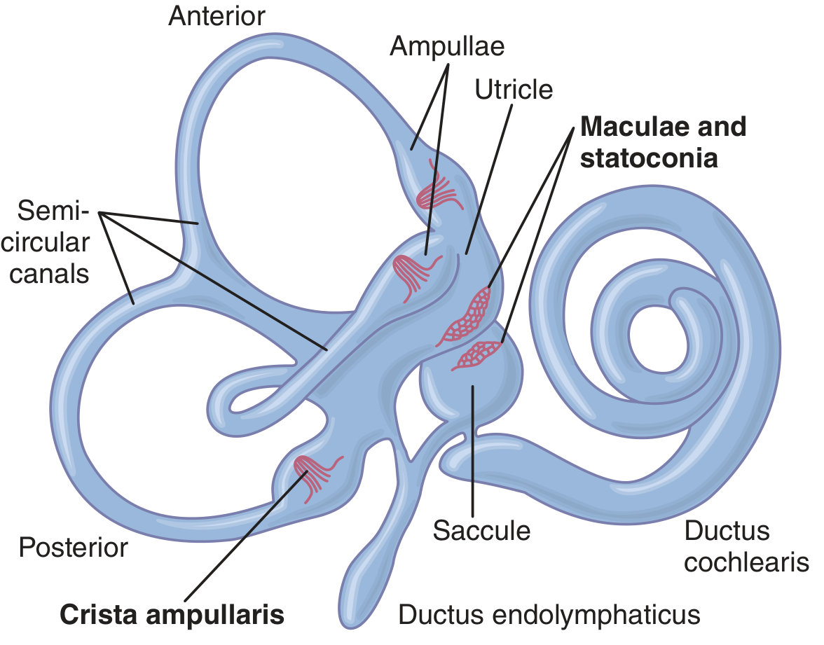
The vestibular apparatus lives in the petrous part of the temporal bone (bony labyrinth). Inside is the membranous labyrinth - the functional part.
It has 5 end organs:
- 3 Semicircular Canals (SCCs): Anterior (Superior), Posterior, Lateral (Horizontal)
- 2 Otolith organs: Utricle + Saccule
The Nerve Supply (Important for MCQs!)
| Division | What it carries |
|---|---|
| Superior vestibular nerve | Lateral SCC + Anterior SCC + Utricle |
| Inferior vestibular nerve | Posterior SCC + Saccule |
- Both divisions have ~25,000 bipolar neurons with cell bodies in Scarpa's ganglion (inside the internal auditory canal).
PART 2: THE HAIR CELL - THE MASTER UNIT
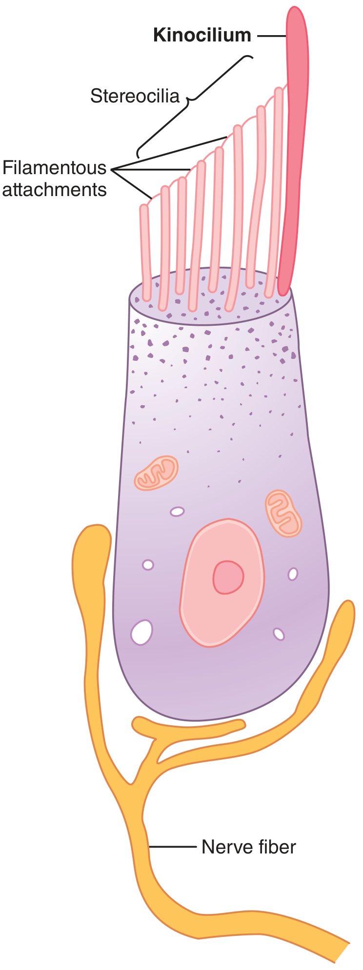
This is the most important thing to understand. Everything in vestibular physiology is based on the hair cell.
Structure:
- Each hair cell has 50-100 stereocilia + 1 kinocilium (the tall one, always on one side)
- Stereocilia are connected to the kinocilium by tiny filamentous (tip-link) attachments
- Stereocilia are graded in height - shorter on one side, taller toward the kinocilium
How it signals (the KEY mechanism):
| Movement | What happens | Effect on nerve |
|---|---|---|
| Stereocilia bend TOWARD kinocilium | Tip-links pull open cation channels → K+/Ca2+ influx → Depolarization | Firing rate INCREASES (excitation) |
| Stereocilia bend AWAY from kinocilium | Tip-links go slack → channels close → Hyperpolarization | Firing rate DECREASES (inhibition) |
Baseline resting firing rate = ~100 impulses/second - this tonic discharge is critical for detecting both increase AND decrease in activity.
Two Types of Hair Cells
| Type I | Type II | |
|---|---|---|
| Shape | Flask-shaped | Cylindrical |
| Innervation | Chalice (cup) of afferent terminal at base | Multiple nerve terminals |
| Effect | Predominantly stimulatory | Predominantly inhibitory |
PART 3: THE OTOLITH ORGANS - DETECTING GRAVITY & LINEAR MOVEMENT
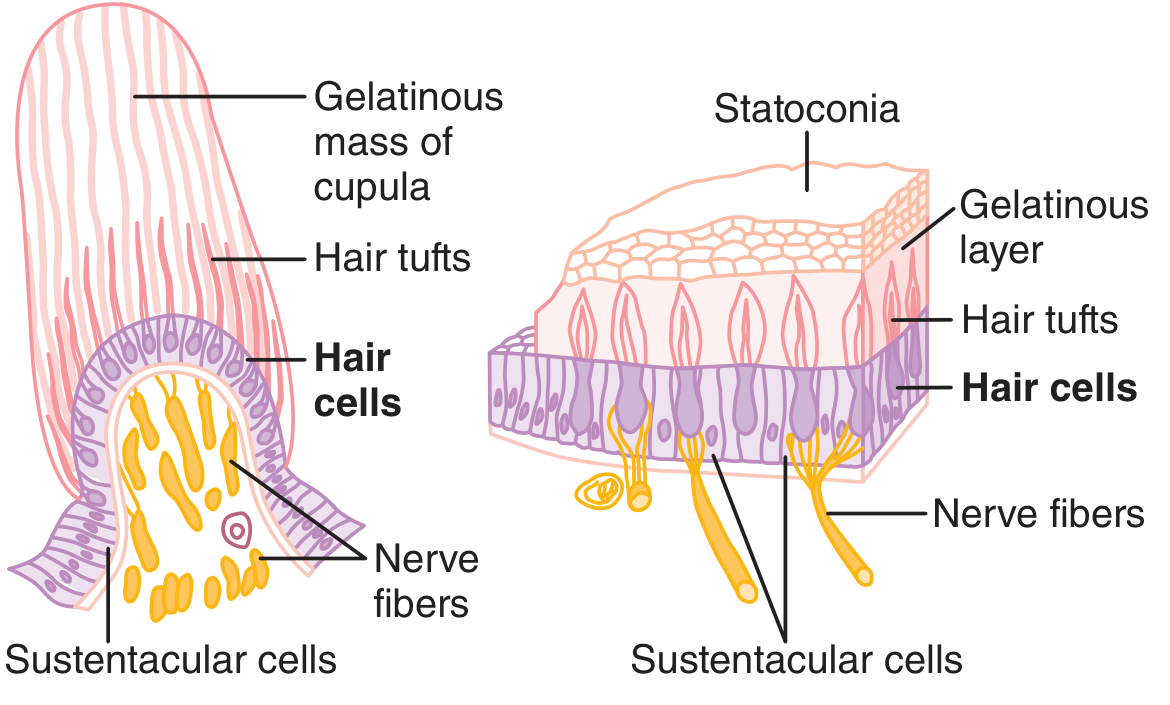
What they detect:
- Linear acceleration (moving in a straight line: car, elevator, gravity)
- Static head tilt (is the head upright or tilted?)
The Macula
- Small sensory area (~2 mm diameter) on the inner surface of utricle and saccule
- Hair cells project into a gelatinous layer
- On top of the gelatin: Statoconia (otoliths) - calcium carbonate crystals, specific gravity 2-3x heavier than surrounding fluid
Utricle vs. Saccule
| Utricle | Saccule | |
|---|---|---|
| Macula orientation | Horizontal plane (floor of utricle) | Vertical plane |
| Best detects | Head upright, horizontal linear acceleration | Head lying down, vertical acceleration (up/down) |
How linear motion is detected:
When the head tilts or accelerates linearly → statoconia (heavy crystals) lag behind due to inertia → they slide over the gelatinous layer → hair cells bend → depolarization signal → brain knows direction of tilt/acceleration.
The Striola
- An imaginary line that almost bisects each otolithic membrane
- Hair cells on either side of the striola have their kinocilia pointing in opposite directions
- This means some hair cells are excited and others inhibited by the same movement
- Result: extremely fine detection of the direction of tilt in all orientations
PART 4: THE SEMICIRCULAR CANALS - DETECTING ROTATION
What they detect:
- Angular (rotational) acceleration - turning the head in any direction
Anatomy (3 canals in 3 planes):
- Lateral (Horizontal): Detects yawing (turning head left/right). Lies horizontal when head tilted 30° forward.
- Anterior (Superior): Detects pitching (nodding up/down). Vertical plane, 45° forward-outward.
- Posterior: Detects rolling (tilting head to shoulder). Vertical plane, 45° backward-outward.
Key pairing (functionally important for nystagmus!)
- Left Lateral ↔ Right Lateral (paired together)
- Left Anterior ↔ Right Posterior (LARP)
- Right Anterior ↔ Left Posterior (RALP)
The Crista Ampullaris
- The sensory end organ inside the ampulla (widened end of each SCC)
- Hair cells embedded in a gelatinous cupula - acts like a hinged gate or swing door
How rotation is detected:
Head rotates → Endolymph LAGS behind (inertia) →
Endolymph flows through canal → Cupula deflects →
Hair cells bend → Signal to brain
Important point: It is the relative movement of endolymph - not the head - that deflects the cupula. The canal rotates, the fluid stays still momentarily.
Kinocilium orientation in SCCs:
- Lateral SCC: Kinocilia face toward utricle (utriculopetal direction = excitatory for lateral canal)
- Anterior and Posterior SCCs: Kinocilia face away from utricle (utriculofugal direction = excitatory for these canals)
Clinical memory trick (Ewald's Laws):
- Lateral canal: Utriculopetal endolymph flow = excitation (TOWARD utricle = more firing)
- Vertical canals (Anterior/Posterior): Utriculofugal endolymph flow = excitation (AWAY from utricle = more firing)
PART 5: THE PUSH-PULL PRINCIPLE (Most Exam-Relevant Concept!)
The SCCs work in functional pairs. When you rotate your head:
- One canal's hair cells are excited (firing goes UP)
- The paired canal on the other side is inhibited (firing goes DOWN)
- The difference in firing rates tells the CNS: direction + speed of rotation
Example - turning head to the RIGHT:
- Right lateral SCC: endolymph flows toward ampulla → cupula deflects utriculopetally → firing INCREASES
- Left lateral SCC: endolymph flows away from ampulla → firing DECREASES
- CNS reads the difference → "head is rotating right"
Why this matters in pathology: A sudden loss of one labyrinth (e.g., left) = left side fires at 0 instead of resting rate (~100/sec). Right side still firing at 100/sec. CNS reads this as "rotating toward the right" even when the head is still → nystagmus with fast phase toward the HEALTHY (right) side.
PART 6: THE VESTIBULO-OCULAR REFLEX (VOR)
The purpose: Keep your vision stable while your head moves. The fovea (small area of sharp vision) must stay pointed at a target even during rapid head movement.
The reflex arc:
Head movement → SCCs detect rotation →
Vestibular nuclei → Via MLF →
Extraocular muscles → Eye moves EQUAL and OPPOSITE to head movement
Result: If head turns RIGHT → eyes move LEFT at the same speed → image stays fixed on the fovea.
If VOR fails: The image "slips" on the retina (retinal slip) → blurred vision during head movement = oscillopsia.
PART 7: CENTRAL VESTIBULAR CONNECTIONS
The vestibular nerve enters the brainstem and synapses at the 4 vestibular nuclei:
| Nucleus | Other name | Main function |
|---|---|---|
| Superior | Bechterew nucleus | Conjugate ocular reflexes (SCCs) |
| Lateral | Deiters nucleus | Ipsilateral vestibulospinal "righting" reflexes |
| Medial | Schwalbe nucleus | Eye, head, neck coordination via MLF |
| Inferior (Descending) | Roller nucleus | Cerebellar connections |
Key Pathways from vestibular nuclei:
- Via MLF (Medial Longitudinal Fasciculus): Controls eye movements, VOR
- Vestibulospinal tract (from Deiters nucleus): Controls posture and muscle tone - keeps you upright
- To Cerebellum: Fine-tuning and coordination
- To Cortex (parietal lobe, deep in Sylvian fissure): Conscious awareness of balance/dizziness
Role of the Cerebellum:
- The flocculonodular lobe is the main cerebellar region for equilibrium (vestibular cerebellum)
- Receives signals from vestibular nuclei and proprioceptors
- Controls equilibrium during sudden changes in direction
- The uvula controls static equilibrium
PART 8: THE THREE INPUTS TO BALANCE
Balance isn't just the inner ear. The brain integrates THREE inputs:
| Input | Detects | Contribution |
|---|---|---|
| Vestibular system | Head movement, gravity, acceleration | Primary signal for motion |
| Vision | Horizon, visual flow | Orientation reference |
| Proprioception | Joint position, muscle stretch | Body position signals |
When inputs conflict: You get dizziness (e.g., watching a moving scene while sitting still = visual-vestibular mismatch).
CNS compensation: After a unilateral vestibular loss, the CNS gradually recalibrates over days-weeks by using the remaining signals. This is vestibular compensation - the basis for vestibular rehabilitation.
PART 9: SUMMARY TABLE FOR QUICK REVISION
| Feature | Semicircular Canals | Otolith Organs |
|---|---|---|
| Organs | Anterior, Posterior, Lateral | Utricle, Saccule |
| End organ | Crista ampullaris | Macula |
| Gelatinous structure | Cupula | Gelatinous layer |
| Special feature | - | Statoconia (CaCO₃ crystals) |
| Detects | Angular (rotational) acceleration | Linear acceleration + static tilt + gravity |
| Stimulus | Endolymph flow deflects cupula | Statoconia slide over hair cells |
| Main reflex | Vestibulo-ocular reflex (VOR) | Postural righting reflexes |
PART 10: MUST-KNOW FACTS FOR MCQs
- Statoconia specific gravity = 2-3x that of surrounding fluid
- Resting baseline firing rate of hair cells = ~100 impulses/second
- Vestibular nerve has ~25,000 bipolar neurons
- Cell bodies of vestibular nerve = Scarpa's ganglion (in internal auditory canal)
- Lateral canal kinocilium points toward utricle (utriculopetal) - opposite to other canals
- Striola = divides macula into two halves with hair cells pointing in opposite directions
- Ewald's 2nd Law: Lateral canal - ampullofugal flow is inhibitory, ampullopetal is excitatory
- Ewald's 2nd Law for vertical canals: reverse (ampullofugal = excitatory)
- Cortical equilibrium center = parietal lobe, deep in Sylvian fissure, opposite to auditory area
Sources: KJ Lee's Essential Otolaryngology, 11th Ed., p. 345-348 | Guyton and Hall Textbook of Medical Physiology, p. 694-698
This is a shared conversation. Sign in to Orris to start your own chat.