

Tenderness from 2 days and how to treat this case
epidermoid cyst scrotum
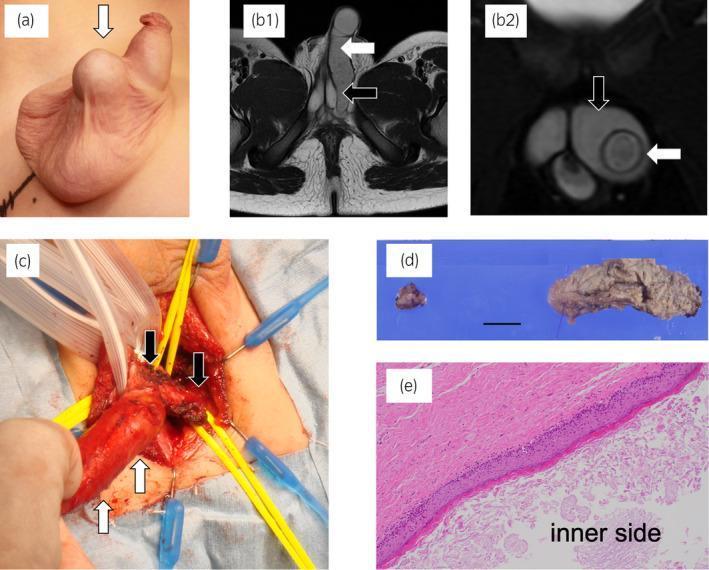
A multi-panel medical figure documenting a case of a scrotal epidermoid cyst in a pediatric patient. (a) A clinical photograph shows a prominent, smooth, midline scrotal mass (white arrow). (b1, b2) T2-weighted axial and coronal MRI scans reveal a tubular, high-signal cystic lesion (white arrows) extending from the scrotum toward the pelvic floor, demonstrating penetration of the penile corpora cavernosa (black arrows). (c) Intraoperative photograph highlights the elongated, reddish-tan tumor (white arrows) being dissected as it traverses the corporal tissue (black arrows). (d) Gross specimen image shows the excised tumor components, including a larger peripheral segment and a smaller central segment, with a 10 mm scale bar for reference. (e) A histopathological micrograph (H&E stain, 100x) displays a cyst wall lined by mature stratified squamous epithelium with focal lymphocytic infiltration, confirming the diagnosis of a benign epidermoid cyst. This case illustrates the rare clinical presentation of an epidermoid cyst penetrating deep into penile erectile structures, requiring precise surgical management to preserve functional anatomy.
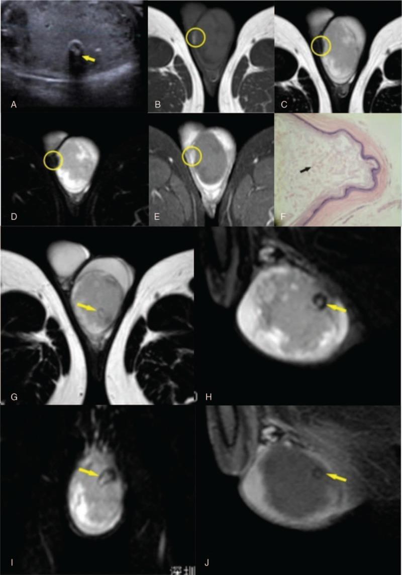
Multi-modal imaging and histopathology of an atypical Testicular Epidermoid Cyst (TEC) in a 26-year-old male. (A) Scrotal ultrasonography displays a large, heterogeneous intratesticular mass with a central focus of hyperechogenicity and significant posterior acoustic shadowing (yellow arrow), suggesting calcification. (B-E) Multi-sequence MRI of the scrotum shows a well-defined mass. A mural nodule (yellow circle) appears hyperintense on T1-weighted imaging (B) and hypointense on T2-weighted (C) and SPAIR (D) sequences. Post-contrast THRIVE sequence (E) demonstrates gadolinium enhancement of the nodule, indicating vascularity. (F) Histopathology (H&E, 20x) reveals a cyst wall with keratinized epithelium and a disorderly internal accumulation of keratin (black arrow), lacking the classic 'onion-skin' arrangement. (G-J) Focused MRI sequences highlight a 'small black ring sign' on T2-weighted and SPAIR images (yellow arrows), which remains non-enhanced on post-contrast THRIVE imaging (J), consistent with a calcified component. This case demonstrates atypical radiological features of TEC, including the absence of alternating concentric rings and the presence of a vascularized mural nodule.
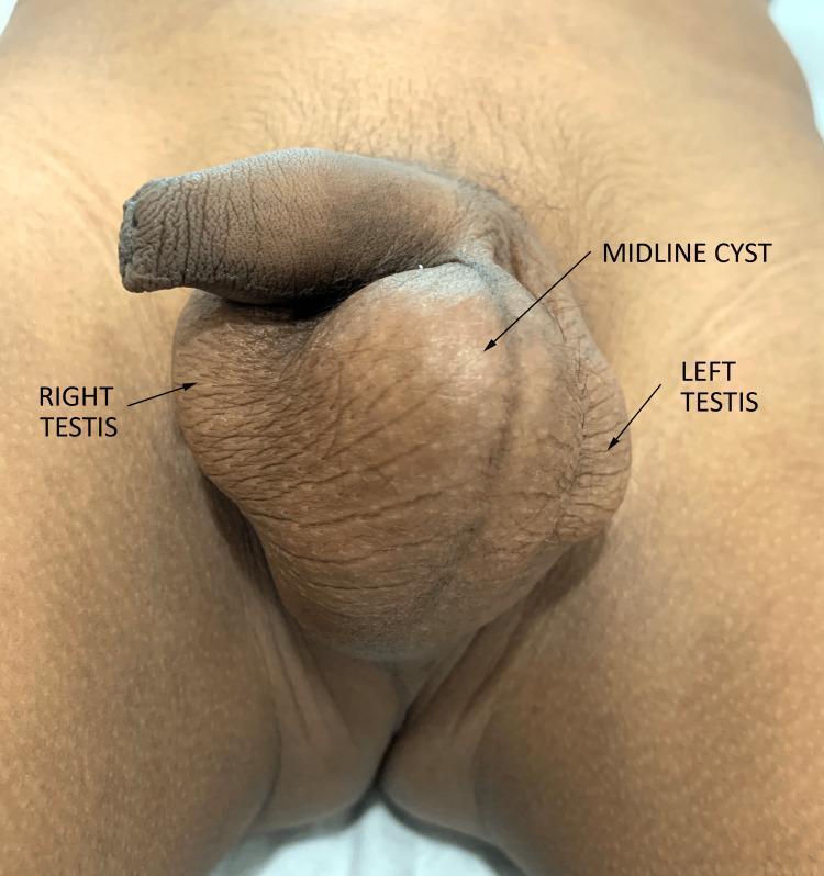
This clinical photograph displays a gross examination of the male genital region featuring a significant midline scrotal swelling. The image identifies a 'MIDLINE CYST' situated centrally within the scrotum, positioned between the 'RIGHT TESTIS' and 'LEFT TESTIS'. The cyst is spherical, prominently larger than the individual testicles, and causes visible stretching of the overlying scrotal skin, resulting in a smoother surface texture compared to the adjacent wrinkled and rugose scrotal folds. The lesion appears discrete and non-inflammatory, with no signs of erythema or ulceration. This visual demonstrates a typical presentation of a large scrotal midline cyst, which in a clinical context could represent an epidermoid cyst or a median raphe cyst. The educational focus is on the anatomical localization of scrotal masses and the differentiation between intratesticular and extratesticular pathology through physical inspection and labeling.
Clinical Assessment
What the Image Shows
Differential Diagnosis
| Diagnosis | Favoring features |
|---|---|
| Inflamed epidermoid cyst | Dome-shaped, tense, well-circumscribed, yellowish center, scrotal location (most common scrotal cyst), sudden onset pain |
| Furuncle / abscess | 2-day tender history, hair-bearing area, fluctuant feel |
| Scrotal calcinosis | Usually multiple, firmer lesions - less likely here |
Pathophysiology
Treatment
- Incision and Drainage (I&D) - This is the indicated procedure for a fluctuant, tender scrotal cyst or abscess. A small sterile incision is made to express the contents. This should be done by a physician/surgeon under sterile conditions.
- Antibiotics - Indicated if there is surrounding cellulitis (spreading redness, warmth, induration beyond the cyst margin). Use an antibiotic covering Staphylococcus aureus:
- Co-amoxiclav (amoxicillin-clavulanate) 625 mg orally three times daily for 5-7 days, OR
- Cefalexin 500 mg four times daily for 5-7 days
- If MRSA is suspected (recurrent/treatment-resistant): Trimethoprim-sulfamethoxazole (TMP-SMX) or Doxycycline
- Warm compresses - Apply a warm, moist cloth to the area 3-4 times daily for 10-15 minutes. This promotes localization and can aid spontaneous drainage.
- Do NOT squeeze or self-drain - The scrotum is highly vascular. Attempting to pop this at home can drive bacteria deeper, cause severe pain, and risk spreading infection.
Red Flag Warning Signs - Seek Emergency Care Immediately If:
- Rapidly spreading redness or swelling
- High fever or chills (suggests systemic infection/sepsis)
- The swelling extends to the perineum or inner thighs (risk of Fournier's gangrene, a life-threatening necrotizing fasciitis)
- Severe, worsening pain that is disproportionate to the size of the lesion
Summary
- See a doctor promptly for formal examination and possible I&D
- Oral antibiotics if cellulitis is present
- Warm soaks at home in the meantime
- Elective surgical excision once the inflammation resolves to prevent recurrence
Disclaimer: This is educational information only. Please consult a urologist, surgeon, or dermatologist for proper diagnosis and treatment. Do not attempt to drain or pop this lesion at home.