
Role of chest X ray in cardiac disease evaluation and and five-step approach for thoracic wall abnormality, cardiac size, pulmonary vascularity, as well as specific sign for specific chamber enlargement and great vessel sign for... Radiology, resident preparing for DNB. Final examination. 10 marks question answer.
cardiac silhouette chest X-ray chamber enlargement cardiac contour
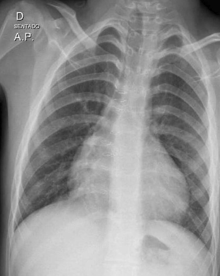
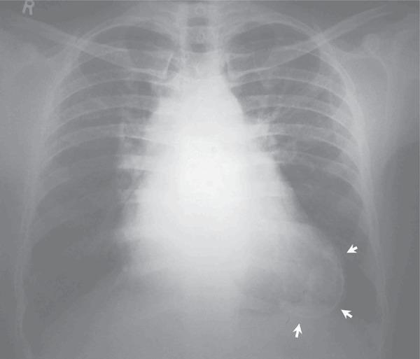
This diagnostic image is an anteroposterior (AP) chest radiograph showing moderate cardiomegaly in a human patient. The cardiac silhouette is enlarged with a cardiothoracic ratio of approximately 0.60, primarily involving the right-sided cardiac chambers as evidenced by the prominence of the atrial and ventricular arches. Notable clinical features include a widened mediastinal contour and progressive cardiac enlargement post-surgical correction. Despite the increased cardiac area, the pulmonary vascular markings appear within normal limits, showing no clear signs of pulmonary venous congestion or edema. The bony structures of the rib cage, the trachea, and the diaphragmatic contours are visible and appear structurally intact. This x-ray is a key educational example of postoperative cardiac monitoring and the visual manifestation of selective chamber enlargement in cardiovascular pathology.
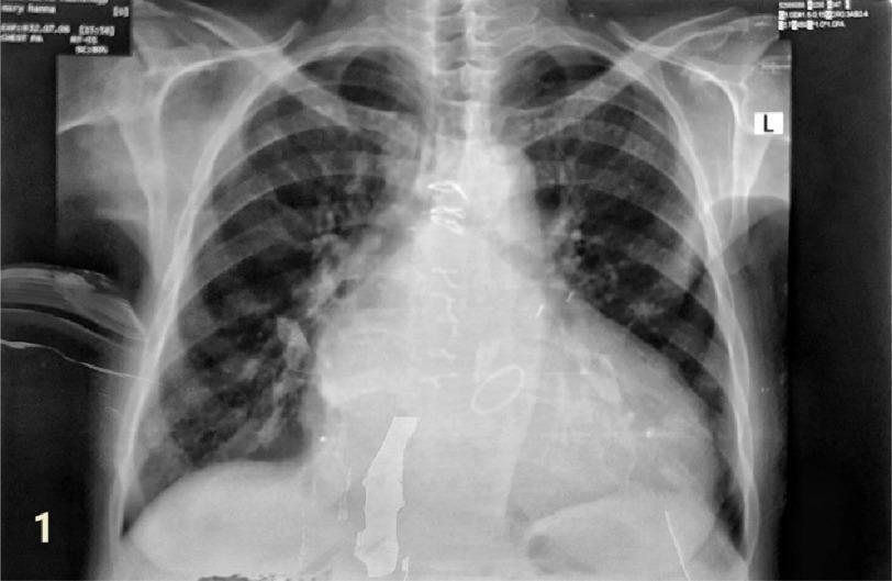
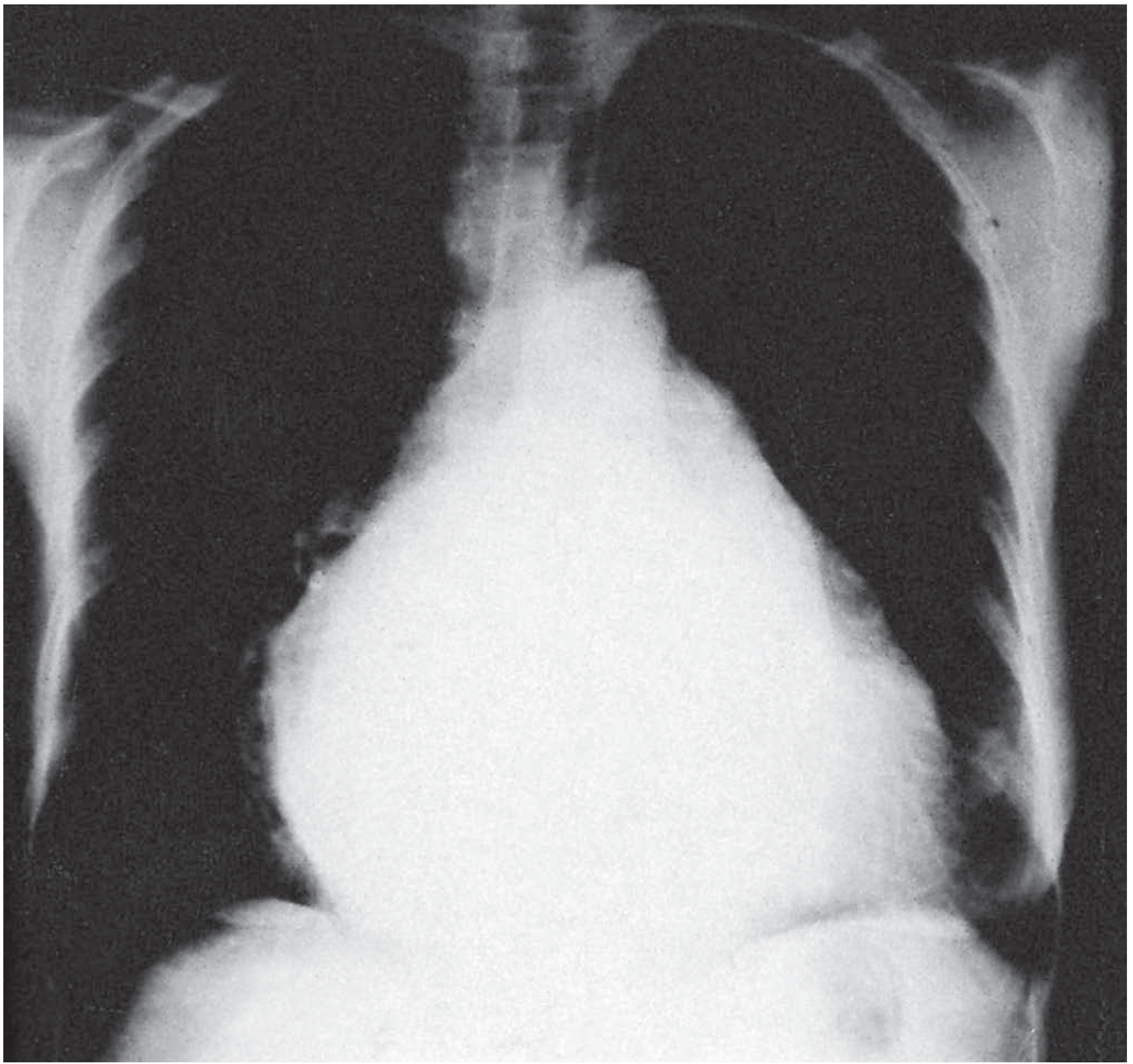
This diagnostic image is a posterior-anterior (PA) chest X-ray demonstrating significant cardiothoracic pathology. The primary finding is marked cardiomegaly with a widened cardiac silhouette. Specific chamber enlargement is visible, including a prominent left atrial contour along the mid-left heart border and signs of biventricular enlargement. In the pulmonary fields, there is evidence of pulmonary venous congestion and interstitial edema, characterized by cephalization of the pulmonary vessels (prominence of upper lobe vessels) and increased hazy opacities throughout both lung fields. Sternal wires and a mitral valve prosthesis are visible, indicating prior cardiac surgery. This image is an educational example of advanced heart failure and valvular heart disease manifestations on radiography, illustrating the relationship between cardiac structural changes and secondary pulmonary vascular congestion.
pulmonary vascularity cephalization chest X-ray heart failure
Chest X-Ray in Cardiac Disease Evaluation: A Systematic Approach
Introduction
Five-Step Systematic Approach to CXR Evaluation in Cardiac Disease
Step 1: Technical Adequacy and Thoracic Wall
- Projection: PA (preferred) vs. AP (overestimates cardiac size)
- Rotation: Medial ends of clavicles should be equidistant from vertebral spinous processes; rotation falsely displaces mediastinum and distorts the cardiac silhouette
- Inspiration: At least 10 posterior ribs should be visible; poor inspiration exaggerates cardiac size
- Penetration: Disc spaces must be faintly visible behind the heart; over- or under-penetration affects assessment of lead integrity and calcifications
- Pectus excavatum: Narrows the AP diameter, compresses and displaces the heart leftward, mimics RV enlargement and falsely enlarges the pulmonary artery segment
- Straight-back syndrome: Loss of thoracic kyphosis compresses the heart
- Scoliosis: Rotates the mediastinum
- Right-sided stomach (situs): Suggests situs inversus/ambiguus; absent IVC shadow + right-sided stomach indicates congenital IVC interruption with azygos continuation
A narrowed AP thoracic diameter is the most common cause of an innocent murmur in young adults.
Step 2: Cardiac Size
- CTR = maximum transverse diameter of the heart / maximum internal diameter of the thorax
- Normal on PA erect CXR: ≤ 0.5
- CTR > 0.5 = cardiomegaly
- A CTR > 0.42 on acute MI CXR is associated with worse in-hospital and long-term outcomes
- Volume overload (e.g., MR, AR, L-to-R shunts): produces greater cardiomegaly than pressure overload alone
- Pressure overload alone (e.g., AS, hypertension): may show normal or mildly enlarged heart with LV hypertrophy without significant dilatation
- Small heart: Seen in COPD, Addison disease, anorexia nervosa, starvation
Step 3: Cardiac Contour (Silhouette and Border Analysis)
- Superior vena cava (SVC) - upper right
- Right atrium (RA) - lower right border
- Aortic knob (aortic arch)
- Pulmonary trunk
- Left atrial appendage (LAA)
- Left ventricle (LV) - forms the bulk of the left heart border
| Chamber | CXR Signs |
|---|---|
| Left Atrium (LA) | Double density sign (retrocardiac opacity); widening of carina >70°; elevation of left main bronchus; posterior esophageal displacement (lateral view); convexity/bulge at LAA segment (3rd mogul sign) |
| Left Atrial Appendage (LAA) | Straightening or convexity of the left heart border just below the pulmonary artery segment ("third mogul/bump") |
| Right Atrium (RA) | Increased convexity and rightward bulge of the right heart border; globular right heart border |
| Left Ventricle (LV) | Downward, outward, and leftward displacement of the cardiac apex; boot-shaped heart in pressure overload |
| Right Ventricle (RV) | Filling of retrosternal space on lateral view; upward and leftward displacement of cardiac apex; RV enlargement rotates the heart leftward making the pulmonary trunk appear more prominent |
| Both Ventricles (biventricular) | Globular cardiomegaly with CTR > 0.5; apex displaced downward and leftward |
- Coeur en sabot ("boot-shaped heart"): Tetralogy of Fallot - elevated apex due to RV hypertrophy with concave pulmonary bay
- "Flask/water bottle" shape: Large pericardial effusion - globular cardiomegaly with sharp cardiac borders
- Double density sign: LA enlargement (visible as a retrocardiac opacity with a distinct second inner density)
- LV aneurysm: Localized bulge along the left cardiac border with a retrosternal double density
- Markedly widened right cardiac contour + straightened left border: Severe MS with secondary TR
- Elevated cardiac silhouette with lucency below (between inferior cardiac border and left hemidiaphragm): Congenital absence of pericardium
Step 4: Pulmonary Vascularity
- Erect PA CXR shows caudal blood flow pattern (gravity-dependent: lower lobes > upper lobes)
- Right descending pulmonary artery: 10-15 mm (males), 9-14 mm (females)
- Arterial-bronchial ratio (ABR): lower lobe arteries almost always larger than accompanying bronchi (ABR ~1.34)
- Vessels taper gradually ("tree-like") from hilum to periphery
A. Abnormalities in Volume:
| Pattern | Cause | CXR Appearance |
|---|---|---|
| Increased PBF (Shunt vascularity) | L-to-R shunts: ASD, VSD, PDA | Uniform bilateral vessel dilatation; right descending pulmonary artery > 15-16 mm; "equalization" of upper and lower lobe vessels |
| Decreased PBF | PS + VSD (ToF), isolated right heart failure | Smaller, shorter pulmonary arteries; more radiolucent lungs; right descending PA < 9 mm |
B. Abnormalities in Distribution (Flow Pattern):
| Pattern | Mechanism | Causes |
|---|---|---|
| Cephalization (redistribution) | Post-capillary PH: pulmonary venous hypertension constricts lower-lobe vessels; upper-lobe vessels dilate | Mitral stenosis, increased LVEDP, severe MR, LV failure |
| Centralization | Pre-capillary PH: pulmonary trunk and central arteries dilate; peripheral arteries prune ("pruning") | Primary PH, Eisenmenger syndrome, recurrent PE, severe emphysema |
| Lateralization | Unilateral obstruction: blood forced into one lung | Massive unilateral PE; jet effect in congenital valvular PS (flow to left lung) |
| Localization | Localized vascular abnormality | Pulmonary arteriovenous fistula |
| Collateralization | Extreme oligemia with bronchial collaterals | Pseudotruncus arteriosus, pulmonary atresia |
- Stage I (PVP 12-18 mmHg): Cephalization - upper lobe vessel dilatation, lower lobe vessel constriction
- Stage II (PVP 18-25 mmHg): Interstitial edema - Kerley B lines (horizontal lines at the costophrenic angles), peribronchial cuffing, hazy hila
- Stage III (PVP >25 mmHg): Alveolar edema - bilateral perihilar "bat-wing/butterfly" opacity
- Acute LV failure: Alveolar edema + normal-sized heart (no time for cardiac remodeling)
- Chronic LV failure: Gross cardiomegaly + cephalization + Kerley B lines + interstitial fibrosis
Step 5: Great Vessels and Other Structures
| Finding | Diagnosis |
|---|---|
| Selective dilatation of ascending aorta | Valvular aortic stenosis (post-stenotic dilatation) |
| Generalized dilatation of entire thoracic aorta | Aortic regurgitation and/or systemic hypertension |
| "3 sign" on aortic knob (upper bulge = pre-stenotic; lower bulge = post-stenotic) + "E sign" on barium esophagogram | Coarctation of the aorta |
| Small aortic knob | ASD, mitral stenosis (LV "crowded out" by RV) |
| Heavy calcification of ascending aortic aneurysm | Luetic (syphilitic) aortitis |
| Widened mediastinum/aortic knob | Aortic dissection, aortic aneurysm |
| Finding | Diagnosis |
|---|---|
| Enlarged pulmonary trunk | RV enlargement (most common); MS; ASD; primary PH |
| Concave/absent pulmonary trunk segment | Tetralogy of Fallot; pulmonary atresia |
Note: Pulmonary trunk prominence is a reliable secondary sign of RV enlargement, EXCEPT in: (1) ToF (RV hypertrophy but hypoplastic pulmonary trunk), (2) idiopathic pulmonary artery dilatation, (3) PDA (normal RV), (4) straight-back syndrome/pectus excavatum/scoliosis (AP compression displaces and falsely enlarges PA).
Summary Table: Chamber-Specific Signs
| Chamber | Key Radiographic Signs |
|---|---|
| LA | Double density sign; widened carina (>70°); elevated left main bronchus; LAA convexity (3rd mogul); posterior esophageal displacement |
| LV | Downward/leftward apex; increased CTR; boot shape (pressure overload) |
| RA | Convex right heart border; rightward bulge |
| RV | Filling of retrosternal space (lateral); apex elevation; pulmonary trunk prominence; cardiac rotation leftward |
| Both | Globular cardiomegaly; CTR > 0.5 |
Devices and Other Findings
- Pacemakers/ICDs: Lead integrity, position (RA, RV, coronary sinus leads), generator position
- LVAD: Inflow cannula in LV, outflow conduit to aorta
- Intra-aortic balloon pump: Tip should be at aortic knob level (1-2 cm below origin of left subclavian artery)
- Calcifications: Mitral annular, valvular (proportional to stenosis severity), coronary artery (almost always atherosclerotic), pericardial
Key Points for DNB Examination
- CTR > 0.5 on erect PA CXR = cardiomegaly; AP CXR overestimates
- Double density sign = LA enlargement (pathognomonic)
- Boot-shaped heart = Tetralogy of Fallot
- "3 sign" / "E sign" = Coarctation of aorta
- Cephalization = post-capillary (venous) PH (mitral stenosis, LV failure)
- Centralization with pruning = pre-capillary (arterial) PH (primary PH, Eisenmenger)
- Ascending aortic dilatation alone = valvular AS (post-stenotic); whole thoracic aorta = AR/hypertension
- Kerley B lines = interstitial edema (stage II pulmonary venous hypertension)
- Acute LV failure: butterfly edema + normal cardiac size (no time for remodeling)
- Pectus excavatum/straight-back = AP narrowing - do not misread as RV enlargement or PA dilatation
Innumerate the structure forming right and left heart border on plain chest radiograph, PA view. Discuss the feature of enlargement of various cardiac chamber on plain radiograph. DNB radiology question answer. For 10 marks.
cardiac borders PA chest radiograph anatomy left right
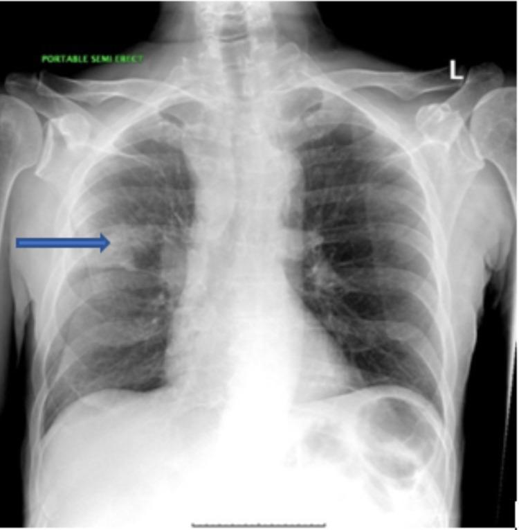
This diagnostic image is a posterior-anterior (PA) chest radiograph demonstrating a focal pulmonary abnormality. A prominent 3 cm radiopaque mass is visible in the right upper lobe, positioned superior to the minor fissure (indicated by a blue horizontal arrow). The mass exhibits irregular borders with an infiltrative appearance extending medially toward the right hilum. The right hilum and right paratracheal soft tissues appear prominent, suggesting possible lymphadenopathy. In contrast, the left lung field is clear. Anatomical landmarks including the trachea, mediastinal contours, and hemidiaphragms are visible, with the right hemidiaphragm normally positioned slightly higher than the left. The cardiac silhouette and skeletal structures of the thorax, including the clavicles and ribs, appear unremarkable. This clinical photograph is highly relevant for medical education regarding the radiological presentation of primary bronchogenic carcinoma, specifically pulmonary adenocarcinoma. It illustrates key diagnostic findings such as lung masses, hilar prominence, and the importance of localized lobe anatomy in chest imaging interpretation.
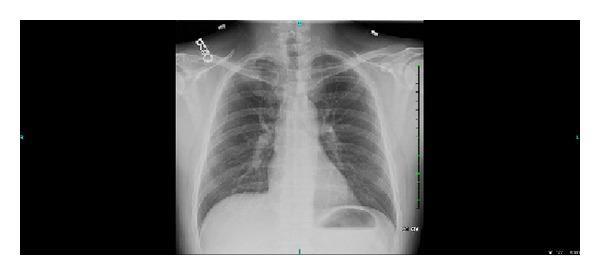
A posterior-anterior (PA) chest X-ray illustrating the thoracic cavity and skeletal structures. The radiograph reveals a distinct anatomical anomaly: a hypoplastic or unusually small right first rib compared to the contralateral side. The primary anatomical landmarks are visible, including the clavicles, rib cage, and vertebral column. The lung fields appear largely clear and radiolucent, with normal bronchovascular markings. The cardiac silhouette is centrally located with well-defined borders and normal size. The diaphragmatic domes are intact, with a clear gastric air bubble visible beneath the left hemidiaphragm. Near the left clavicle, a radiopaque marker or artifact is noted. Clinically, such variations in the first rib anatomy are significant in the context of thoracic outlet syndrome or venous compression (Paget-Schroetter syndrome). This diagnostic image serves as an educational example of rib morphology variations and their potential relationship to vascular pathologies in the thoracic inlet.
left atrial enlargement double density sign chest X-ray mitral stenosis
right ventricular enlargement boot shaped heart tetralogy of Fallot chest radiograph
cardiomegaly biventricular enlargement PA chest X-ray heart failure
This diagnostic image is a posterior-anterior (PA) chest X-ray demonstrating significant cardiothoracic pathology. The primary finding is marked cardiomegaly with a widened cardiac silhouette. Specific chamber enlargement is visible, including a prominent left atrial contour along the mid-left heart border and signs of biventricular enlargement. In the pulmonary fields, there is evidence of pulmonary venous congestion and interstitial edema, characterized by cephalization of the pulmonary vessels (prominence of upper lobe vessels) and increased hazy opacities throughout both lung fields. Sternal wires and a mitral valve prosthesis are visible, indicating prior cardiac surgery. This image is an educational example of advanced heart failure and valvular heart disease manifestations on radiography, illustrating the relationship between cardiac structural changes and secondary pulmonary vascular congestion.
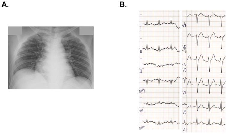
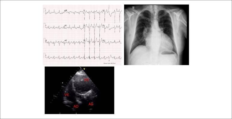
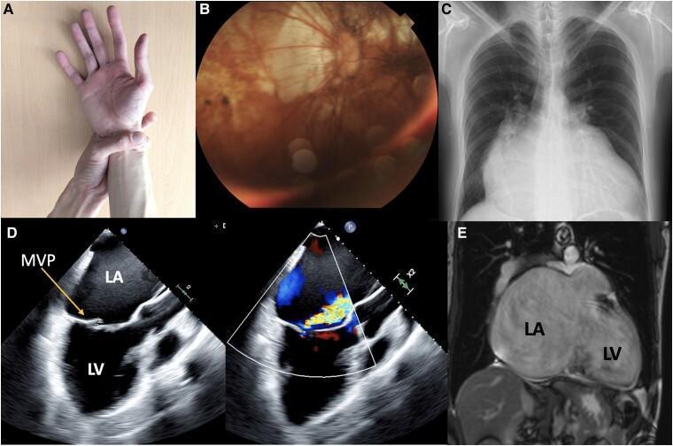
This composite clinical diagnostic image includes a chest X-ray (A) and a 12-lead electrocardiogram (B). Panel A shows a posterior-anterior (PA) view of the thorax, demonstrating a markedly enlarged cardiac silhouette consistent with cardiomegaly. The lung fields exhibit increased bronchovascular markings and subtle bilateral interstitial opacities, characteristic of mild pulmonary congestion. Panel B displays a 12-lead ECG strip showing a regular sinus rhythm. Notably, there is an absence of ST-segment elevation or significant ischemic changes across the limb leads (I, II, III, aVR, aVL, aVF) and precordial leads (V1-V6), which clinically helps rule out acute ST-elevation myocardial infarction (STEMI) in the context of acute heart failure. The primary educational focus is the correlation between radiological findings of biventricular enlargement/congestive heart failure and the corresponding baseline electrical activity of the heart.
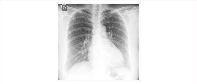
This diagnostic image is a posterior-anterior (PA) chest X-ray of a human adult. The primary finding is significant global cardiomegaly, evidenced by a widened cardiac silhouette that occupies more than 50% of the thoracic diameter, suggesting multi-chamber enlargement. Additionally, the pulmonary vasculature is prominently increased, indicating pulmonary venous hypertension or congestion. This is characterized by increased vascular density and vessel visibility extending into the peripheral lung fields. The hilar regions appear enlarged and dense, consistent with vascular engorgement. The costophrenic angles appear relatively clear on this view, and the bony structures of the thorax are intact. This clinical photograph is used in medical education to illustrate the radiological hallmarks of congestive heart failure or advanced valvular heart disease, such as mitral regurgitation or chronic biventricular dilation.
Cardiac Borders on PA CXR and Chamber Enlargement Signs
Part I: Structures Forming the Cardiac Borders on PA CXR
RIGHT CARDIAC BORDER (from above downward)
| Structure | Level | Notes |
|---|---|---|
| 1. Superior vena cava (SVC) | Upper right border | Forms the right paratracheal stripe; straight or gently convex margin |
| 2. Ascending aorta | Upper-mid right | Partially contributes in elderly/hypertensive patients; curves into the arch |
| 3. Right atrium (RA) | Lower right border | Forms the entire lower right cardiac border; a gently convex curvilinear margin extending from the SVC to the right cardiophrenic angle |
The right cardiophrenic angle is formed by the RA and the right hemidiaphragm. The inferior vena cava (IVC) enters the RA at this level and may be faintly visible as a short oblique shadow.
LEFT CARDIAC BORDER (from above downward)
| Structure | Level | Notes |
|---|---|---|
| 1. Aortic knob (aortic arch) | Uppermost left | The transverse arch of the aorta as it turns from ascending to descending; forms a distinct rounded convexity. Normally measures < 3.5 cm in width |
| 2. Main pulmonary artery (pulmonary trunk) | Just below aortic knob | A gentle convexity below the aortic knob; the "pulmonary bay" or "pulmonary segment" |
| 3. Left atrial appendage (LAA) | Between PA and LV | Normally inapparent or produces only a slight straight/flat segment just below the pulmonary trunk |
| 4. Left ventricle (LV) | Lower left border | Forms the bulk of the left heart border - a smooth, gently convex margin tapering to the cardiac apex. The apex normally points downward and to the left, resting on the left hemidiaphragm |
These four structures from top to bottom on the left border are sometimes called the "four moguls": aortic knob, pulmonary trunk, LAA, and LV.
Diaphragmatic Borders
- Right: RV forms most of the inferior cardiac surface; right hemidiaphragm level
- Left: LV apex rests on left hemidiaphragm
Part II: Radiographic Features of Chamber Enlargement
1. LEFT ATRIUM (LA) ENLARGEMENT
- Double density sign - The enlarged LA projects as a second opacity (inner density) behind and to the right of the normal right heart border, visible as two distinct curvilinear contours superimposed on the right side of the heart. This is the most specific sign of LA enlargement.
- Widening of the subcarinal angle (>70°) - The carina is splayed as the enlarged LA pushes the left main bronchus superiorly. Normal carinal angle is ~60-70°; >70° is abnormal.
- Elevation of left main bronchus - The left main bronchus is elevated and may appear horizontal; the right main bronchus descends at a steeper angle by comparison.
- Left atrial appendage (LAA) bulge / Third mogul sign - Enlargement of the LAA produces a convexity or straightening of the left heart border just below the pulmonary artery segment. In mitral regurgitation and mitral stenosis, this produces the characteristic "third mogul" or bump on the left border.
- Posterior displacement of esophagus - Best seen on lateral view with barium; enlarged LA displaces the barium-filled esophagus posteriorly and to the right.
2. LEFT VENTRICLE (LV) ENLARGEMENT
- Elongation and downward displacement of the cardiac apex - The apex is displaced downward, outward, and to the left ("drooping" or "sagging" apex); the heart appears more globular
- Increased CTR (>0.5) - LV dilatation is the most common cause of overall cardiomegaly
- Rounding of the left cardiac border - The normally flat or gently convex left border becomes more curved and prominent
- Upward displacement of the aortic knob - In aortic regurgitation, the markedly dilated LV may push the heart upward, elongating the ascending aorta
- Volume overload (MR, AR, VSD): significant cardiomegaly with enlarged, rounded left border and dilated LV
- Pressure overload alone (AS, hypertension): LV hypertrophy with concentric remodeling; CTR may be near normal despite significant hypertrophy; only mild or no enlargement of the cardiac silhouette
3. RIGHT VENTRICLE (RV) ENLARGEMENT
- Upward and leftward displacement of the cardiac apex - The enlarged RV pushes the LV upward and posteriorly, elevating and rotating the apex (cardiac apex appears "uplifted")
- Prominence of the pulmonary trunk/main pulmonary artery - When the RV enlarges, the heart rotates leftward on its long axis, bringing the pulmonary trunk into a more lateral and prominent position (appears larger than it actually is)
- Coeur en sabot ("boot-shaped heart") - Classic in Tetralogy of Fallot: RV hypertrophy elevates the apex + concave/absent pulmonary artery segment = boot or wooden shoe shape
- Filling of the retrosternal airspace on lateral view - Enlarged RV fills the normally air-filled retrosternal space; the RV-sternum contact extends more than one-third of the way up the sternum
4. RIGHT ATRIUM (RA) ENLARGEMENT
- Increased convexity and rightward bulging of the right heart border - The right border becomes more convex and projects further to the right of the spine; CTR may be asymmetrically increased on the right side
- Increased height of the right cardiac border - The right atrial arc appears taller and more prominent
- Globular right heart border - Prominent, bulging right cardiac contour extending from SVC to cardiophrenic angle
- Dilated right cardiac border + straightened left border - In severe mitral stenosis with secondary tricuspid regurgitation, the massively enlarged RA dominates the right side while the LA and pulmonary trunk contribute to the straightened left border
5. BIVENTRICULAR ENLARGEMENT
- Global cardiomegaly - CTR > 0.5, with heart enlarged in all dimensions
- Apex displaced downward AND the right border projects markedly to the right
- Found in dilated cardiomyopathy, advanced valvular disease, end-stage heart failure
Summary Table: Chamber-Specific Signs on PA CXR
| Chamber | Key PA View Signs | Additional/Lateral Signs |
|---|---|---|
| LA | Double density sign; subcarinal angle >70°; elevated left main bronchus; LAA convexity (3rd mogul) | Posterior esophageal displacement on barium |
| LAA | Straightening or convexity below pulmonary artery segment | - |
| LV | Apex displaced downward/laterally; rounding of left border; increased CTR | LV projects posterior to IVC on lateral |
| RV | Elevated apex; prominent pulmonary trunk; boot shape (ToF) | Fills retrosternal space on lateral |
| RA | Convex, rightward bulge of right border; increased height of right arc | - |
| Biventricular | Global cardiomegaly (CTR >0.5); all borders enlarged | - |
Important Diagnostic Contour Signs
| Sign | Chamber/Structure | Diagnosis |
|---|---|---|
| Double density sign | LA | LA enlargement (mitral stenosis, MR) |
| Third mogul/bump | LAA | LAA enlargement (mitral valve disease) |
| Coeur en sabot (boot shape) | RV + concave pulmonary bay | Tetralogy of Fallot |
| Globular/water-bottle shape | All chambers | Pericardial effusion |
| Widened right border + straight left border | RA + LA | Severe MS with TR |
| LV aneurysm bulge | LV | LV aneurysm (post-MI) - localized bulge on left border |
| Elevated cardiac silhouette + sub-diaphragmatic lucency | - | Congenital absence of pericardium |
Measurement: Cardiothoracic Ratio (CTR)
- CTR = Maximum transverse cardiac diameter / Maximum internal thoracic diameter (at same level)
- Normal on erect PA CXR: ≤ 0.5
- CTR > 0.5 = cardiomegaly
- AP CXR overestimates cardiac size due to magnification and beam divergence - CTR assessment is unreliable on AP films
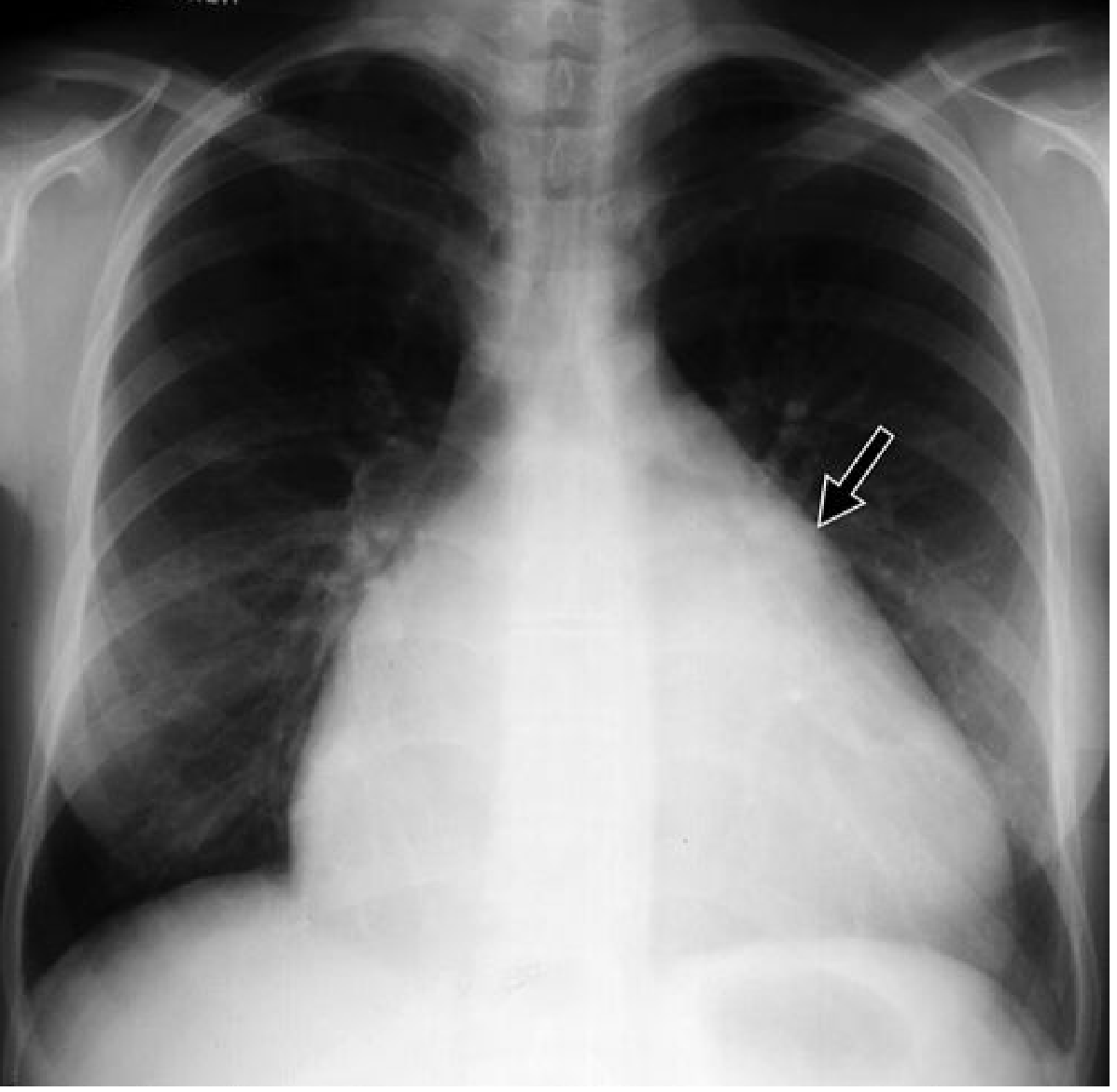
Create a PDF reference in 2 or 3 pages card on cardiac chamber enlargement signs for rapid exam review
ls -la /tmp/workspace/ 2>/dev/null || mkdir -p /tmp/workspace && echo "workspace ready"
mkdir -p /tmp/workspace/cardiac-xray-card && python3 << 'PYEOF' from reportlab.platypus import ( SimpleDocTemplate, Table, TableStyle, Paragraph, Spacer, HRFlowable, KeepTogether ) from reportlab.lib.styles import getSampleStyleSheet, ParagraphStyle from reportlab.lib import colors from reportlab.lib.units import mm, cm from reportlab.lib.pagesizes import A4 from reportlab.lib.enums import TA_CENTER, TA_LEFT, TA_JUSTIFY from reportlab.platypus import PageBreak OUTPUT = "/tmp/workspace/cardiac-xray-card/Cardiac_Chamber_Enlargement_CXR.pdf" # ── Colour palette (medical/academic feel) ────────────────────────── C_NAVY = colors.HexColor("#0A2540") C_BLUE = colors.HexColor("#1565C0") C_SKY = colors.HexColor("#E3F0FF") C_TEAL = colors.HexColor("#006D77") C_TEAL_LT = colors.HexColor("#D9F0F2") C_AMBER = colors.HexColor("#B45309") C_AMBER_LT= colors.HexColor("#FEF3C7") C_RED = colors.HexColor("#B91C1C") C_RED_LT = colors.HexColor("#FEE2E2") C_GREEN = colors.HexColor("#166534") C_GREEN_LT= colors.HexColor("#DCFCE7") C_PURPLE = colors.HexColor("#6B21A8") C_PURPLE_LT=colors.HexColor("#F3E8FF") C_GREY_LT = colors.HexColor("#F1F5F9") C_GREY = colors.HexColor("#64748B") C_WHITE = colors.white C_BLACK = colors.HexColor("#1E293B") W, H = A4 # 595 x 842 pt MARGIN = 14*mm doc = SimpleDocTemplate( OUTPUT, pagesize=A4, leftMargin=MARGIN, rightMargin=MARGIN, topMargin=12*mm, bottomMargin=12*mm, title="Cardiac Chamber Enlargement – CXR Signs", author="DNB Radiology Quick Reference" ) styles = getSampleStyleSheet() def S(name, **kw): return ParagraphStyle(name, parent=styles["Normal"], **kw) # ── Custom styles ──────────────────────────────────────────────────── sTitle = S("sTitle", fontSize=17, leading=21, textColor=C_WHITE, fontName="Helvetica-Bold", alignment=TA_CENTER, spaceAfter=2) sSubtitle = S("sSubtitle", fontSize=9, leading=12, textColor=colors.HexColor("#CBD5E1"), fontName="Helvetica", alignment=TA_CENTER) sSec = S("sSec", fontSize=10, leading=13, textColor=C_WHITE, fontName="Helvetica-Bold", alignment=TA_CENTER) sBullet = S("sBullet", fontSize=8.2, leading=11.5, textColor=C_BLACK, fontName="Helvetica", leftIndent=6, spaceAfter=1) sBold = S("sBold", fontSize=8.2, leading=11.5, textColor=C_BLACK, fontName="Helvetica-Bold") sNote = S("sNote", fontSize=7.5, leading=10.5, textColor=C_GREY, fontName="Helvetica-Oblique", alignment=TA_JUSTIFY) sKeyHdr = S("sKeyHdr", fontSize=9, leading=12, textColor=C_WHITE, fontName="Helvetica-Bold", alignment=TA_CENTER) sKeyCell = S("sKeyCell", fontSize=8, leading=11, textColor=C_BLACK, fontName="Helvetica") sKeyCellB = S("sKeyCellB", fontSize=8, leading=11, textColor=C_BLACK, fontName="Helvetica-Bold") sFooter = S("sFooter", fontSize=7, leading=9, textColor=C_GREY, fontName="Helvetica-Oblique", alignment=TA_CENTER) COL_W = (W - 2*MARGIN) # usable width def bullet(text, bold_prefix=None): if bold_prefix: return Paragraph(f"<b>\u2022 {bold_prefix}</b> {text}", sBullet) return Paragraph(f"\u2022 {text}", sBullet) def sp(h=3): return Spacer(1, h) def hr(color=C_BLUE, thickness=0.5): return HRFlowable(width="100%", thickness=thickness, color=color, spaceAfter=3, spaceBefore=3) # ═══════════════════════════════════════════════════════════════════ # PAGE 1 # ═══════════════════════════════════════════════════════════════════ elems = [] # ── HEADER BANNER ─────────────────────────────────────────────────── header_data = [[ Paragraph("CARDIAC CHAMBER ENLARGEMENT", sTitle), Paragraph("CXR Signs · Rapid Exam Reference · DNB Radiology", sSubtitle), ]] hdr_tbl = Table([[Paragraph("CARDIAC CHAMBER ENLARGEMENT", sTitle)], [Paragraph("CXR Signs · PA View · DNB Radiology Quick Reference", sSubtitle)]], colWidths=[COL_W]) hdr_tbl.setStyle(TableStyle([ ('BACKGROUND', (0,0), (-1,-1), C_NAVY), ('ROWPADDING', (0,0), (-1,-1), 6), ('TOPPADDING', (0,0), (-1, 0), 10), ('BOTTOMPADDING',(0,-1),(-1,-1), 8), ('ROUNDEDCORNERS', [5]), ])) elems.append(hdr_tbl) elems.append(sp(5)) # ── CARDIAC BORDERS TABLE ──────────────────────────────────────────── borders_title = Table([[Paragraph("CARDIAC BORDERS ON PA CXR", sSec)]], colWidths=[COL_W]) borders_title.setStyle(TableStyle([ ('BACKGROUND', (0,0),(-1,-1), C_TEAL), ('ROWPADDING', (0,0),(-1,-1), 5), ('ROUNDEDCORNERS',[4]), ])) elems.append(borders_title) elems.append(sp(4)) # two-column layout for borders def cell(content, bg=C_WHITE, pad=6): t = Table([[content]], colWidths=[(COL_W-4)/2]) t.setStyle(TableStyle([ ('BACKGROUND',(0,0),(-1,-1), bg), ('BOX',(0,0),(-1,-1), 0.5, C_TEAL), ('ROWPADDING',(0,0),(-1,-1), pad), ('VALIGN',(0,0),(-1,-1),'TOP'), ])) return t RIGHT_content = [ Paragraph("<b>RIGHT CARDIAC BORDER</b> (Top → Bottom)", S("rh", parent=sBold, textColor=C_TEAL, fontSize=8.5)), sp(3), bullet("Superior Vena Cava (SVC)", "1."), bullet("Ascending Aorta — partial (elderly)", "2."), bullet("Right Atrium (RA) — entire lower right border", "3."), sp(2), Paragraph("RA → right cardiophrenic angle (IVC entry point)", sNote), ] LEFT_content = [ Paragraph("<b>LEFT CARDIAC BORDER</b> (Top → Bottom)", S("lh", parent=sBold, textColor=C_BLUE, fontSize=8.5)), sp(3), bullet("Aortic Knob (Aortic Arch)", "1."), bullet("Main Pulmonary Artery (Pulmonary Trunk)", "2."), bullet("Left Atrial Appendage (LAA) — normally flat/inconspicuous", "3."), bullet("Left Ventricle (LV) — bulk of left border + apex", "4."), sp(2), Paragraph("Mnemonic: <b>A – P – L – L</b> (Aorta · PA · LAA · LV)", sNote), ] def multi_cell(items, bg, border_col, w): rows = [[i] for i in items] t = Table(rows, colWidths=[w]) t.setStyle(TableStyle([ ('BACKGROUND',(0,0),(-1,-1), bg), ('BOX',(0,0),(-1,-1), 0.8, border_col), ('LEFTPADDING',(0,0),(-1,-1), 7), ('RIGHTPADDING',(0,0),(-1,-1), 7), ('TOPPADDING',(0,0),(-1,-1), 2), ('BOTTOMPADDING',(0,0),(-1,-1), 2), ('TOPPADDING',(0,0),(-1,0), 7), ('BOTTOMPADDING',(0,-1),(-1,-1), 7), ('ROUNDEDCORNERS',[4]), ])) return t HW = (COL_W - 5) / 2 borders_row = Table( [[multi_cell(RIGHT_content, C_TEAL_LT, C_TEAL, HW), multi_cell(LEFT_content, C_SKY, C_BLUE, HW)]], colWidths=[HW, HW], hAlign='LEFT' ) borders_row.setStyle(TableStyle([ ('LEFTPADDING',(0,0),(-1,-1), 0), ('RIGHTPADDING',(0,0),(-1,-1), 0), ('VALIGN',(0,0),(-1,-1),'TOP'), ('COLPADDING', (0,0),(0,-1), 0), ('INNERGRID',(0,0),(-1,-1), 0, C_WHITE), ])) elems.append(borders_row) elems.append(sp(6)) # ── CHAMBER ENLARGEMENT SECTIONS ──────────────────────────────────── def chamber_block(title, color_hdr, color_bg, color_border, signs_pa, signs_extra, causes): title_row = Table([[Paragraph(title, sSec)]], colWidths=[COL_W]) title_row.setStyle(TableStyle([ ('BACKGROUND',(0,0),(-1,-1), color_hdr), ('ROWPADDING',(0,0),(-1,-1), 4), ('ROUNDEDCORNERS',[4]), ])) # Three sub-columns: PA Signs | Additional Signs | Causes cw = [(COL_W - 4) * 0.42, (COL_W - 4) * 0.32, (COL_W - 4) * 0.26] pa_items = [Paragraph("<b>PA VIEW SIGNS</b>", S("ph", parent=sBold, textColor=color_hdr, fontSize=7.8))] + \ [sp(2)] + [bullet(s) for s in signs_pa] ex_items = [Paragraph("<b>LATERAL / EXTRA SIGNS</b>", S("eh", parent=sBold, textColor=color_hdr, fontSize=7.8))] + \ [sp(2)] + [bullet(s) for s in signs_extra] ca_items = [Paragraph("<b>CAUSES</b>", S("ch", parent=sBold, textColor=color_hdr, fontSize=7.8))] + \ [sp(2)] + [bullet(s) for s in causes] def sub_cell(items, w, bg): rows = [[i] for i in items] t = Table(rows, colWidths=[w]) t.setStyle(TableStyle([ ('BACKGROUND',(0,0),(-1,-1), bg), ('LEFTPADDING',(0,0),(-1,-1), 6), ('RIGHTPADDING',(0,0),(-1,-1), 6), ('TOPPADDING',(0,0),(-1,-1), 2), ('BOTTOMPADDING',(0,0),(-1,-1), 2), ('TOPPADDING',(0,0),(0,0), 5), ('BOTTOMPADDING',(0,-1),(0,-1), 5), ])) return t inner = Table( [[sub_cell(pa_items, cw[0], bg=color_bg), sub_cell(ex_items, cw[1], bg=color_bg), sub_cell(ca_items, cw[2], bg=colors.HexColor("#F8FAFC"))]], colWidths=cw ) inner.setStyle(TableStyle([ ('BOX',(0,0),(-1,-1), 0.8, color_border), ('INNERGRID',(0,0),(-1,-1), 0.4, color_border), ('VALIGN',(0,0),(-1,-1),'TOP'), ('LEFTPADDING',(0,0),(-1,-1), 0), ('RIGHTPADDING',(0,0),(-1,-1), 0), ('TOPPADDING',(0,0),(-1,-1), 0), ('BOTTOMPADDING',(0,0),(-1,-1), 0), ('ROUNDEDCORNERS',[4]), ])) return [title_row, sp(3), inner, sp(5)] # ── LEFT ATRIUM ────────────────────────────────────────────────────── elems += chamber_block( "LEFT ATRIUM (LA) ENLARGEMENT", C_BLUE, C_SKY, C_BLUE, signs_pa=[ "Double density sign — retrocardiac opacity (2nd inner curvilinear shadow) on right side of heart", "Widening of subcarinal (carinal) angle > 70°", "Elevation of left main bronchus (horizontal left bronchus)", "LAA convexity — straightening or bulge below pulmonary artery = Third mogul sign", "Increased CTR if massive LA", ], signs_extra=[ "Posterior esophageal displacement (barium swallow / lateral CXR)", "Splaying of carina on lateral view", "Posterior displacement of descending aorta", ], causes=["Mitral stenosis", "Mitral regurgitation", "LV failure", "Atrial fibrillation (chronic)"] ) # ── LEFT VENTRICLE ─────────────────────────────────────────────────── elems += chamber_block( "LEFT VENTRICLE (LV) ENLARGEMENT", C_PURPLE, C_PURPLE_LT, C_PURPLE, signs_pa=[ "Cardiac apex displaced downward, outward and to the left (drooping apex)", "Rounding and increased convexity of left cardiac border", "Increased CTR > 0.5 (volume overload > pressure overload)", "Localized left border bulge = LV aneurysm (post-MI)", "Boot-shaped heart in pressure overload (AS)", ], signs_extra=[ "LV projects posterior to IVC on lateral view", "Posterior cardiac border extends > 1.8 cm behind IVC on lateral", "Volume overload: gross cardiomegaly; pressure overload: near-normal size", ], causes=["Aortic regurgitation", "Mitral regurgitation", "Dilated CMP", "Aortic stenosis", "Hypertension", "Post-MI aneurysm"] ) elems.append(PageBreak()) # ═══════════════════════════════════════════════════════════════════ # PAGE 2 # ═══════════════════════════════════════════════════════════════════ # Repeat compact header hdr2 = Table([[Paragraph("CARDIAC CHAMBER ENLARGEMENT — CXR SIGNS | Page 2", sTitle)]], colWidths=[COL_W]) hdr2.setStyle(TableStyle([ ('BACKGROUND',(0,0),(-1,-1), C_NAVY), ('ROWPADDING',(0,0),(-1,-1), 7), ('ROUNDEDCORNERS',[5]), ])) elems.append(hdr2) elems.append(sp(5)) # ── RIGHT VENTRICLE ────────────────────────────────────────────────── elems += chamber_block( "RIGHT VENTRICLE (RV) ENLARGEMENT", C_AMBER, C_AMBER_LT, C_AMBER, signs_pa=[ "Cardiac apex elevated and displaced upward/leftward", "Prominent pulmonary trunk (cardiac rotation brings PA laterally)", "Coeur en sabot (boot-shaped heart) — RV hypertrophy + concave PA bay = Tetralogy of Fallot", "Widened right cardiac contour if RV + RA both enlarged", "Reduced pulmonary vascularity (if associated PS/ToF)", ], signs_extra=[ "Retrosternal airspace obliteration on lateral view (most sensitive sign)", "RV-sternum contact > 1/3 of sternal height on lateral", "Diaphragmatic surface occupied by RV (not LV) on lateral", ], causes=["Pulmonary hypertension", "Cor pulmonale (COPD)", "ASD / VSD", "Pulmonary stenosis", "Tetralogy of Fallot", "Tricuspid regurgitation"] ) # ── RIGHT ATRIUM ───────────────────────────────────────────────────── elems += chamber_block( "RIGHT ATRIUM (RA) ENLARGEMENT", C_RED, C_RED_LT, C_RED, signs_pa=[ "Increased convexity and rightward bulging of right heart border", "Increased height of right cardiac arc", "Right cardiophrenic angle becomes rounded and widened", "Globular right heart border extending from SVC to cardiophrenic angle", "Massive RA + straightened left border = severe MS with secondary TR", ], signs_extra=[ "Dilated right cardiac border without pulmonary artery segment enlargement = tricuspid stenosis", "Ebstein anomaly: massively dilated RA with decreased pulmonary vascularity", ], causes=["Tricuspid regurgitation", "Tricuspid stenosis", "Right heart failure", "ASD", "Ebstein anomaly", "Severe MS → TR"] ) # ── BIVENTRICULAR ──────────────────────────────────────────────────── elems += chamber_block( "BIVENTRICULAR ENLARGEMENT", C_GREEN, C_GREEN_LT, C_GREEN, signs_pa=[ "Global cardiomegaly — CTR > 0.5", "All cardiac borders enlarged: right border bulges right, left border bulges left", "Apex displaced downward AND rightward border prominent", "Flask / globular shape in dilated cardiomyopathy", "Pulmonary venous congestion + Kerley B lines (CHF)", ], signs_extra=[ "Combined retrosternal filling + posterior cardiac extension on lateral", "Pericardial effusion mimics biventricular enlargement — water-bottle shape with sharp borders", ], causes=["Dilated cardiomyopathy", "Advanced valvular disease (MR + AR)", "End-stage CHF", "Myocarditis", "Viral cardiomyopathy"] ) elems.append(sp(4)) elems.append(hr(C_GREY, thickness=0.8)) elems.append(sp(3)) # ── QUICK-REFERENCE DIAGNOSTIC SIGNS TABLE ────────────────────────── qr_title = Table([[Paragraph("PATHOGNOMONIC & CLASSIC CXR SIGNS — QUICK REFERENCE", sSec)]], colWidths=[COL_W]) qr_title.setStyle(TableStyle([ ('BACKGROUND',(0,0),(-1,-1), C_NAVY), ('ROWPADDING',(0,0),(-1,-1), 5), ('ROUNDEDCORNERS',[4]), ])) elems.append(qr_title) elems.append(sp(4)) qr_data = [ [Paragraph("SIGN", sKeyHdr), Paragraph("STRUCTURE", sKeyHdr), Paragraph("DIAGNOSIS", sKeyHdr)], ["Double density sign", "Left atrium", "LA enlargement (MS, MR)"], ["Third mogul / LAA bump", "Left atrial appendage", "Mitral valve disease"], ["Widened subcarinal angle > 70°", "Left atrium / Carina", "LA enlargement"], ["Coeur en sabot (boot shape)", "RV hypertrophy + absent PA", "Tetralogy of Fallot"], ["Water-bottle / flask shape", "Pericardial sac", "Pericardial effusion"], ["Drooping/downward apex", "Left ventricle", "LV dilatation (AR, MR, DCM)"], ["Elevated uplifted apex", "Right ventricle pushing LV up", "RV enlargement"], ["Localized left border bulge", "Left ventricle wall", "LV aneurysm (post-MI)"], ["Widened Rt border + straight Lt border", "RA enlargement + LA/PA", "Severe MS → TR"], ["3-sign / E-sign on aorta/esophagus", "Aortic arch", "Coarctation of aorta"], ["Retrosternal space obliteration", "Right ventricle (lateral)", "RV enlargement"], ["Pruning of peripheral vessels", "Pulmonary arteries", "Pre-capillary PH (Eisenmenger)"], ["Kerley B lines", "Interstitial / lymphatics", "Pulmonary venous hypertension Gr II"], ["Butterfly / bat-wing edema", "Alveoli", "Acute LV failure / alveolar oedema"], ] def qr_fmt(row_idx, text): if row_idx == 0: return Paragraph(text, sKeyHdr) elif row_idx % 2 == 0: return Paragraph(text, S(f"ev{row_idx}", parent=sKeyCell, backColor=C_GREY_LT)) else: return Paragraph(text, sKeyCell) qr_rows = [] for i, row in enumerate(qr_data): if i == 0: qr_rows.append([Paragraph(c, sKeyHdr) for c in row]) else: qr_rows.append([Paragraph(c, sKeyCell) for c in row]) cw3 = [COL_W * 0.35, COL_W * 0.28, COL_W * 0.37] qr_tbl = Table(qr_rows, colWidths=cw3, repeatRows=1) row_styles = [ ('BACKGROUND', (0,0), (-1,0), C_NAVY), ('TEXTCOLOR', (0,0), (-1,0), C_WHITE), ('FONTNAME', (0,0), (-1,0), 'Helvetica-Bold'), ('FONTSIZE', (0,0), (-1,-1), 8), ('ROWPADDING', (0,0), (-1,-1), 4), ('VALIGN', (0,0), (-1,-1), 'MIDDLE'), ('INNERGRID', (0,0), (-1,-1), 0.3, C_GREY), ('BOX', (0,0), (-1,-1), 0.8, C_NAVY), ('ROUNDEDCORNERS',[4]), ] for i in range(1, len(qr_rows)): bg = C_GREY_LT if i % 2 == 0 else C_WHITE row_styles.append(('BACKGROUND', (0,i), (-1,i), bg)) qr_tbl.setStyle(TableStyle(row_styles)) elems.append(qr_tbl) elems.append(sp(5)) elems.append(PageBreak()) # ═══════════════════════════════════════════════════════════════════ # PAGE 3 — CTR + Pulmonary Vascularity + Memory Aids # ═══════════════════════════════════════════════════════════════════ hdr3 = Table([[Paragraph("CARDIAC CHAMBER ENLARGEMENT — CXR SIGNS | Page 3", sTitle)]], colWidths=[COL_W]) hdr3.setStyle(TableStyle([ ('BACKGROUND',(0,0),(-1,-1), C_NAVY), ('ROWPADDING',(0,0),(-1,-1), 7), ('ROUNDEDCORNERS',[5]), ])) elems.append(hdr3) elems.append(sp(5)) # ── CTR BOX ────────────────────────────────────────────────────────── ctr_title = Table([[Paragraph("CARDIOTHORACIC RATIO (CTR)", sSec)]], colWidths=[COL_W]) ctr_title.setStyle(TableStyle([ ('BACKGROUND',(0,0),(-1,-1), C_TEAL), ('ROWPADDING',(0,0),(-1,-1), 5), ('ROUNDEDCORNERS',[4]), ])) elems.append(ctr_title) elems.append(sp(3)) ctr_data = [ [Paragraph("<b>CTR Formula</b>", sBold), Paragraph("Max transverse cardiac diameter ÷ Max internal thoracic diameter (at same level)", sKeyCell)], [Paragraph("<b>Normal (erect PA)</b>", sBold), Paragraph("≤ 0.5", sKeyCell)], [Paragraph("<b>Cardiomegaly</b>", sBold), Paragraph("> 0.5 on PA erect CXR", sKeyCell)], [Paragraph("<b>Caution — AP CXR</b>", sBold), Paragraph("AP view OVERESTIMATES cardiac size due to magnification; CTR assessment unreliable on AP", sKeyCell)], [Paragraph("<b>Clinical note</b>", sBold), Paragraph("CTR > 0.42 on acute MI CXR is associated with worse in-hospital and long-term outcome", sKeyCell)], [Paragraph("<b>Volume vs Pressure</b>", sBold), Paragraph("Volume overload (AR, MR) → greater cardiomegaly; Pressure overload alone (AS) → near-normal size", sKeyCell)], ] ctr_tbl = Table(ctr_data, colWidths=[COL_W*0.28, COL_W*0.72]) ctr_tbl.setStyle(TableStyle([ ('BOX',(0,0),(-1,-1), 0.8, C_TEAL), ('INNERGRID',(0,0),(-1,-1), 0.3, C_TEAL_LT), ('BACKGROUND',(0,0),(0,-1), C_TEAL_LT), ('BACKGROUND',(1,0),(-1,-1), C_WHITE), ('ROWPADDING',(0,0),(-1,-1), 5), ('VALIGN',(0,0),(-1,-1),'MIDDLE'), ('ROUNDEDCORNERS',[4]), ])) elems.append(ctr_tbl) elems.append(sp(6)) # ── PULMONARY VASCULARITY TABLE ─────────────────────────────────────── pv_title = Table([[Paragraph("PULMONARY VASCULARITY PATTERNS IN CARDIAC DISEASE", sSec)]], colWidths=[COL_W]) pv_title.setStyle(TableStyle([ ('BACKGROUND',(0,0),(-1,-1), C_PURPLE), ('ROWPADDING',(0,0),(-1,-1), 5), ('ROUNDEDCORNERS',[4]), ])) elems.append(pv_title) elems.append(sp(3)) pv_data = [ [Paragraph("<b>Pattern</b>", sKeyHdr), Paragraph("<b>Mechanism</b>", sKeyHdr), Paragraph("<b>CXR Appearance</b>", sKeyHdr), Paragraph("<b>Causes</b>", sKeyHdr)], ["Cephalization\n(Redistribution)", "Post-capillary PH → lower lobe vessel constriction", "Upper lobe vessels dilated; lower lobe vessels constricted", "Mitral stenosis, LV failure, increased LVEDP"], ["Centralization", "Pre-capillary PH → central PA dilation, peripheral pruning", "Dilated pulmonary trunk + central PAs; peripheral pruning", "Primary PH, Eisenmenger, recurrent PE, severe COPD"], ["Increased PBF\n(Shunt vascularity)", "L-to-R shunt → increased pulmonary flow", "Uniform bilateral vessel dilatation; Rt PA > 16 mm", "ASD, VSD, PDA"], ["Decreased PBF", "RV outflow obstruction / right heart failure", "Small, short pulmonary arteries; lucent lungs", "Tetralogy of Fallot, isolated right heart failure"], ["Lateralization", "Unilateral obstruction", "Asymmetric PBF; one lung oligemic", "Massive unilateral PE; congenital valvular PS"], ] pv_rows = [] for i, row in enumerate(pv_data): pv_rows.append([Paragraph(str(c), sKeyHdr if i==0 else sKeyCell) for c in row]) cw4 = [COL_W*0.18, COL_W*0.24, COL_W*0.30, COL_W*0.28] pv_tbl = Table(pv_rows, colWidths=cw4, repeatRows=1) pv_styles = [ ('BACKGROUND',(0,0),(-1,0), C_PURPLE), ('TEXTCOLOR',(0,0),(-1,0), C_WHITE), ('FONTNAME',(0,0),(-1,0), 'Helvetica-Bold'), ('FONTSIZE',(0,0),(-1,-1), 7.5), ('ROWPADDING',(0,0),(-1,-1), 4), ('VALIGN',(0,0),(-1,-1),'TOP'), ('INNERGRID',(0,0),(-1,-1), 0.3, C_GREY), ('BOX',(0,0),(-1,-1), 0.8, C_PURPLE), ('ROUNDEDCORNERS',[4]), ] for i in range(1, len(pv_rows)): bg = C_GREY_LT if i % 2 == 0 else C_WHITE pv_styles.append(('BACKGROUND', (0,i), (-1,i), bg)) pv_tbl.setStyle(TableStyle(pv_styles)) elems.append(pv_tbl) elems.append(sp(6)) # ── MEMORY AIDS ────────────────────────────────────────────────────── mem_title = Table([[Paragraph("MEMORY AIDS & HIGH-YIELD EXAM FACTS", sSec)]], colWidths=[COL_W]) mem_title.setStyle(TableStyle([ ('BACKGROUND',(0,0),(-1,-1), C_AMBER), ('ROWPADDING',(0,0),(-1,-1), 5), ('ROUNDEDCORNERS',[4]), ])) elems.append(mem_title) elems.append(sp(3)) mem_data = [ ["Left border (top → bottom)", "A – P – L – L (Aorta · Pulmonary trunk · LAA · LV)"], ["Right border (top → bottom)", "S – A – R (SVC · Ascending Aorta · RA)"], ["LA enlargement signs", "Double density · Carina >70° · Elevated left bronchus · 3rd mogul · Posterior esophageal displacement"], ["RV enlargement best sign", "Retrosternal space obliteration on LATERAL view"], ["Boot-shaped heart", "RV hypertrophy + concave PA bay = Tetralogy of Fallot"], ["3-sign on aorta", "Coarctation of aorta (pre + post-stenotic dilatation)"], ["Ascending aorta dilatation only", "Valvular aortic stenosis (post-stenotic)"], ["Whole thoracic aorta enlarged", "Aortic regurgitation and/or systemic hypertension"], ["Water-bottle / flask heart", "Pericardial effusion — sharp cardiac borders (no lung markings effaced)"], ["Acute LV failure", "Alveolar oedema (butterfly pattern) + NORMAL-sized heart"], ["Chronic LV failure", "Gross cardiomegaly + cephalization + Kerley B lines"], ["Eisenmenger syndrome", "Centralization with pruning + enlarged pulmonary trunk"], ["Ebstein anomaly", "Massive RA + decreased PBF ('wall-to-wall' heart)"], ["PA > 16 mm (right descending)", "Suggests increased pulmonary blood flow / L-to-R shunt"], ] mem_rows = [[Paragraph(f"<b>{r[0]}</b>", S("mk", parent=sBold, fontSize=7.8, textColor=C_AMBER)), Paragraph(r[1], S("mv", parent=sKeyCell, fontSize=7.8))] for r in mem_data] mem_tbl = Table(mem_rows, colWidths=[COL_W*0.30, COL_W*0.70]) mem_styles = [ ('BOX',(0,0),(-1,-1), 0.8, C_AMBER), ('INNERGRID',(0,0),(-1,-1), 0.3, C_AMBER_LT), ('BACKGROUND',(0,0),(0,-1), C_AMBER_LT), ('ROWPADDING',(0,0),(-1,-1), 4), ('VALIGN',(0,0),(-1,-1),'TOP'), ('ROUNDEDCORNERS',[4]), ] for i in range(len(mem_rows)): bg = C_WHITE if i % 2 == 0 else C_GREY_LT mem_styles.append(('BACKGROUND',(1,i),(1,i), bg)) mem_tbl.setStyle(TableStyle(mem_styles)) elems.append(mem_tbl) elems.append(sp(6)) elems.append(hr(C_GREY, 0.5)) elems.append(sp(3)) # ── FOOTER ─────────────────────────────────────────────────────────── footer_tbl = Table([[ Paragraph("Sources: Fuster & Hurst's The Heart 15e · Grainger & Allison's Diagnostic Radiology · Braunwald's Heart Disease", sFooter), Paragraph("DNB Radiology Rapid Review Card", sFooter), ]], colWidths=[COL_W*0.72, COL_W*0.28]) footer_tbl.setStyle(TableStyle([ ('VALIGN',(0,0),(-1,-1),'MIDDLE'), ('LEFTPADDING',(0,0),(-1,-1), 0), ('RIGHTPADDING',(0,0),(-1,-1), 0), ])) elems.append(footer_tbl) # ── BUILD ───────────────────────────────────────────────────────────── doc.build(elems) print(f"PDF created: {OUTPUT}") PYEOF
python3 << 'PYEOF' from reportlab.platypus import ( SimpleDocTemplate, Table, TableStyle, Paragraph, Spacer, HRFlowable, PageBreak ) from reportlab.lib.styles import getSampleStyleSheet, ParagraphStyle from reportlab.lib import colors from reportlab.lib.units import mm from reportlab.lib.pagesizes import A4 from reportlab.lib.enums import TA_CENTER, TA_LEFT, TA_JUSTIFY OUTPUT = "/tmp/workspace/cardiac-xray-card/Cardiac_Chamber_Enlargement_CXR.pdf" # ── Colour palette ────────────────────────────────────────────────── C_NAVY = colors.HexColor("#0A2540") C_BLUE = colors.HexColor("#1565C0") C_SKY = colors.HexColor("#E3F0FF") C_TEAL = colors.HexColor("#006D77") C_TEAL_LT = colors.HexColor("#D9F0F2") C_AMBER = colors.HexColor("#B45309") C_AMBER_LT = colors.HexColor("#FEF3C7") C_RED = colors.HexColor("#B91C1C") C_RED_LT = colors.HexColor("#FEE2E2") C_GREEN = colors.HexColor("#166534") C_GREEN_LT = colors.HexColor("#DCFCE7") C_PURPLE = colors.HexColor("#6B21A8") C_PURP_LT = colors.HexColor("#F3E8FF") C_GREY_LT = colors.HexColor("#F1F5F9") C_GREY = colors.HexColor("#64748B") C_WHITE = colors.white C_BLACK = colors.HexColor("#1E293B") W, H = A4 MARGIN = 14*mm COL_W = W - 2*MARGIN doc = SimpleDocTemplate( OUTPUT, pagesize=A4, leftMargin=MARGIN, rightMargin=MARGIN, topMargin=12*mm, bottomMargin=12*mm, title="Cardiac Chamber Enlargement – CXR Signs", author="DNB Radiology Quick Reference" ) # ── Base style helpers (no parent kwarg conflict) ──────────────────── base = getSampleStyleSheet()["Normal"] def mkS(name, fontSize=8, leading=11, fontName="Helvetica", textColor=C_BLACK, alignment=TA_LEFT, leftIndent=0, spaceAfter=1, spaceBefore=0): return ParagraphStyle(name, fontSize=fontSize, leading=leading, fontName=fontName, textColor=textColor, alignment=alignment, leftIndent=leftIndent, spaceAfter=spaceAfter, spaceBefore=spaceBefore) sTitle = mkS("sTitle", 17, 22, "Helvetica-Bold", C_WHITE, TA_CENTER, spaceAfter=2) sSub = mkS("sSub", 8, 11, "Helvetica", colors.HexColor("#CBD5E1"), TA_CENTER) sSec = mkS("sSec", 9.5,13, "Helvetica-Bold", C_WHITE, TA_CENTER) sBold = mkS("sBold", 8.2,11.5,"Helvetica-Bold",C_BLACK) sNorm = mkS("sNorm", 8.2,11.5,"Helvetica", C_BLACK, leftIndent=6) sNote = mkS("sNote", 7.5,10.5,"Helvetica-Oblique", C_GREY, TA_JUSTIFY) sHdr = mkS("sHdr", 8, 11, "Helvetica-Bold", C_WHITE, TA_CENTER) sCell = mkS("sCell", 7.8,11, "Helvetica", C_BLACK) sCellB = mkS("sCellB", 7.8,11, "Helvetica-Bold", C_BLACK) sFooter = mkS("sFooter", 7, 9, "Helvetica-Oblique", C_GREY, TA_CENTER) def sp(h=3): return Spacer(1, h) def hr(c=C_GREY, t=0.5): return HRFlowable(width="100%", thickness=t, color=c, spaceAfter=3, spaceBefore=3) def bul(txt): return Paragraph(f"\u2022 {txt}", sNorm) def pbul(b,r): return Paragraph(f"<b>\u2022 {b}</b> {r}", sNorm) def banner(text, bg, sub=None): rows = [[Paragraph(text, sTitle)]] if sub: rows.append([Paragraph(sub, sSub)]) t = Table(rows, colWidths=[COL_W]) t.setStyle(TableStyle([ ('BACKGROUND',(0,0),(-1,-1), bg), ('TOPPADDING',(0,0),(-1,0), 9), ('BOTTOMPADDING',(0,-1),(-1,-1), 8), ('ROWPADDING',(0,0),(-1,-1), 4), ])) return t def sec_hdr(txt, bg): t = Table([[Paragraph(txt, sSec)]], colWidths=[COL_W]) t.setStyle(TableStyle([ ('BACKGROUND',(0,0),(-1,-1), bg), ('ROWPADDING',(0,0),(-1,-1), 5), ])) return t def make_inner(pa_items, ex_items, ca_items, bg, border): cw = [(COL_W-4)*0.42, (COL_W-4)*0.32, (COL_W-4)*0.26] def col(items, w): rows = [[i] for i in items] t = Table(rows, colWidths=[w]) t.setStyle(TableStyle([ ('BACKGROUND',(0,0),(-1,-1), bg), ('LEFTPADDING',(0,0),(-1,-1), 6), ('RIGHTPADDING',(0,0),(-1,-1), 6), ('TOPPADDING',(0,0),(-1,-1), 2), ('BOTTOMPADDING',(0,0),(-1,-1), 2), ('TOPPADDING',(0,0),(0,0), 5), ('BOTTOMPADDING',(0,-1),(0,-1), 5), ('VALIGN',(0,0),(-1,-1),'TOP'), ])) return t t = Table([[col(pa_items, cw[0]), col(ex_items, cw[1]), col(ca_items, cw[2])]], colWidths=cw) t.setStyle(TableStyle([ ('BOX',(0,0),(-1,-1), 0.8, border), ('INNERGRID',(0,0),(-1,-1), 0.4, border), ('VALIGN',(0,0),(-1,-1),'TOP'), ('LEFTPADDING',(0,0),(-1,-1), 0), ('RIGHTPADDING',(0,0),(-1,-1), 0), ('TOPPADDING',(0,0),(-1,-1), 0), ('BOTTOMPADDING',(0,0),(-1,-1), 0), ])) return t def sub_label(txt, c): return Paragraph(f"<b>{txt}</b>", mkS(f"sl_{txt[:4]}", 7.8, 11, "Helvetica-Bold", c)) def chamber_section(title, hdr_col, bg_col, brd_col, pa, ex, ca): elems = [] elems.append(sec_hdr(title, hdr_col)) elems.append(sp(3)) pa_col = [sub_label("PA VIEW SIGNS", hdr_col), sp(2)] + [bul(s) for s in pa] ex_col = [sub_label("LATERAL / EXTRA SIGNS", hdr_col), sp(2)] + [bul(s) for s in ex] ca_col = [sub_label("CAUSES", hdr_col), sp(2)] + [bul(s) for s in ca] elems.append(make_inner(pa_col, ex_col, ca_col, bg_col, brd_col)) elems.append(sp(5)) return elems # ═══════════════════════════════════════════ # PAGE 1 # ═══════════════════════════════════════════ E = [] E.append(banner("CARDIAC CHAMBER ENLARGEMENT",C_NAVY, "CXR Signs · PA View · DNB Radiology Rapid Exam Reference")) E.append(sp(5)) # CARDIAC BORDERS E.append(sec_hdr("CARDIAC BORDERS ON PA CXR", C_TEAL)) E.append(sp(3)) HW = (COL_W-5)/2 def border_col(items, bg, brd, w): rows = [[i] for i in items] t = Table(rows, colWidths=[w]) t.setStyle(TableStyle([ ('BACKGROUND',(0,0),(-1,-1), bg), ('BOX',(0,0),(-1,-1), 0.8, brd), ('LEFTPADDING',(0,0),(-1,-1), 8), ('RIGHTPADDING',(0,0),(-1,-1), 8), ('TOPPADDING',(0,0),(-1,-1), 2), ('BOTTOMPADDING',(0,0),(-1,-1), 2), ('TOPPADDING',(0,0),(0,0), 7), ('BOTTOMPADDING',(0,-1),(0,-1), 7), ('VALIGN',(0,0),(-1,-1),'TOP'), ])) return t right_items = [ Paragraph("<b>RIGHT CARDIAC BORDER</b> (Top \u2192 Bottom)", mkS("rh",8.5,12,"Helvetica-Bold",C_TEAL)), sp(3), Paragraph("\u2460 <b>Superior Vena Cava (SVC)</b>", sNorm), Paragraph("\u2461 <b>Ascending Aorta</b> — partial (elderly / hypertensive)", sNorm), Paragraph("\u2462 <b>Right Atrium (RA)</b> — entire lower right border", sNorm), sp(2), Paragraph("RA forms right cardiophrenic angle; IVC enters here", sNote), ] left_items = [ Paragraph("<b>LEFT CARDIAC BORDER</b> (Top \u2192 Bottom)", mkS("lh",8.5,12,"Helvetica-Bold",C_BLUE)), sp(3), Paragraph("\u2460 <b>Aortic Knob</b> (transverse aortic arch)", sNorm), Paragraph("\u2461 <b>Main Pulmonary Artery</b> (pulmonary trunk)", sNorm), Paragraph("\u2462 <b>Left Atrial Appendage (LAA)</b> — normally flat/inapparent", sNorm), Paragraph("\u2463 <b>Left Ventricle (LV)</b> — bulk of left border + apex", sNorm), sp(2), Paragraph("Mnemonic left border: <b>A \u2013 P \u2013 L \u2013 L</b> (Aorta · PA · LAA · LV)", sNote), ] brd_row = Table([[border_col(right_items,C_TEAL_LT,C_TEAL,HW), border_col(left_items, C_SKY, C_BLUE,HW)]], colWidths=[HW,HW]) brd_row.setStyle(TableStyle([ ('LEFTPADDING',(0,0),(-1,-1),0),('RIGHTPADDING',(0,0),(-1,-1),0), ('TOPPADDING',(0,0),(-1,-1),0),('BOTTOMPADDING',(0,0),(-1,-1),0), ('VALIGN',(0,0),(-1,-1),'TOP'), ])) E.append(brd_row) E.append(sp(6)) E += chamber_section( "LEFT ATRIUM (LA) ENLARGEMENT", C_BLUE, C_SKY, C_BLUE, pa=[ "Double density sign — retrocardiac 2nd opacity on right side of heart (most specific)", "Widening of subcarinal (carinal) angle > 70°", "Elevation of left main bronchus (horizontal left bronchus)", "LAA convexity / 3rd mogul sign — bulge/straightening below PA segment", "Straightening or rightward bulge of right heart border if massive LA", ], ex=[ "Posterior esophageal displacement (barium / lateral)", "Splaying of carina on lateral view", "Posterior displacement of descending aorta", "Double density best seen on well-penetrated PA", ], ca=["Mitral stenosis", "Mitral regurgitation", "LV failure (chronic)", "Atrial fibrillation (chronic)"] ) E += chamber_section( "LEFT VENTRICLE (LV) ENLARGEMENT", C_PURPLE, C_PURP_LT, C_PURPLE, pa=[ "Cardiac apex displaced downward, outward, leftward (drooping apex)", "Rounding and increased convexity of left cardiac border", "Increased CTR > 0.5 (volume overload causes more cardiomegaly than pressure overload)", "Localized left border bulge = LV aneurysm (post-MI)", "Boot-shaped heart: pressure overload hypertrophy + small PA", ], ex=[ "LV projects posterior to IVC on lateral view", "Posterior cardiac border > 1.8 cm behind IVC", "Pressure overload (AS, HTN): near-normal cardiac size despite hypertrophy", ], ca=["Aortic regurgitation", "Mitral regurgitation", "Dilated CMP", "Hypertension", "Aortic stenosis", "Post-MI aneurysm"] ) E.append(PageBreak()) # ═══════════════════════════════════════════ # PAGE 2 # ═══════════════════════════════════════════ E.append(banner("CARDIAC CHAMBER ENLARGEMENT — CXR SIGNS | Page 2", C_NAVY)) E.append(sp(5)) E += chamber_section( "RIGHT VENTRICLE (RV) ENLARGEMENT", C_AMBER, C_AMBER_LT, C_AMBER, pa=[ "Cardiac apex elevated and displaced upward/leftward (RV pushes LV up)", "Prominent pulmonary trunk — cardiac rotation brings PA laterally", "Coeur en sabot (boot-shaped heart) = RV hypertrophy + concave PA bay (Tetralogy of Fallot)", "Widened right border if RV + RA both enlarged", "Oligemic lung fields if associated PS/ToF", ], ex=[ "Retrosternal airspace obliteration on lateral view — MOST SENSITIVE sign of RV enlargement", "RV-sternum contact > 1/3 of sternal height on lateral", "Diaphragmatic surface = RV (not LV) on lateral", ], ca=["Pulmonary hypertension", "Cor pulmonale (COPD)", "ASD / VSD", "PS / Tetralogy of Fallot", "Tricuspid regurgitation", "Eisenmenger syndrome"] ) E += chamber_section( "RIGHT ATRIUM (RA) ENLARGEMENT", C_RED, C_RED_LT, C_RED, pa=[ "Increased convexity and rightward bulging of right heart border", "Increased height of right cardiac arc", "Right cardiophrenic angle becomes rounded and widened", "Globular right border extending from SVC to cardiophrenic angle", "Massive RA + straightened left border = severe MS with secondary TR", ], ex=[ "Dilated right border without PA enlargement = tricuspid stenosis", "Ebstein anomaly: massively enlarged RA + severely decreased PBF (wall-to-wall heart)", ], ca=["Tricuspid regurgitation", "Tricuspid stenosis", "Right heart failure", "ASD", "Ebstein anomaly", "Severe MS → TR"] ) E += chamber_section( "BIVENTRICULAR ENLARGEMENT", C_GREEN, C_GREEN_LT, C_GREEN, pa=[ "Global cardiomegaly — CTR > 0.5", "All cardiac borders enlarged: Rt border bulges right, Lt border bulges left", "Apex displaced downward AND right border prominent", "Flask / globular shape in dilated CMP", "Pulmonary venous congestion + Kerley B lines (CHF)", ], ex=[ "Combined retrosternal filling + posterior cardiac extension on lateral", "Pericardial effusion mimics: water-bottle shape, sharp borders, rapid size change", ], ca=["Dilated cardiomyopathy", "Advanced MR + AR", "End-stage CHF", "Myocarditis"] ) E.append(sp(4)) E.append(hr(C_GREY, 0.8)) E.append(sp(3)) # QUICK-REFERENCE TABLE E.append(sec_hdr("PATHOGNOMONIC & CLASSIC CXR SIGNS — QUICK REFERENCE", C_NAVY)) E.append(sp(4)) signs = [ ["Double density sign", "Left atrium", "LA enlargement (MS, MR)"], ["3rd mogul / LAA bump", "Left atrial appendage", "Mitral valve disease (MS/MR)"], ["Subcarinal angle > 70°", "Carina splayed by LA", "LA enlargement"], ["Coeur en sabot (boot heart)", "RV hypertrophy + absent PA", "Tetralogy of Fallot"], ["Water-bottle / flask heart", "Pericardial sac", "Pericardial effusion"], ["Drooping / downward apex", "LV dilatation", "AR, MR, DCM"], ["Elevated / uplifted apex", "RV pushing LV upward", "RV enlargement"], ["Localized left border bulge", "LV wall", "LV aneurysm (post-MI)"], ["Widened Rt + straight Lt border","RA + LA/PA", "Severe MS \u2192 TR"], ["3-sign / E-sign (aorta)", "Aortic arch", "Coarctation of aorta"], ["Retrosternal obliteration", "RV (lateral view)", "RV enlargement (most sensitive)"], ["Pruning of peripheral PAs", "Pulmonary arteries", "Pre-capillary PH / Eisenmenger"], ["Kerley B lines", "Interstitial / lymphatics","PVH Grade II (18–25 mmHg)"], ["Butterfly / bat-wing oedema", "Alveoli (perihilar)", "Acute LV failure"], ] hdr_row = [Paragraph(h, sHdr) for h in ["SIGN", "STRUCTURE", "DIAGNOSIS"]] all_rows = [hdr_row] + [[Paragraph(c, sCell) for c in r] for r in signs] cw3 = [COL_W*0.33, COL_W*0.28, COL_W*0.39] t = Table(all_rows, colWidths=cw3, repeatRows=1) ts = [ ('BACKGROUND',(0,0),(-1,0), C_NAVY), ('FONTNAME',(0,0),(-1,0),'Helvetica-Bold'), ('FONTSIZE',(0,0),(-1,-1), 7.8), ('ROWPADDING',(0,0),(-1,-1), 4), ('VALIGN',(0,0),(-1,-1),'MIDDLE'), ('INNERGRID',(0,0),(-1,-1), 0.3, C_GREY), ('BOX',(0,0),(-1,-1), 0.8, C_NAVY), ] for i in range(1, len(all_rows)): ts.append(('BACKGROUND',(0,i),(-1,i), C_GREY_LT if i%2==0 else C_WHITE)) t.setStyle(TableStyle(ts)) E.append(t) E.append(sp(5)) E.append(PageBreak()) # ═══════════════════════════════════════════ # PAGE 3 # ═══════════════════════════════════════════ E.append(banner("CARDIAC CHAMBER ENLARGEMENT — CXR SIGNS | Page 3", C_NAVY)) E.append(sp(5)) # CTR E.append(sec_hdr("CARDIOTHORACIC RATIO (CTR)", C_TEAL)) E.append(sp(3)) ctr_rows = [ ["<b>Formula</b>", "Max transverse cardiac diameter \u00f7 Max internal thoracic diameter (at same level)"], ["<b>Normal (erect PA)</b>", "\u2264 0.5"], ["<b>Cardiomegaly</b>", "> 0.5"], ["<b>AP CXR caution</b>", "AP view OVERESTIMATES cardiac size (magnification); CTR assessment unreliable on AP"], ["<b>Acute MI</b>", "CTR > 0.42 on acute MI CXR associated with worse in-hospital and long-term outcome"], ["<b>Volume vs Pressure</b>", "Volume overload (AR, MR) \u2192 significant cardiomegaly; Pressure overload alone (AS, HTN) \u2192 near-normal size despite hypertrophy"], ] ctr_tbl = Table([[Paragraph(r[0],sCell), Paragraph(r[1],sCell)] for r in ctr_rows], colWidths=[COL_W*0.26, COL_W*0.74]) ctr_ts = [ ('BOX',(0,0),(-1,-1), 0.8, C_TEAL), ('INNERGRID',(0,0),(-1,-1), 0.3, C_TEAL_LT), ('BACKGROUND',(0,0),(0,-1), C_TEAL_LT), ('ROWPADDING',(0,0),(-1,-1), 5), ('VALIGN',(0,0),(-1,-1),'MIDDLE'), ] for i in range(len(ctr_rows)): ctr_ts.append(('BACKGROUND',(1,i),(1,i), C_GREY_LT if i%2==0 else C_WHITE)) ctr_tbl.setStyle(TableStyle(ctr_ts)) E.append(ctr_tbl) E.append(sp(6)) # PULMONARY VASCULARITY E.append(sec_hdr("PULMONARY VASCULARITY PATTERNS IN CARDIAC DISEASE", C_PURPLE)) E.append(sp(3)) pv_data = [ ["Cephalization\n(Redistribution)", "Post-capillary PH; lower lobe vessel constriction", "Upper lobe vessels dilated; lower lobe constricted", "MS, LV failure, increased LVEDP"], ["Centralization", "Pre-capillary PH; central PA dilatation", "Enlarged trunk + central PAs; peripheral pruning", "Primary PH, Eisenmenger, recurrent PE, COPD"], ["Increased PBF\n(Shunt vascularity)", "L-to-R shunt; increased pulmonary flow", "Uniform bilateral vessel dilatation; Rt PA > 16 mm", "ASD, VSD, PDA"], ["Decreased PBF", "RV outflow obstruction / right heart failure", "Small, short PAs; lucent lung fields", "Tetralogy of Fallot, right heart failure"], ["Lateralization", "Unilateral PA obstruction", "Asymmetric PBF; one lung oligemic", "Massive unilateral PE; valvular PS"], ] pv_hdr = [Paragraph(h, sHdr) for h in ["Pattern","Mechanism","CXR Appearance","Causes"]] pv_rows = [pv_hdr] + [[Paragraph(c, sCell) for c in r] for r in pv_data] cw4 = [COL_W*0.17, COL_W*0.24, COL_W*0.31, COL_W*0.28] pv_tbl = Table(pv_rows, colWidths=cw4, repeatRows=1) pv_ts = [ ('BACKGROUND',(0,0),(-1,0), C_PURPLE), ('FONTNAME',(0,0),(-1,0),'Helvetica-Bold'), ('FONTSIZE',(0,0),(-1,-1), 7.5), ('ROWPADDING',(0,0),(-1,-1), 4), ('VALIGN',(0,0),(-1,-1),'TOP'), ('INNERGRID',(0,0),(-1,-1), 0.3, C_GREY), ('BOX',(0,0),(-1,-1), 0.8, C_PURPLE), ] for i in range(1, len(pv_rows)): pv_ts.append(('BACKGROUND',(0,i),(-1,i), C_GREY_LT if i%2==0 else C_WHITE)) pv_tbl.setStyle(TableStyle(pv_ts)) E.append(pv_tbl) E.append(sp(6)) # MEMORY AIDS E.append(sec_hdr("MEMORY AIDS & HIGH-YIELD EXAM FACTS", C_AMBER)) E.append(sp(3)) mem = [ ["Left border (top\u2192bottom)", "A \u2013 P \u2013 L \u2013 L (Aorta \xb7 Pulmonary trunk \xb7 LAA \xb7 LV)"], ["Right border (top\u2192bottom)", "S \u2013 A \u2013 R (SVC \xb7 Ascending Aorta \xb7 RA)"], ["LA enlargement signs (5)", "Double density \xb7 Carina >70\xb0 \xb7 Elevated left bronchus \xb7 3rd mogul \xb7 Posterior esoph. displacement"], ["RV enlargement — best sign", "Retrosternal airspace obliteration on LATERAL view"], ["Boot-shaped heart", "RV hypertrophy + concave PA bay = Tetralogy of Fallot"], ["3-sign on aorta / E-sign esophagus", "Coarctation of aorta"], ["Ascending aorta dilatation ONLY", "Valvular aortic stenosis (post-stenotic dilatation)"], ["Whole thoracic aorta enlarged", "Aortic regurgitation and/or systemic hypertension"], ["Water-bottle / flask heart", "Pericardial effusion — sharp borders, rapid change in size"], ["Acute LV failure", "Butterfly oedema + NORMAL-sized heart (no time for remodelling)"], ["Chronic LV failure", "Gross cardiomegaly + cephalization + Kerley B lines"], ["Ebstein anomaly", "Massive RA enlargement + severely decreased PBF ('wall-to-wall heart')"], ["PA trunk prominent — exceptions", "NOT RV if: ToF (PA hypoplasia), idiopathic PA dilatation, PDA, pectus excavatum / straight-back syndrome"], ["Right desc. PA > 16 mm", "Suggests increased pulmonary blood flow / L-to-R shunt"], ] mem_rows = [[Paragraph(f"<b>{r[0]}</b>", mkS(f"mk{i}",7.8,11,"Helvetica-Bold",C_AMBER)), Paragraph(r[1], sCell)] for i,r in enumerate(mem)] mem_tbl = Table(mem_rows, colWidths=[COL_W*0.30, COL_W*0.70]) mem_ts = [ ('BOX',(0,0),(-1,-1), 0.8, C_AMBER), ('INNERGRID',(0,0),(-1,-1), 0.3, C_AMBER_LT), ('BACKGROUND',(0,0),(0,-1), C_AMBER_LT), ('ROWPADDING',(0,0),(-1,-1), 4), ('VALIGN',(0,0),(-1,-1),'TOP'), ] for i in range(len(mem_rows)): mem_ts.append(('BACKGROUND',(1,i),(1,i), C_WHITE if i%2==0 else C_GREY_LT)) mem_tbl.setStyle(TableStyle(mem_ts)) E.append(mem_tbl) E.append(sp(6)) E.append(hr(C_GREY, 0.5)) E.append(sp(3)) # FOOTER ft = Table([[ Paragraph("Sources: Fuster & Hurst's The Heart 15e \xb7 Grainger & Allison's Diagnostic Radiology \xb7 Braunwald's Heart Disease", sFooter), Paragraph("DNB Radiology Rapid Review \xb7 For educational use only", sFooter), ]], colWidths=[COL_W*0.70, COL_W*0.30]) ft.setStyle(TableStyle([('VALIGN',(0,0),(-1,-1),'MIDDLE'), ('LEFTPADDING',(0,0),(-1,-1),0), ('RIGHTPADDING',(0,0),(-1,-1),0)])) E.append(ft) doc.build(E) print(f"SUCCESS: {OUTPUT}") PYEOF
Cardiac Chamber Enlargement CXR
PDF Document · PDF
- Cardiac borders on PA CXR (right: SVC / Ascending Aorta / RA; left: Aortic knob / PA trunk / LAA / LV) with the mnemonic A-P-L-L
- Left Atrium enlargement: all 5 PA signs (double density, carina >70°, elevated left bronchus, 3rd mogul, LAA bulge) + lateral signs
- Left Ventricle enlargement: drooping apex, rounded border, LV aneurysm bulge, volume vs pressure overload distinction
- Right Ventricle: boot-shaped heart, elevated apex, prominent PA trunk, retrosternal obliteration on lateral
- Right Atrium: globular right border, Ebstein anomaly pattern, severe MS-TR pattern
- Biventricular enlargement: global cardiomegaly vs pericardial effusion distinction
- Quick-reference table of 14 pathognomonic/classic signs (double density, boot heart, water-bottle, 3-sign, butterfly oedema, etc.)
- CTR formula, normal values, AP vs PA caveat, volume vs pressure overload rule
- Pulmonary vascularity table: cephalization / centralization / shunt / decreased PBF / lateralization - each with mechanism, CXR appearance, and causes
- Memory aids table: 14 high-yield exam facts for rapid last-minute review
Radiographic features in case of rheumatic heart disease, DNB radiology question answer 10 marks.
Radiographic Features in Rheumatic Heart Disease
Introduction
- Mitral valve - most commonly affected (>50% isolated; >95% affected in combined disease)
- Aortic valve (often combined with mitral)
- Tricuspid valve (usually functional, secondary to left-sided disease)
- Pulmonary valve (rarely involved)
A. MITRAL STENOSIS (MS) — Most Classic RHD on CXR
Cardiac Silhouette Changes
- Double density sign: The enlarged LA creates a second curvilinear opacity behind the right heart border (retrocardiac density), best seen on a well-penetrated PA film
- Third mogul sign / LAA bulge: The left atrial appendage (LAA) produces a convexity or straightening of the left heart border just below the pulmonary artery segment
- Widening of subcarinal angle > 70°: The enlarged LA splays the carina
- Elevation of left main bronchus: The left main bronchus becomes horizontal
- Posterior esophageal displacement: Seen on lateral view with barium (earliest and most sensitive sign of LA enlargement); enlarged LA displaces the esophagus posteriorly
- Apex elevated and displaced upward/leftward
- Prominent pulmonary trunk segment on the left cardiac border
- Filling of retrosternal space on lateral view
- In advanced disease: massive cardiomegaly with RV dominance
- The aortic knob is characteristically small (inconspicuous) in MS
- The reduced cardiac output and leftward cardiac rotation (from RV enlargement) make the aorta fold on itself and become inconspicuous
- Meanwhile the pulmonary trunk is brought laterally and appears more prominent
- Increased convexity and rightward bulge of the right heart border
- Markedly widened right cardiac contour with straightened left cardiac border
Pulmonary Vascular Changes
| Grade | PVP | CXR Finding |
|---|---|---|
| Grade I | 12-18 mmHg | Cephalization - upper lobe vessel dilatation; lower lobe vessel constriction |
| Grade II | 18-25 mmHg | Interstitial oedema - Kerley B lines, peribronchial cuffing, hazy hila |
| Grade III | >25 mmHg | Alveolar oedema - bilateral perihilar "bat-wing/butterfly" opacity |
- Kerley B lines: Short (1-2 cm), horizontal, dense lines at the costophrenic angles (bases); represent distended lymphatics in interlobular septa - most common
- Kerley A lines: Longer (4-6 cm), oblique lines radiating toward the hila from the upper zones
- Kerley C lines: Fine reticular pattern throughout both lungs (summation of Kerley B lines seen en face)
- Enlarged pulmonary trunk and central pulmonary arteries
- Peripheral pruning (centralization pattern)
- Right descending pulmonary artery > 16 mm
Calcification
- Mitral valve calcification: Dense calcification visible over the mitral valve area (visible fluoroscopically; seen on CXR when heavy)
- Mitral annular calcification: Curvilinear "C" or "J" shaped opacity in the mitral valve region
- The extent of calcification correlates with severity of stenosis
Long-Standing/Advanced MS
- Pulmonary haemosiderosis: Fine nodular ("miliary") opacities throughout the lungs due to repeated intraalveolar haemorrhage and haemosiderin deposition
- Pulmonary ossification: Dense nodular opacities (ossified blood) in chronic severe MS
- Pleural effusion: Bilateral (or predominantly right-sided) small pleural effusions in decompensated MS
B. MITRAL REGURGITATION (MR)
CXR Features:
- LA enlargement: Same signs as MS (double density, 3rd mogul, elevated left bronchus) - but often the LAA enlargement producing the 3rd mogul is more prominent in MR than in pure MS
- LV enlargement: This distinguishes MR from MS
- Cardiac apex displaced downward, outward, and leftward (drooping apex)
- Rounding of lower left cardiac border
- Increased CTR - volume overload produces significant cardiomegaly
- Combined LA + LV enlargement = biventricular/biatrial enlargement in advanced chronic MR
- Pulmonary venous hypertension with cephalization
- Generally less severe pulmonary congestion than MS for equivalent LA size (the LA is more compliant in chronic MR)
- Pulmonary oedema in acute/decompensated MR (e.g., ruptured chordae) - normal-sized heart with acute pulmonary oedema
- Mitral valve/annular calcification may be present in rheumatic MR
C. AORTIC STENOSIS (AS)
CXR Features:
- Normal or near-normal cardiac size (pressure overload → concentric hypertrophy without significant dilatation)
- When LV failure occurs: LV dilatation with drooping apex and increased CTR
- Post-stenotic dilatation of the ascending aorta: A localised, selective dilatation of the ascending aorta (not involving the arch or descending aorta) - the jet effect from the stenotic valve causes turbulence and focal dilatation
- This is the most specific sign of valvular AS on CXR
- Entire thoracic aorta enlarged = AR or hypertension (not isolated AS)
- Aortic valve calcification: Seen on penetrated CXR as dense calcification in the aortic valve area (just above and to the right of the spine, at the level of the 3rd right costal cartilage)
- Calcification is proportional to severity of stenosis
- Best identified on lateral CXR or fluoroscopy
- Normal in compensated AS
- Pulmonary venous hypertension (cephalization, Kerley B lines) when LV failure supervenes
D. AORTIC REGURGITATION (AR)
CXR Features:
- Significant LV enlargement (volume overload): Cardiac apex displaced downward and laterally; rounding of left cardiac border
- Significant overall cardiomegaly - among the largest hearts seen on CXR
- "Cor bovinum" (ox heart) in severe chronic AR
- Generalised dilatation of the entire thoracic aorta (ascending + arch + descending) - distinguishes AR from AS (which shows selective ascending aorta dilatation only)
- Prominent aortic knob
- The degree of aortic dilatation is proportional to LV size
- Normal in compensated AR
- Pulmonary venous hypertension when LV failure develops
E. TRICUSPID VALVE DISEASE
CXR Features - Tricuspid Regurgitation (TR):
- Gross right atrial enlargement: Markedly widened, convex right cardiac border
- Straightened left cardiac border: As the RA dominates the right side, the left border straightens (from LA + PA + LAA)
- In severe MS → functional TR: the massive RA on the right + the LA enlargement + PA prominence on the left produces the classic "double-convexity" or "wall-to-wall" appearance
CXR Features - Tricuspid Stenosis (TS):
- Dilated RA without enlarged PA segment (RA enlargement without pulmonary arterial hypertension) - characteristic pattern
- SVC and azygos vein dilatation (systemic venous hypertension)
Summary Table: CXR Features by Valve Lesion in RHD
| Feature | MS | MR | AS | AR | TR |
|---|---|---|---|---|---|
| LA enlargement | +++ | +++ | - | - | - |
| LV enlargement | - | ++ | -(early) ++ (late) | +++ | - |
| RV enlargement | ++ | + | - | - | + |
| RA enlargement | +(secondary) | +(secondary) | - | - | +++ |
| Aortic knob | Small | Normal | Post-stenotic dil. (ascending) | Entire aorta enlarged | - |
| Pulmonary trunk | Enlarged | Enlarged | Normal → enlarged (late) | Normal → enlarged (late) | Normal |
| Pulmonary vascularity | Cephalization → oedema | Cephalization | Normal → cephalization (late) | Normal → cephalization (late) | Normal → decreased |
| Kerley B lines | +++ | + | - (late) | - (late) | - |
| Valve calcification | Mitral | Mitral annular | Aortic | Aortic | - |
| CTR | Variable | ++ | Normal (early) | +++ | + |
Key Radiographic Clues for DNB Exam
- Small aortic knob + prominent pulmonary trunk + LA enlargement + cephalization = Mitral stenosis (classic triad)
- Double density sign + 3rd mogul + elevated left bronchus = LA enlargement (MS/MR)
- Kerley B lines = Pulmonary venous hypertension Grade II (18-25 mmHg) - classic in MS
- Post-stenotic dilatation of ascending aorta alone = Valvular AS
- Entire thoracic aorta enlarged + large LV = AR (cor bovinum)
- Massive RA + straightened left border = Severe MS → functional TR
- Pulmonary haemosiderosis + ossification = Long-standing severe MS
- RA enlargement WITHOUT PA enlargement = Organic tricuspid stenosis
Create a PDF quick-reference card on RHD radiographic signs for rapid exam review with added CXR findings with representative images
mitral stenosis chest X-ray left atrial enlargement double density pulmonary hypertension
aortic stenosis chest X-ray post-stenotic dilatation ascending aorta calcification
aortic regurgitation chest X-ray cardiomegaly LV enlargement whole aorta
pulmonary venous hypertension Kerley B lines cephalization interstitial oedema
rheumatic heart disease cardiomegaly valvular heart disease chest radiograph
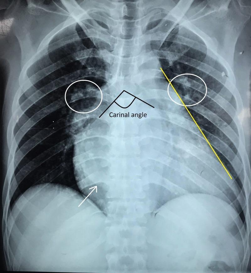
This posteroanterior (PA) chest radiograph illustrates significant cardiomegaly with a cardiothoracic ratio of approximately 70%, commonly associated with advanced valvular disease such as rheumatic mitral stenosis. Annotations highlight pathognomonic radiological signs of left atrial enlargement, including a widened carinal angle (>90 degrees) and a double right heart border (indicated by the white arrow). The left heart border shows characteristic 'straightening' (yellow line) due to the enlargement of the left atrial appendage. Furthermore, the lung fields exhibit cephalization of the pulmonary vasculature (white circles), also known as the 'Antler sign' or 'upturned moustache sign,' which reflects pulmonary venous hypertension and redistribution of blood flow to the upper lobes. The cardiac apex is displaced laterally and downward, suggesting concurrent left ventricular enlargement. This diagnostic image serves as a clinical example of chronic heart failure and congestive changes secondary to severe mitral valve pathology.
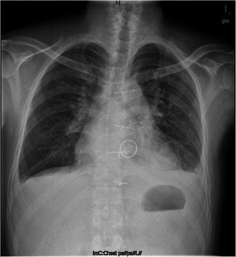
This diagnostic image is a posterior-anterior (PA) chest radiograph showcasing a postoperative state following double valve replacement. The thoracic cavity reveals several key surgical features: radio-opaque sternal wires are visible along the midline, indicating a recent median sternotomy. Within the cardiac silhouette, two prosthetic valves are identified by their distinct radiographic densities and positions. A mechanical valve, characterized by high radio-opacity, is located in the mitral valve position. Superior and slightly anterior to this is a bioprosthetic valve in the pulmonary position, visualized as a less dense, thin radio-opaque ring. The heart appears moderately enlarged (cardiomegaly), and there is a visible increase in bronchovascular markings within the lung fields, consistent with postoperative changes or underlying rheumatic heart disease pathology. Key anatomical landmarks include well-defined clavicles, ribs, and a visible gastric air bubble below the left hemi-diaphragm. This image serves as an educational example of postoperative imaging for complex valvular heart disease and the radiographic differentiation between mechanical and biological prostheses.
mkdir -p /tmp/workspace/rhd-card/imgs && cd /tmp/workspace/rhd-card/imgs && \ curl -sf "https://cdn.orris.care/cdss_images/b7639f7527a4e7eb07d4d3695b2d5168eabaa4508675bb32216e91af05c96f6e.png" -o ms_massive_la.png && \ curl -sf "https://cdn.orris.care/cdss_images/pmc_clinical_VQA_572f92f49f3d27452e2a8a6d347198348f26fde284903e0e7a366cb20ec87bde.jpg" -o ms_annotated.jpg && \ curl -sf "https://cdn.orris.care/cdss_images/7e2cc66468687a7d7ebefeb8b08f54ae2e8a669601c4f24e80c5b68c0b67a898.png" -o as_cxr.png && \ curl -sf "https://cdn.orris.care/cdss_images/pmc_clinical_VQA_53118074199d4fb38c824e50ff1d3d2d904be832653d53de873d45c8214c64d1.jpg" -o ms_cxr2.jpg && \ curl -sf "https://cdn.orris.care/cdss_images/pmc_clinical_VQA_44750b671d6dae74888c4709e32cef29ff345aa3bcd38a28ff2ffdcce31763e1.jpg" -o rhd_double_valve.jpg && \ curl -sf "https://cdn.orris.care/cdss_images/6d77d5c1c95851b65dc8a643c91c4c1b25d602c989db1dd55413cbd96bc2020b.png" -o mr_laa.png && \ ls -la
python3 << 'PYEOF' from reportlab.platypus import ( SimpleDocTemplate, Table, TableStyle, Paragraph, Spacer, HRFlowable, PageBreak, Image, KeepTogether ) from reportlab.lib.styles import getSampleStyleSheet, ParagraphStyle from reportlab.lib import colors from reportlab.lib.units import mm from reportlab.lib.pagesizes import A4 from reportlab.lib.enums import TA_CENTER, TA_LEFT, TA_RIGHT, TA_JUSTIFY import os OUTPUT = "/tmp/workspace/rhd-card/RHD_Radiographic_Signs_CXR.pdf" IMGS = "/tmp/workspace/rhd-card/imgs" W, H = A4 MARGIN = 13*mm CW = W - 2*MARGIN # usable column width # ── Colours ────────────────────────────────────────────────────────── CN = colors.HexColor("#0A2540") # navy CB = colors.HexColor("#1A56DB") # blue CSK = colors.HexColor("#EBF5FF") # sky-blue light CT = colors.HexColor("#0E7490") # teal CTL = colors.HexColor("#CFFAFE") # teal light CAM = colors.HexColor("#92400E") # amber CAL = colors.HexColor("#FEF3C7") # amber light CRD = colors.HexColor("#991B1B") # red CRL = colors.HexColor("#FEE2E2") # red light CGN = colors.HexColor("#065F46") # green CGL = colors.HexColor("#D1FAE5") # green light CPP = colors.HexColor("#5B21B6") # purple CPL = colors.HexColor("#EDE9FE") # purple light CGR = colors.HexColor("#4B5563") # grey CGL2 = colors.HexColor("#F3F4F6") # light grey bg CW2 = colors.white doc = SimpleDocTemplate( OUTPUT, pagesize=A4, leftMargin=MARGIN, rightMargin=MARGIN, topMargin=11*mm, bottomMargin=11*mm, title="Rheumatic Heart Disease – Radiographic Signs", author="DNB Radiology Quick Reference" ) # ── Style factory ───────────────────────────────────────────────────── def mk(name, sz=8, lead=11, fn="Helvetica", tc=colors.HexColor("#1E293B"), align=TA_LEFT, li=0, sa=1, sb=0): return ParagraphStyle(name, fontSize=sz, leading=lead, fontName=fn, textColor=tc, alignment=align, leftIndent=li, spaceAfter=sa, spaceBefore=sb) sTitle = mk("sTitle", 16,21,"Helvetica-Bold",CW2, TA_CENTER, sa=1) sSub = mk("sSub", 8,11,"Helvetica", colors.HexColor("#93C5FD"), TA_CENTER) sSec = mk("sSec", 9.5,13,"Helvetica-Bold",CW2, TA_CENTER) sBold = mk("sBold", 8.2,11.5,"Helvetica-Bold",colors.HexColor("#1E293B")) sNorm = mk("sNorm", 8.2,11.5,"Helvetica", colors.HexColor("#1E293B"), li=5) sNote = mk("sNote", 7.5,10.5,"Helvetica-Oblique",CGR, TA_JUSTIFY) sHdr = mk("sHdr", 8,11,"Helvetica-Bold", CW2, TA_CENTER) sCell = mk("sCell", 7.8,10.8,"Helvetica", colors.HexColor("#1E293B")) sCellB = mk("sCellB", 7.8,10.8,"Helvetica-Bold",colors.HexColor("#1E293B")) sCaption= mk("sCaption",7,9.5,"Helvetica-Oblique",CGR, TA_CENTER) sFooter = mk("sFooter",6.8,9,"Helvetica-Oblique",CGR, TA_CENTER) sWarn = mk("sWarn", 7.8,11,"Helvetica-Bold",CRD) def sp(h=3): return Spacer(1,h) def hr(c=CGR,t=0.5): return HRFlowable(width="100%",thickness=t,color=c,spaceAfter=3,spaceBefore=3) def bul(t): return Paragraph(f"\u2022 {t}", sNorm) def banner(txt, bg, sub=None): rows = [[Paragraph(txt, sTitle)]] if sub: rows.append([Paragraph(sub, sSub)]) t = Table(rows, colWidths=[CW]) t.setStyle(TableStyle([ ('BACKGROUND',(0,0),(-1,-1), bg), ('TOPPADDING',(0,0),(-1,0), 9), ('BOTTOMPADDING',(0,-1),(-1,-1), 8), ('ROWPADDING',(0,0),(-1,-1), 4), ])) return t def sec(txt, bg): t = Table([[Paragraph(txt, sSec)]], colWidths=[CW]) t.setStyle(TableStyle([ ('BACKGROUND',(0,0),(-1,-1), bg), ('ROWPADDING',(0,0),(-1,-1), 5), ])) return t def img_box(path, w, h_max, caption=""): if not os.path.exists(path): return Paragraph(f"[Image not available]", sCaption) from PIL import Image as PILImage pi = PILImage.open(path) pw, ph = pi.size scale = min(w/pw, h_max/ph) iw, ih = pw*scale, ph*scale items = [Image(path, width=iw, height=ih)] if caption: items.append(sp(2)) items.append(Paragraph(caption, sCaption)) rows = [[i] for i in items] t = Table(rows, colWidths=[w]) t.setStyle(TableStyle([ ('ALIGN',(0,0),(-1,-1),'CENTER'), ('TOPPADDING',(0,0),(-1,-1),2), ('BOTTOMPADDING',(0,0),(-1,-1),2), ])) return t def col_box(items, bg, brd, w): rows = [[i] for i in items] t = Table(rows, colWidths=[w]) t.setStyle(TableStyle([ ('BACKGROUND',(0,0),(-1,-1), bg), ('BOX',(0,0),(-1,-1), 0.8, brd), ('LEFTPADDING',(0,0),(-1,-1), 7), ('RIGHTPADDING',(0,0),(-1,-1), 7), ('TOPPADDING',(0,0),(-1,-1), 2), ('BOTTOMPADDING',(0,0),(-1,-1), 2), ('TOPPADDING',(0,0),(0,0), 6), ('BOTTOMPADDING',(0,-1),(0,-1), 6), ('VALIGN',(0,0),(-1,-1),'TOP'), ])) return t def lbl(txt, c): return Paragraph(f"<b>{txt}</b>", mk(f"lb_{txt[:6]}", 8, 11, "Helvetica-Bold", c)) # ══════════════════════════════════════════════════════ # PAGE 1 – Mitral Stenosis (the prototypical RHD film) # ══════════════════════════════════════════════════════ E = [] E.append(banner( "RHEUMATIC HEART DISEASE — RADIOGRAPHIC SIGNS", CN, "CXR Quick Reference · DNB Radiology Rapid Exam Review" )) E.append(sp(5)) # Intro strip intro_rows = [ [Paragraph("<b>Valve frequency in RHD:</b>", sCellB), Paragraph("Mitral (>50% isolated) \u2192 Aortic \u2192 Tricuspid \u2192 Pulmonary (rare)", sCell)], [Paragraph("<b>CXR role:</b>", sCellB), Paragraph("Assess chamber enlargement, pulmonary vascularity, calcification, great vessel changes", sCell)], ] intro = Table(intro_rows, colWidths=[CW*0.22, CW*0.78]) intro.setStyle(TableStyle([ ('BOX',(0,0),(-1,-1), 0.8, CB), ('INNERGRID',(0,0),(-1,-1), 0.3, CSK), ('BACKGROUND',(0,0),(0,-1), CSK), ('BACKGROUND',(1,0),(-1,-1), CW2), ('ROWPADDING',(0,0),(-1,-1), 5), ('VALIGN',(0,0),(-1,-1),'MIDDLE'), ])) E.append(intro) E.append(sp(6)) # ── MITRAL STENOSIS ────────────────────────────────────────────────── E.append(sec("MITRAL STENOSIS (MS) — The Prototypical RHD Radiograph", CT)) E.append(sp(4)) IW = CW * 0.44 # image column TW = CW * 0.53 # text column GAP = CW - IW - TW ms_signs = [ lbl("LEFT ATRIUM ENLARGEMENT (hallmark):", CT), sp(2), bul("Double density sign — retrocardiac 2nd opacity on right"), bul("Third mogul / LAA bump — convexity below PA segment"), bul("Subcarinal angle widening > 70\u00b0 (splayed carina)"), bul("Elevation & horizontalisation of left main bronchus"), bul("Posterior esophageal displacement (lateral / barium)"), sp(3), lbl("RIGHT VENTRICLE ENLARGEMENT (from PAH):", CT), sp(2), bul("Elevated & leftward apex"), bul("Prominent pulmonary trunk segment (left border)"), bul("Retrosternal filling on lateral (most sensitive)"), sp(3), lbl("SMALL AORTIC KNOB:", CT), sp(2), bul("Inconspicuous aortic knob — reduced CO + LV rotation"), bul("ASD + MS: small aortic knob + enlarged PA = classic"), sp(3), lbl("GREAT VESSELS:", CT), sp(2), bul("Enlarged pulmonary trunk + central PAs (PAH)"), bul("Peripheral pruning (pre-capillary component)"), ] ms_img = col_box([ img_box(f"{IMGS}/ms_massive_la.png", IW-14, 88, "Fig 1. Massive LA enlargement in longstanding MS.\nHeart fills the cardiac silhouette.\n(Bailey & Love's Surgery 28e, Fig 59.14)"), sp(4), img_box(f"{IMGS}/ms_annotated.jpg", IW-14, 88, "Fig 2. Annotated MS CXR: double density (arrow),\nstraightened left border (LAA), cephalization\n(circles), widened carina."), ], CTL, CT, IW) ms_txt = col_box(ms_signs, CTL, CT, TW) ms_row = Table([[ms_img, ms_txt]], colWidths=[IW, TW]) ms_row.setStyle(TableStyle([ ('LEFTPADDING',(0,0),(-1,-1),0),('RIGHTPADDING',(0,0),(-1,-1),0), ('TOPPADDING',(0,0),(-1,-1),0),('BOTTOMPADDING',(0,0),(-1,-1),0), ('VALIGN',(0,0),(-1,-1),'TOP'), ('COLPADDING',(0,0),(0,-1),0), ])) E.append(ms_row) E.append(sp(5)) # ── PULMONARY VENOUS HYPERTENSION GRADES ───────────────────────────── E.append(sec("PULMONARY VENOUS HYPERTENSION (PVH) GRADES IN MS", CB)) E.append(sp(3)) pvh = [ [Paragraph("<b>Grade</b>",sHdr), Paragraph("<b>PVP</b>",sHdr), Paragraph("<b>CXR Findings</b>",sHdr), Paragraph("<b>Key Signs</b>",sHdr)], ["I — Redistribution","12–18 mmHg", "Upper lobe vessel dilatation; lower lobe constriction", "Cephalization / upper lobe diversion"], ["II — Interstitial oedema","18–25 mmHg", "Peribronchial cuffing; septal lines; hazy hila", "Kerley A, B, C lines; \u2018antler sign\u2019"], ["III — Alveolar oedema",">25 mmHg", "Bilateral perihilar consolidation", "Butterfly / bat-wing pattern"], ["Chronic MS","Longstanding", "Pulmonary haemosiderosis; ossification nodules", "Fine nodular opacities (miliary haemosiderin)"], ] pvh_rows = [[Paragraph(str(c),sHdr if i==0 else sCell) for c in r] for i,r in enumerate(pvh)] cw4 = [CW*0.16, CW*0.14, CW*0.41, CW*0.29] pvh_tbl = Table(pvh_rows, colWidths=cw4, repeatRows=1) pvh_ts = [ ('BACKGROUND',(0,0),(-1,0),CB), ('FONTNAME',(0,0),(-1,0),'Helvetica-Bold'), ('FONTSIZE',(0,0),(-1,-1),7.8), ('ROWPADDING',(0,0),(-1,-1),4), ('INNERGRID',(0,0),(-1,-1),0.3,CGR), ('BOX',(0,0),(-1,-1),0.8,CB), ('VALIGN',(0,0),(-1,-1),'MIDDLE'), ] for i in range(1,len(pvh_rows)): pvh_ts.append(('BACKGROUND',(0,i),(-1,i), CSK if i%2==0 else CW2)) pvh_tbl.setStyle(TableStyle(pvh_ts)) E.append(pvh_tbl) E.append(sp(4)) # Kerley lines box kerley = [ Paragraph("<b>KERLEY LINES — Mnemonics</b>", mk("kh",8,11,"Helvetica-Bold",CAM)), sp(2), bul("<b>Kerley B</b> — Short (1–2 cm), HORIZONTAL, at BASES (costophrenic angles); distended interlobular lymphatics — Most common in MS"), bul("<b>Kerley A</b> — Long (4–6 cm), oblique, radiating toward hila from upper zones; deeper lymphatics"), bul("<b>Kerley C</b> — Fine reticular network throughout both lungs (summation of B lines en face)"), ] kerley_box = col_box(kerley, CAL, CAM, CW) E.append(kerley_box) E.append(PageBreak()) # ══════════════════════════════════════════════════════ # PAGE 2 – MR, AS, AR # ══════════════════════════════════════════════════════ E.append(banner("RHEUMATIC HEART DISEASE — RADIOGRAPHIC SIGNS | Page 2", CN)) E.append(sp(5)) # ── MITRAL REGURGITATION ───────────────────────────────────────────── E.append(sec("MITRAL REGURGITATION (MR)", CGN)) E.append(sp(3)) HW = (CW - 4) / 2 mr_left_items = [ lbl("CARDIAC SILHOUETTE:", CGN), sp(2), bul("LA enlargement: double density, 3rd mogul, elevated left bronchus"), bul("LAA bulge is often MORE prominent in MR vs. pure MS"), bul("LV enlargement (DISTINGUISHES MR from MS):"), Paragraph(" \u2192 Drooping apex (downward + leftward + outward)", sNorm), Paragraph(" \u2192 Rounding of lower left cardiac border", sNorm), Paragraph(" \u2192 Increased CTR (volume overload)", sNorm), bul("Biventricular / biatrial enlargement in advanced chronic MR"), sp(3), lbl("PULMONARY VASCULARITY:", CGN), sp(2), bul("Pulmonary venous hypertension with cephalization"), bul("Less severe pulmonary oedema than MS (more compliant LA)"), bul("Acute MR (ruptured chordae): pulmonary oedema + NORMAL-sized heart"), sp(3), lbl("KEY DISTINCTION FROM MS:", CGN), sp(2), Paragraph("<b>MS</b>: LA + RV enlarged; LV small; small aortic knob", sNorm), Paragraph("<b>MR</b>: LA + LV enlarged; larger overall cardiomegaly", sNorm), ] mr_right_items = [ img_box(f"{IMGS}/mr_laa.png", HW-14, 105, "Fig 3. Mitral regurgitation: LAA\nenlargement (arrow); subcarinal opacity\nand elevated left main bronchus from LA\nenlargement visible.\n(Grainger & Allison Fig 14.33)"), ] mr_row = Table([[ col_box(mr_left_items, CGL, CGN, HW), col_box(mr_right_items, CGL, CGN, HW), ]], colWidths=[HW, HW]) mr_row.setStyle(TableStyle([ ('LEFTPADDING',(0,0),(-1,-1),0),('RIGHTPADDING',(0,0),(-1,-1),0), ('TOPPADDING',(0,0),(-1,-1),0),('BOTTOMPADDING',(0,0),(-1,-1),0), ('VALIGN',(0,0),(-1,-1),'TOP'), ])) E.append(mr_row) E.append(sp(5)) # ── AORTIC STENOSIS ────────────────────────────────────────────────── E.append(sec("AORTIC STENOSIS (AS) — Almost Always with Mitral Disease in RHD", CRD)) E.append(sp(3)) as_left = [ lbl("CARDIAC SILHOUETTE:", CRD), sp(2), bul("Normal / near-normal cardiac size — pressure overload causes concentric hypertrophy WITHOUT dilatation"), bul("LV dilatation + drooping apex only when LV failure develops"), sp(3), lbl("AORTIC SIGNS (most specific):", CRD), sp(2), bul("Post-stenotic dilatation of ASCENDING AORTA ONLY — jet effect from stenotic valve → turbulence → focal dilatation"), bul("Selective ascending aorta dilatation = VALVULAR AS (not arch/descending)"), bul("Entire thoracic aorta enlarged = AR or hypertension (NOT isolated AS)"), sp(3), lbl("CALCIFICATION:", CRD), sp(2), bul("Aortic valve calcification: dense opacity at aortic valve area (3rd right costal cartilage level)"), bul("Best on lateral CXR / fluoroscopy"), bul("Degree of calcification ∝ severity of stenosis"), sp(3), lbl("PULMONARY VASCULARITY:", CRD), sp(2), bul("Normal in compensated AS"), bul("Cephalization + Kerley B lines when LV fails"), ] as_right = [ img_box(f"{IMGS}/as_cxr.png", HW-14, 100, "Fig 4. Aortic stenosis CXR.\nNote selective post-stenotic dilatation\nof the ascending aorta.\n(Bailey & Love's Surgery 28e, Fig 59.19)"), ] as_row = Table([[ col_box(as_left, CRL, CRD, HW), col_box(as_right, CRL, CRD, HW), ]], colWidths=[HW, HW]) as_row.setStyle(TableStyle([ ('LEFTPADDING',(0,0),(-1,-1),0),('RIGHTPADDING',(0,0),(-1,-1),0), ('TOPPADDING',(0,0),(-1,-1),0),('BOTTOMPADDING',(0,0),(-1,-1),0), ('VALIGN',(0,0),(-1,-1),'TOP'), ])) E.append(as_row) E.append(sp(5)) # ── AORTIC REGURGITATION ───────────────────────────────────────────── E.append(sec("AORTIC REGURGITATION (AR) — Usually Combined with Mitral Disease", CPP)) E.append(sp(3)) ar_items = [ lbl("CARDIAC SILHOUETTE:", CPP), sp(2), bul("Significant LV enlargement (volume overload): apex displaced downward + laterally; rounding of left border"), bul("Largest hearts on CXR — 'Cor bovinum' (ox heart) in severe chronic AR"), bul("Increased CTR proportional to AR severity"), sp(3), lbl("AORTIC SIGNS:", CPP), sp(2), bul("Generalised dilatation of ENTIRE thoracic aorta (ascending + arch + descending) — distinguishes from AS"), bul("Prominent aortic knob"), bul("Degree of aortic dilatation proportional to LV size"), sp(3), lbl("PULMONARY VASCULARITY:", CPP), sp(2), bul("Normal in compensated AR; cephalization + oedema when LV fails"), ] ar_box = col_box(ar_items, CPL, CPP, CW) E.append(ar_box) E.append(PageBreak()) # ══════════════════════════════════════════════════════ # PAGE 3 – Tricuspid, Summary table, Memory aids, Images # ══════════════════════════════════════════════════════ E.append(banner("RHEUMATIC HEART DISEASE — RADIOGRAPHIC SIGNS | Page 3", CN)) E.append(sp(5)) # ── TRICUSPID DISEASE ──────────────────────────────────────────────── E.append(sec("TRICUSPID VALVE DISEASE IN RHD", CAM)) E.append(sp(3)) HW2 = (CW-4)/2 tr_items = [ lbl("TRICUSPID REGURGITATION (TR) — Usually Functional / Secondary:", CAM), sp(2), bul("Gross right atrial enlargement: markedly convex, rightward bulging right border"), bul("Straightened left cardiac border (from LA + PA prominence on left)"), bul("Severe MS \u2192 functional TR: massive RA on right + LA/PA on left = 'double convexity'"), bul("SVC and azygos vein dilatation (systemic venous hypertension)"), bul("When TR relieves LV back-pressure: paradoxical improvement of pulmonary congestion"), sp(3), lbl("TRICUSPID STENOSIS (TS) — Organic, Rare:", CAM), sp(2), bul("RA enlargement WITHOUT pulmonary artery enlargement — characteristic pattern"), bul("SVC + azygos vein dilatation"), bul("No pulmonary venous hypertension (obstruction is pre-pulmonary)"), bul("Hepatomegaly + ascites shadows on CXR (systemic congestion)"), ] tv_img_items = [ img_box(f"{IMGS}/rhd_double_valve.jpg", HW2-14, 95, "Fig 5. Post double-valve replacement in RHD.\nMechanical valve (mitral position, high opacity)\nand bioprosthetic valve (pulmonary/aortic,\nless dense ring) with sternal wires.\n(RHD post-operative state)"), ] tr_row = Table([[ col_box(tr_items, CAL, CAM, HW2), col_box(tv_img_items, CAL, CAM, HW2), ]], colWidths=[HW2, HW2]) tr_row.setStyle(TableStyle([ ('LEFTPADDING',(0,0),(-1,-1),0),('RIGHTPADDING',(0,0),(-1,-1),0), ('TOPPADDING',(0,0),(-1,-1),0),('BOTTOMPADDING',(0,0),(-1,-1),0), ('VALIGN',(0,0),(-1,-1),'TOP'), ])) E.append(tr_row) E.append(sp(5)) # ── MASTER COMPARISON TABLE ─────────────────────────────────────────── E.append(sec("COMPARATIVE CXR FEATURES — ALL RHD VALVULAR LESIONS", CN)) E.append(sp(3)) comp = [ ["Feature", "MS", "MR", "AS", "AR", "TR (functional)"], ["LA enlargement", "+++", "+++", "\u2014", "\u2014", "\u2014"], ["LV enlargement", "\u2014", "++", "+ (late)", "+++", "\u2014"], ["RV enlargement", "++", "+", "\u2014", "\u2014", "+"], ["RA enlargement", "+ (2\u00b0)", "+ (2\u00b0)", "\u2014", "\u2014", "+++"], ["Aortic knob", "Small", "Normal","Ascending dil.", "Whole aorta dil.", "Normal"], ["Pulmonary trunk", "Enlarged","Enlarged","Normal","Normal", "Normal"], ["PVH / Cephalization", "+++", "++", "+ (late)","+ (late)", "\u2014"], ["Kerley B lines", "+++", "+", "\u2014", "\u2014", "\u2014"], ["Calcification", "Mitral", "Mitral annular","Aortic","Aortic", "\u2014"], ["CTR", "++", "+++", "Normal", "+++", "+"], ["Classic silhouette", "Straight L border","Big LV apex","Normal","Cor bovinum","Wide Rt border"], ] col_colors = [CN, CT, CGN, CRD, CPP, CAM] comp_hdrs = [Paragraph(str(c), sHdr) for c in comp[0]] comp_rows = [comp_hdrs] for r in comp[1:]: comp_rows.append([Paragraph(str(c), sCell) for c in r]) cw6 = [CW*0.20, CW*0.13, CW*0.13, CW*0.13, CW*0.13, CW*0.28] comp_tbl = Table(comp_rows, colWidths=cw6, repeatRows=1) comp_ts = [ ('FONTSIZE',(0,0),(-1,-1), 7.5), ('ROWPADDING',(0,0),(-1,-1), 3.5), ('INNERGRID',(0,0),(-1,-1), 0.3, CGR), ('BOX',(0,0),(-1,-1), 0.9, CN), ('VALIGN',(0,0),(-1,-1),'MIDDLE'), ('FONTNAME',(0,0),(-1,0),'Helvetica-Bold'), ] # Header row colours for j, c in enumerate(col_colors): comp_ts.append(('BACKGROUND',(j,0),(j,0), c)) # Alternating row bg for i in range(1, len(comp_rows)): comp_ts.append(('BACKGROUND',(0,i),(-1,i), CGL2 if i%2==0 else CW2)) # Bold first column for i in range(1, len(comp_rows)): comp_ts.append(('FONTNAME',(0,i),(0,i),'Helvetica-Bold')) comp_tbl.setStyle(TableStyle(comp_ts)) E.append(comp_tbl) E.append(sp(5)) # ── MEMORY AIDS ────────────────────────────────────────────────────── E.append(sec("HIGH-YIELD MEMORY AIDS & EXAM CLUES", CAM)) E.append(sp(3)) mem = [ ["Small aortic knob + prominent PA + LA enlargement + cephalization", "= Mitral Stenosis (classic triad)"], ["Double density sign + 3rd mogul + elevated left bronchus + widened carina", "= LA enlargement (MS/MR)"], ["MS: LA + RV enlarged; LV SMALL; small aortic knob", "vs. MR: LA + LV enlarged; bigger cardiomegaly"], ["Post-stenotic dilatation of ASCENDING AORTA ONLY", "= Valvular AS (not arch/descending)"], ["ENTIRE thoracic aorta enlarged + large LV", "= AR (cor bovinum)"], ["RA enlargement WITHOUT PA segment enlargement", "= Organic tricuspid stenosis"], ["Massive RA + straightened left border", "= Severe MS \u2192 functional TR"], ["Normal-sized heart + pulmonary oedema (acute)", "= Acute MR (ruptured chordae); also acute LV failure"], ["Pulmonary haemosiderosis + ossification nodules", "= Long-standing severe MS"], ["Improvement of pulmonary congestion with worsening RHF", "Paradox: TR relieves pulmonary back-pressure in MS"], ] mem_rows = [[Paragraph(f"<b>\u25ba {r[0]}</b>", mk(f"mm{i}",7.8,11,"Helvetica-Bold",CAM)), Paragraph(r[1], sCell)] for i,r in enumerate(mem)] mem_tbl = Table(mem_rows, colWidths=[CW*0.52, CW*0.48]) mem_ts = [ ('BOX',(0,0),(-1,-1), 0.8, CAM), ('INNERGRID',(0,0),(-1,-1), 0.3, CAL), ('ROWPADDING',(0,0),(-1,-1), 4), ('VALIGN',(0,0),(-1,-1),'TOP'), ] for i in range(len(mem_rows)): mem_ts.append(('BACKGROUND',(0,i),(0,i), CAL)) mem_ts.append(('BACKGROUND',(1,i),(1,i), CW2 if i%2==0 else CGL2)) mem_tbl.setStyle(TableStyle(mem_ts)) E.append(mem_tbl) E.append(sp(5)) E.append(hr(CGR, 0.6)) E.append(sp(3)) ft = Table([[ Paragraph("Sources: Fuster & Hurst's The Heart 15e · Bailey & Love's Surgery 28e · Grainger & Allison's Diagnostic Radiology · Braunwald's Heart Disease", sFooter), Paragraph("DNB Radiology Rapid Review | For educational use only", sFooter), ]], colWidths=[CW*0.72, CW*0.28]) ft.setStyle(TableStyle([ ('VALIGN',(0,0),(-1,-1),'MIDDLE'), ('LEFTPADDING',(0,0),(-1,-1),0),('RIGHTPADDING',(0,0),(-1,-1),0), ])) E.append(ft) # BUILD doc.build(E) print(f"SUCCESS: {OUTPUT}") PYEOF