
Whiplash injury
whiplash injury cervical spine
whiplash injury cervical spine mechanism
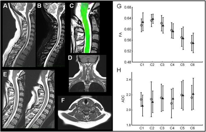
A multi-panel medical figure presenting diagnostic imaging and quantitative data for Whiplash-Associated Disorder (WAD). Panels A-F show anatomical and functional MRI of the cervical spine: (A) Sagittal T2-weighted and (B) STIR sequences; (C) DTI-tractography highlighting spinal cord fibers; (D) Coronal T1-weighted view of the neck; (E) Dynamic T2-weighted sagittal imaging assessing cervical stability; and (F) Axial T1-weighted view of the neck musculature. These images demonstrate an absence of traumatic lesions, fractures, or disc herniation. Panels G and H are comparison charts displaying Diffusion Tensor Imaging (DTI) metrics from cervical levels C1 to C6. Graph G measures Fractional Anisotropy (FA), and Graph H measures the Apparent Diffusion Coefficient (ADC). In both, open circles represent the acute phase and filled triangles represent the chronic phase of WAD. The data indicates no significant change in spinal cord microstructural integrity over time. The figure serves as a clinical example of structural findings in WAD patients at different injury stages.
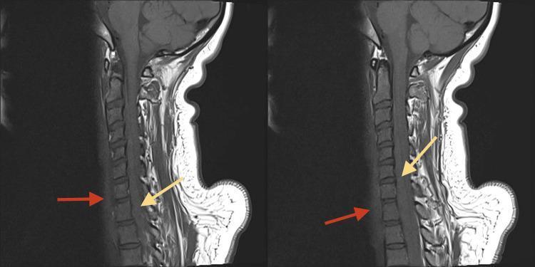
This diagnostic image consists of two side-by-side sagittal T1-weighted MRI scans of the cervical spine. The imaging demonstrates acute post-traumatic findings, specifically identifying prevertebral and intraspinal hemorrhage. Red arrows point to a prevertebral hematoma, visualized as a region of slight hyperintensity in the prevertebral soft tissues anterior to the C5-T1 vertebral bodies. Yellow arrows indicate an intraspinal epidural hematoma, which appears as a hyperintense signal within the spinal canal, posterior to the vertebral bodies, causing mass effect and moderate compression of the cervical spinal cord. The anatomical region spans the lower cervical and upper thoracic levels. These features are characteristic of acute whiplash or high-energy trauma, highlighting the risk of multi-compartment bleeding. This content is intended for intermediate to advanced medical education, focusing on neuroradiology and emergency trauma assessment of spinal cord compression and ligamentous injury.
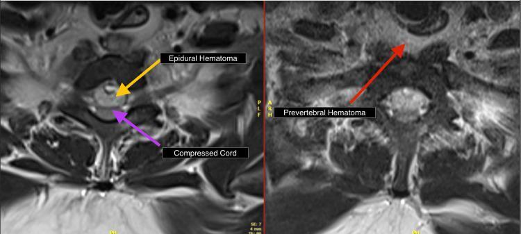
This diagnostic image consists of two side-by-side axial T2-weighted MRI (T2WI) views of the cervical-thoracic spine. The left panel demonstrates a large intraspinal epidural hematoma, visualized as an area of heterogeneous hyperintensity within the spinal canal. A yellow arrow points to the hematoma, while a purple arrow indicates the resulting anterior compression of the spinal cord, which appears flattened and displaced. The right panel highlights the prevertebral region, where a red arrow identifies a prevertebral hematoma manifesting as hyperintense fluid collection anterior to the vertebral body. These findings illustrate significant soft tissue trauma, specifically secondary to whiplash or spinal injury, leading to cord compromise. The image serves as an educational tool for identifying acute post-traumatic spinal hematomas and assessing their spatial relationship with the spinal cord and vertebral column.
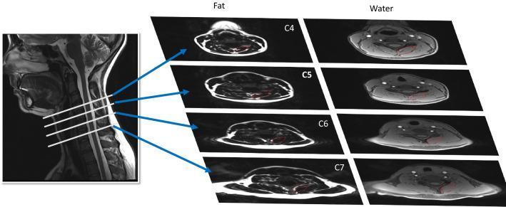
This diagnostic image demonstrates a comparative fat/water MRI study of the deep cervical extensor muscles (multifidus and semispinalis cervicis) across the C4 to C7 vertebral levels. On the left, a sagittal T2-weighted MRI of the cervical spine serves as a reference, with horizontal white lines and blue arrows indicating the specific axial slice locations. To the right, two columns of axial images illustrate the results of a 2-point Dixon sequence: the 'Fat' column highlights high-signal (bright) areas of muscle fat infiltration (MFI), while the 'Water' column displays corresponding water-signal images. Red outlines on the axial views delineate the manual segmentation of the dorsal muscle groups for quantitative analysis. This visual material is used in clinical research to assess muscle composition changes in patients following whiplash-associated disorders (WAD). The presentation emphasizes the anatomical distribution of intramuscular adipose tissue, which is a key biomarker for cervical spine pathology and functional recovery post-injury.
Whiplash Injury
Definition
- Scott-Brown's Otorhinolaryngology, Vol. 2, p. 1158
- Bradley and Daroff's Neurology in Clinical Practice, p. 552
Epidemiology
-
Incidence: 70-300 per 100,000 population
-
Approximately 1 million new cases per year in the United States
-
In the UK, ~250,000 patients per year cost an estimated £3 billion
-
Rear-end collisions account for 85% of whiplash injuries
-
Compulsory seat belt laws paradoxically increased legal submissions by 268% in the UK
-
Scott-Brown's Otorhinolaryngology, Vol. 2, p. 1158
Biomechanics
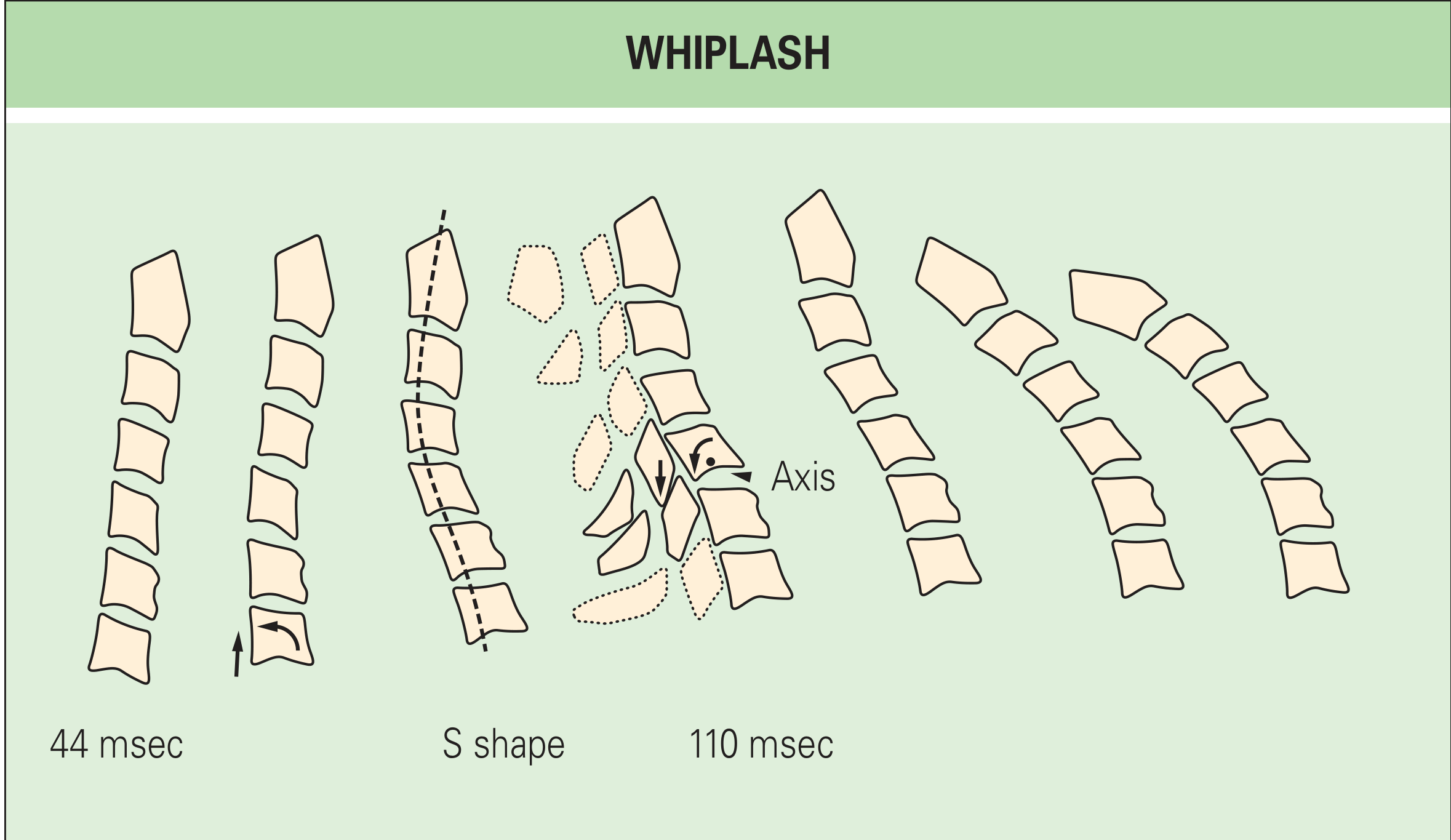
- Rear-end impact thrusts the lower cervical spine upward and forward
- The neck is compressed from below
- The lower cervical segments extend while the upper segments remain relatively flexed - creating an "S" shape at 50-100 msec
- At ~100 msec, the C6 vertebra rotates about an abnormally high axis of rotation; the vertebral body separates anteriorly from C7 and the inferior articular process chisels into the superior articular process below
- All segments then undergo progressive extension, and finally the head is thrown forward (flexion phase)
Pathology
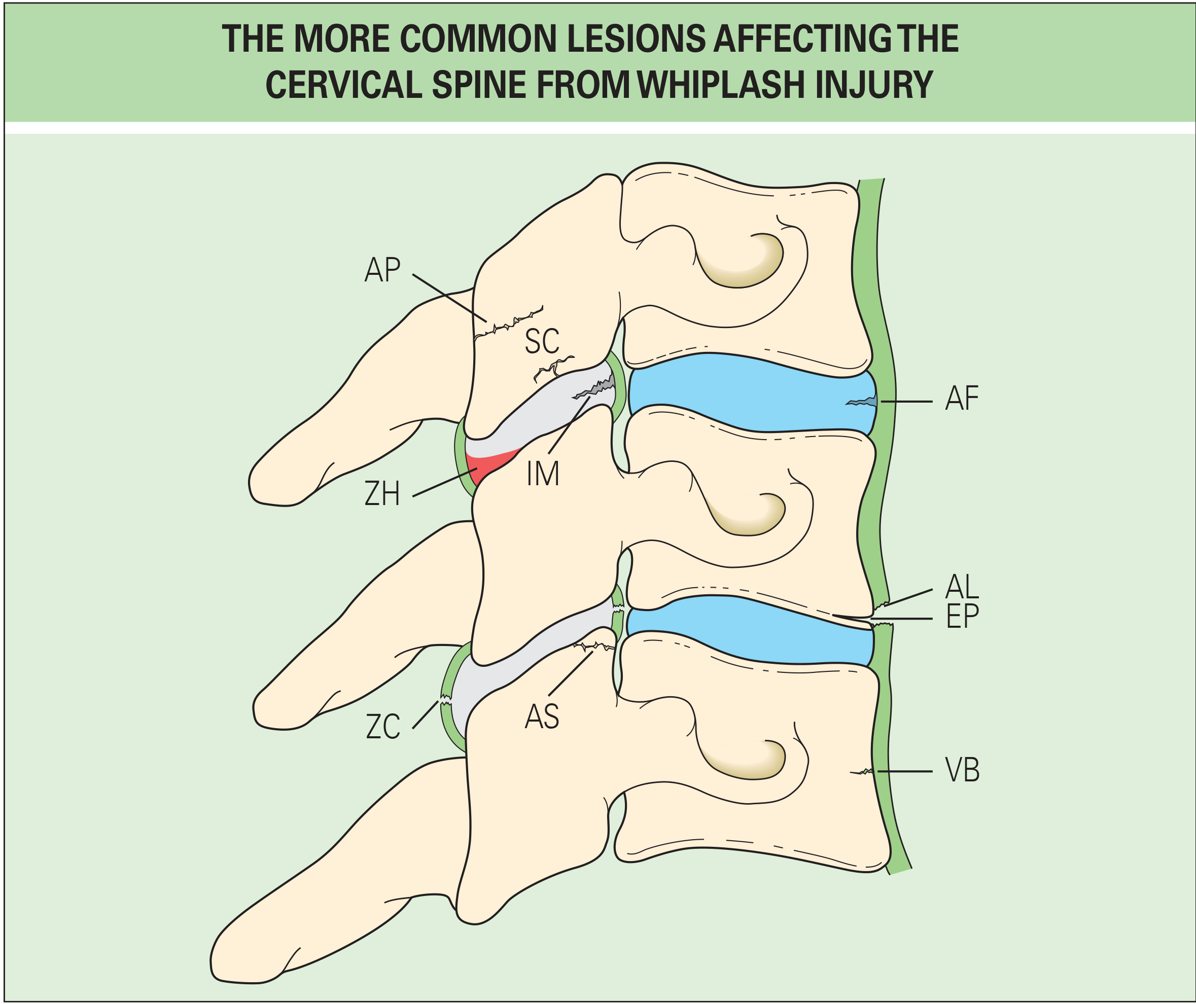
| Structure | Injury |
|---|---|
| Zygapophyseal joint | Hemarthrosis (ZH), capsule rupture (ZC), articular surface fracture (AS) |
| Intraarticular meniscus | Contusion (IM) |
| Subchondral plate | Fracture (SC) |
| Articular pillar | Fracture (AP) |
| Intervertebral disc | Annulus fibrosus tear (AF) |
| Anterior longitudinal ligament | Tear (AL) |
| Vertebral endplate | Avulsion/fracture (EP) |
| Vertebral body | Fracture (VB) |
- Rheumatology, 2-Volume Set (Elsevier, 2022), p. 77
Quebec Task Force Classification (1995) - WAD Grading
| Grade | Symptoms and Signs |
|---|---|
| 0 | No symptoms |
| I | Complaint of pain on motion; no physical signs |
| II | Pain on motion + physical signs (decreased ROM, point tenderness) |
| III | Grade II + neurological signs (weakness, sensory loss, absent reflexes, long-tract signs) |
| IV | Fracture or dislocation |
- Scott-Brown's Otorhinolaryngology, Vol. 2, p. 1158
Clinical Features
| Symptom | Approximate Prevalence |
|---|---|
| Neck stiffness | 96% |
| Neck pain | 94% |
| Headache | 44% |
| Interscapular pain | 35% |
| Sleeping problems | 35% |
| Signs of stress | 30% |
| Numbness/paraesthesia | 22% |
| Vertigo | 15% |
| Memory problems | 15% |
| Eye symptoms | 12% |
| Hearing symptoms | 13% |
ENT/Audiovestibular Manifestations
-
Subjective hearing loss: ~13% of patients; objective audiometric loss (grade I-II) is rare
-
Tinnitus: particularly in grade I and II injuries
-
Vertigo/dizziness: one of the more persistent symptoms in late whiplash syndrome; 15-20% of severe cases develop late whiplash syndrome with headache, vertigo, instability, nausea, tinnitus, and hearing loss
-
Speech discrimination: 30% of whiplash patients show abnormalities in speech-in-noise testing vs. 5% of controls, despite a normal audiogram
-
Grade III-IV injuries may cause intralaybyrinthine trauma similar to head injury
-
Audiovestibular symptoms typically resolve within 5 months
-
Scott-Brown's Otorhinolaryngology, Vol. 2, pp. 1158-1160
Neurological/Pain Features (Bradley & Daroff)
-
~20% of patients complain of cognitive symptoms after whiplash; cognitive dysfunction in minor injuries is likely functional
-
The influence of compensation and legal context is notable: studies from Lithuania (where personal injury insurance is uncommon) showed significantly less symptomatology at 1 year compared to similar accidents in the United States
-
About 40% of patients report complete recovery at 2 years; ~45% continue to have major complaints at 2 years
-
Bradley and Daroff's Neurology in Clinical Practice, p. 552
Imaging
- Plain radiographs: Insensitive for soft tissue injuries; useful only for fractures/dislocations
- CT: Better for bony detail
- MRI: Investigation of choice for soft tissue, disc, ligamentous, and spinal cord injuries. DTI (Diffusion Tensor Imaging) can assess spinal cord microstructural integrity and muscle fat infiltration of deep cervical extensors - useful for chronic WAD research
Prognosis
- Grade I-II: 97-100% spontaneous recovery within 1 year
- Overall (grades I-IV): most recover within 8 weeks
- 14-50% develop chronic WAD
- Up to 50% may have minor residual symptoms for up to 2 years
- Degenerative MRI changes can progress over 10 years, but >50% of patients improve symptomatically (no correlation between imaging and symptoms)
-
High initial pain intensity (strongest predictor)
-
Severity of initial trauma
-
Double collision (rear-end followed by front-end)
-
Serious vehicle damage
-
Pre-existing mental health problems
-
High WAD grade
-
Not recovered by 6 months (associated with long-term difficulties)
-
Scott-Brown's Otorhinolaryngology, Vol. 2, p. 1160
Prognosis by Grade (Bradley & Daroff)
| Finding | Grade I | Grade II | Grade III |
|---|---|---|---|
| Neck pain >6 months | 44% | 81% | Up to 90% |
| Headache >6 months | 37% | 37% | 70% |
Treatment
Acute Whiplash
- Active approaches - activation, encouragement to resume normal activities
- Multimodal interventions
- Simple reassurance about the benign nature
-
Cervical collars (soft collars immobilize and may worsen outcomes)
-
Rest
-
Passive physical modalities
-
Rheumatology, 2-Volume Set (Elsevier, 2022)
Chronic Whiplash
- Percutaneous radiofrequency neurotomy of the medial branches supplying the zygapophyseal joints - the single most promising treatment for constant, debilitating pain (palliative but valuable)
- Exercise therapy for chronic neck pain
- Management of comorbidities: anxiety, sleep disorders, depression should be actively addressed
For ENT Manifestations
- Hearing losses and tinnitus: treated on their own merits
- Dizziness/vertigo: vestibular rehabilitation exercises are effective even when neck pain limits movement
- Psychological support for stress disorders and anxiety
Forensic Significance
- P.C. Dikshit's Textbook of Forensic Medicine and Toxicology
Summary
Recent evidence note: A 2025 systematic review (PMID 39791243) on clinical guidelines for neck pain found substantial variation in recommendations for cervical mobilization/manipulation and screening for vascular complications - clinicians should screen appropriately before manual therapy for post-whiplash neck pain.
Vertebrae involved in whiplash injury
cervical vertebrae levels whiplash injury C5 C6 diagram
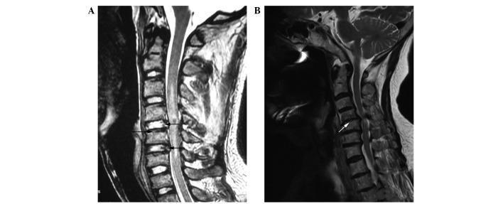
This diagnostic image consists of two sagittal T2-weighted magnetic resonance imaging (MRI) scans of the cervical spine, demonstrating a comparison between traumatic injury and degenerative disease. Panel A shows a control patient with cervical vertebrae fractures and dislocation at the C4 and C5 levels; however, the intervertebral discs at C4-5 and C5-6 maintain high T2 signal intensity, indicating healthy, non-degenerated nuclei pulposi. Black arrows point toward the experimental harvest sites. Panel B displays the cervical spondylosis group, characterized by multilevel disc herniation and significant signal loss (darkening) at the C4-5, C5-6, and C6-7 levels, which is diagnostic of advanced intervertebral disc degeneration (Thompson grades 3-5). A white arrow highlights a subtotal vertebrectomy site. The image illustrates the contrast between acute mechanical trauma (fracture/dislocation) with preserved disc integrity versus the progressive pathological changes of cervical spondylosis, including canal stenosis and decreased disc height.
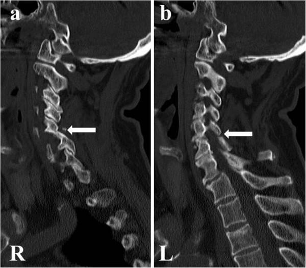
This diagnostic image consists of two sagittal computed tomography (CT) scans of the human cervical spine, bone window, labeled (a) 'R' for the right side and (b) 'L' for the left side. The images demonstrate the cervical vertebrae from the skull base down to the upper thoracic region. The primary pathology is a bilateral facet joint dislocation at the C5-C6 level, indicated by white horizontal arrows in both panels. This finding represents a significant post-traumatic cervical spine injury characterized by the displacement of the inferior articular processes of C5 anterior to the superior articular processes of C6, resulting in vertebral malalignment and disruption of the normal posterior spinal curvature. The vertebral bodies at other levels maintain a generally rectangular shape with preserved intervertebral disc spaces. This visual serves as a critical educational example of high-grade spinal instability and segmental dislocation following cervical trauma, highlighting the importance of sagittal reconstruction in identifying facet joint 'jump' or lock.
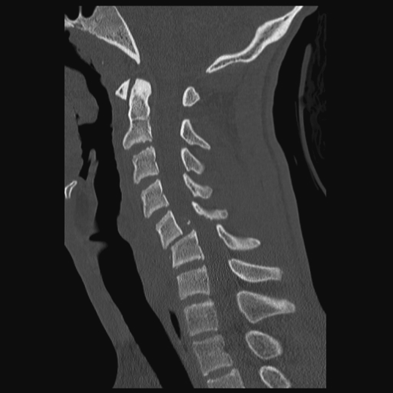
**Imaging Modality:** Computed Tomography (CT), sagittal reformatted view. **Anatomical Region:** Cervical spine extending from the craniocervical junction to the upper thoracic levels. **Observed Pathology:** Traumatic cervical spine injury characterized by a high-grade anterolisthesis of C5 on C6. There is a complete disruption of the normal spinolaminar line and posterior vertebral line, consistent with a bilateral facet dislocation or severe fracture-subluxation. **Characteristic Visual Features:** - **Skeletal Alignment:** Significant anterior displacement of the C5 vertebral body relative to C6, resulting in "off-set" alignment. - **Canal Dimensions:** Severe narrowing and compromise of the central spinal canal diameter at the C5-C6 level due to the osseous translation. - **Associated Findings:** Disruption of the anterior and posterior longitudinal ligament planes; prevertebral soft tissue swelling is suggested anterior to the mid-cervical vertebrae. The remaining cervical segments (C1-C4 and C7) appear relatively aligned. **Key Diagnostic Features:** Traumatic spondylolisthesis with acute spinal canal stenosis, secondary to a distractive-flexion injury mechanism. This represents a highly unstable cervical spine fracture-dislocation pattern.
Vertebrae Involved in Whiplash Injury
The Key Principle: Not a Single Level
Phases and Vertebral Levels Involved
Phase 1 - S-Shape Deformation (0-100 msec after impact)
| Cervical Region | Movement | Vulnerable Levels |
|---|---|---|
| Lower cervical (C4-C7) | Extension - moves first | C5-C6, C6-C7 most stressed |
| Upper cervical (C1-C4) | Relative flexion | C2-C3, C3-C4 |
- The anterior end of the vertebral body to separate from the vertebral body below
- The inferior articular process to chisel into the superior articular process of the segment below
- Rheumatology, 2-Volume Set (Elsevier, 2022), p. 77
Phase 2 - Full Extension (>100 msec)
Vertebral Levels by Injury Type
Most Commonly Injured Levels
| Level | Why It Is Vulnerable | Typical Injury |
|---|---|---|
| C5-C6 | Maximum rotational stress during S-phase; most mobile lower cervical segment | Zygapophyseal joint injury, disc herniation, facet dislocation |
| C6-C7 | Lower cervical extension zone; adjacent to C6 pivot point | Disc herniation, endplate avulsion, annulus tear |
| C2-C3 | Hyperflexion-hyperextension at upper cervical; axis fracture | Hangman's fracture (C2 pedicle/pars fracture), odontoid fracture |
| C3-C4 | Upper-lower cervical transition zone | Soft tissue injury, zygapophyseal joint capsule tear |
Forensic Context (Road Traffic Accidents)
-
C2-C3 - fracture-dislocation from hyperextension/hyperflexion
-
C3-C4 - similar mechanism
-
P.C. Dikshit's Textbook of Forensic Medicine and Toxicology
Structures Injured at Each Level
| Structure | Level Typically Affected |
|---|---|
| Zygapophyseal joint capsule (ZC) | C2-C3, C3-C4, C5-C6 |
| Zygapophyseal joint hemarthrosis (ZH) | C5-C6, C6-C7 |
| Intraarticular meniscus contusion (IM) | C4-C5, C5-C6 |
| Annulus fibrosus tear (AF) | C5-C6, C6-C7 |
| Vertebral endplate avulsion (EP) | C5-C6, C6-C7 |
| Anterior longitudinal ligament tear (AL) | Any lower cervical level |
| Articular pillar fracture (AP) | C5, C6 |
CT Imaging Example
Summary Table
| Cervical Level | Phase of Injury | Primary Structures at Risk |
|---|---|---|
| C0-C1 (Occipitoatlantal) | Severe/high-energy | Ligamentous injury, atlantoaxial instability |
| C1-C2 (Atlantoaxial) | High-energy flexion | Odontoid fracture, transverse ligament rupture |
| C2-C3 | Hyperflexion/extension | Hangman's fracture, fracture-dislocation |
| C3-C4 | S-shape upper zone | Zygapophyseal joint tear, soft tissue |
| C4-C5 | Transition zone | Disc, zygapophyseal joint |
| C5-C6 | ★ Most commonly injured | Facet dislocation, disc herniation, ALL tear, endplate fracture |
| C6-C7 | ★ Second most common | Disc herniation, annulus tear, endplate avulsion |
| C7-T1 | Extension terminus | Less common; cervicothoracic junction stress |
Key point: C5-C6 is the single most vulnerable level in whiplash because it sits at the pivot of the S-shaped deformity, is the most mobile lower cervical segment, and undergoes the greatest abnormal rotation during the injury sequence. - Rheumatology, 2-Volume Set (Elsevier, 2022)