
Pls generate 3d model of ear anatomy
Detailed 3D model of human ear anatomy, showing cross-section with labeled parts including outer ear (pinna/auricle, ear canal), middle ear (tympanic membrane/eardrum, malleus, incus, stapes ossicles, Eustachian tube), and inner ear (cochlea, semicircular canals, vestibule, auditory nerve). Medical illustration style, photorealistic 3D rendering, vibrant colors to distinguish anatomical structures, transparent cutaway view, educational diagram
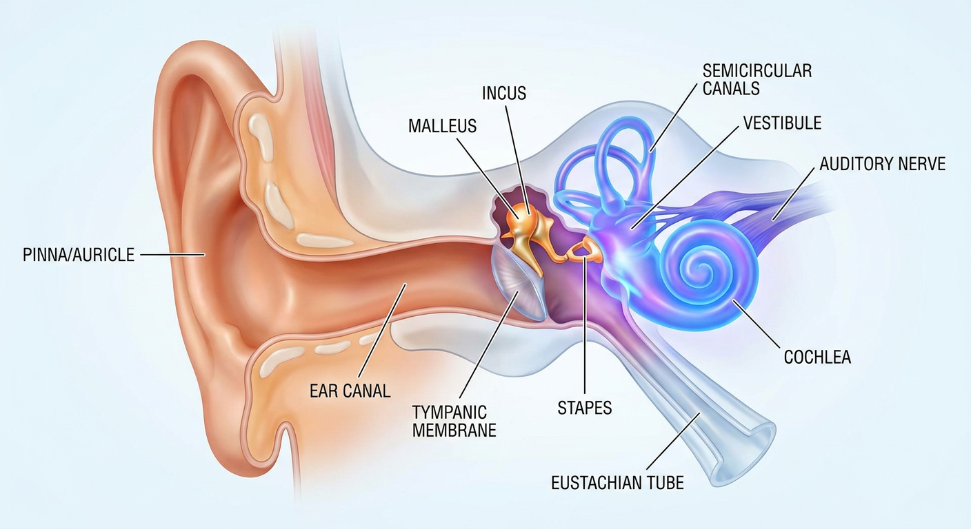
- Pinna (auricle) - the visible external structure
- External auditory canal - channels sound inward
- Tympanic membrane (eardrum) - vibrates in response to sound
- Ossicles: malleus, incus, and stapes - the three smallest bones in the body, transmitting vibrations
- Eustachian tube - equalizes pressure between the middle ear and throat
- Cochlea - snail-shaped structure that converts vibrations into nerve signals
- Semicircular canals - three fluid-filled loops responsible for balance and spatial orientation
- Vestibule - connects the cochlea and semicircular canals
- Vestibulocochlear nerve (CN VIII) - carries auditory and balance signals to the brain
Create a detailed study guide on ear anatomy
Ear Anatomy - Detailed Study Guide
Overview
1. The Temporal Bone
| Component | Location/Role |
|---|---|
| Squamous | Forms the lateral skull; provides muscle attachments |
| Mastoid | Contains mastoid air cells; houses the mastoid antrum |
| Petrous | Pyramid-shaped; contains the inner ear structures |
| Tympanic | Forms the majority of the bony external auditory canal (EAC) |
-
Temporal line: ~5 mm inferior to the lowest level of the middle fossa floor
-
MacEwen triangle (suprameatal triangle): depressed cribriform area posterior to the spine of Henle; marks the mastoid antrum
-
Stylomastoid foramen: anterior limit of the digastric groove; facial nerve exits here
-
Arcuate eminence: prominence of the superior semicircular canal on the middle fossa floor
-
Cummings Otolaryngology, Chapter 126
2. The External (Outer) Ear
2a. Auricle (Pinna)
- Keratinizing squamous epithelium over an elastic cartilage framework
- Perichondrium is tightly bound to the lateral surface, loosely bound medially
- Sebaceous glands and hair follicles are in the subcutaneous layer
- The lobule (earlobe) is cartilage-free and contains adipose tissue
2b. External Auditory Canal (EAC)
- ~2.5 cm in adults; runs from the tympanic membrane to the external meatus
- Lateral 1/3: cartilaginous skeleton - contains hair follicles, sebaceous and apocrine glands
- Medial 2/3: bony wall (formed by tympanic part of temporal bone) - thin squamous epithelium, no glands, no hair follicles
- Isthmus: the bony-cartilaginous junction - narrowest part of the canal
- Produced in the cartilaginous canal from glandular secretions + sloughed epithelium
- Hydrophobic, slightly acidic (pH 6.0 - 6.5)
- Normal flora of the EAC: predominantly gram-positive (S. auricularis, S. epidermidis)
- Fissures of Santorini: transverse slits in cartilaginous canal - allow spread of infection/tumors to surrounding soft tissues
- Foramen of Huschke: defect in anterior bony canal (incomplete ossification) - allows disease spread to parotid gland's deep lobe
- Cummings Otolaryngology, Chapter 130
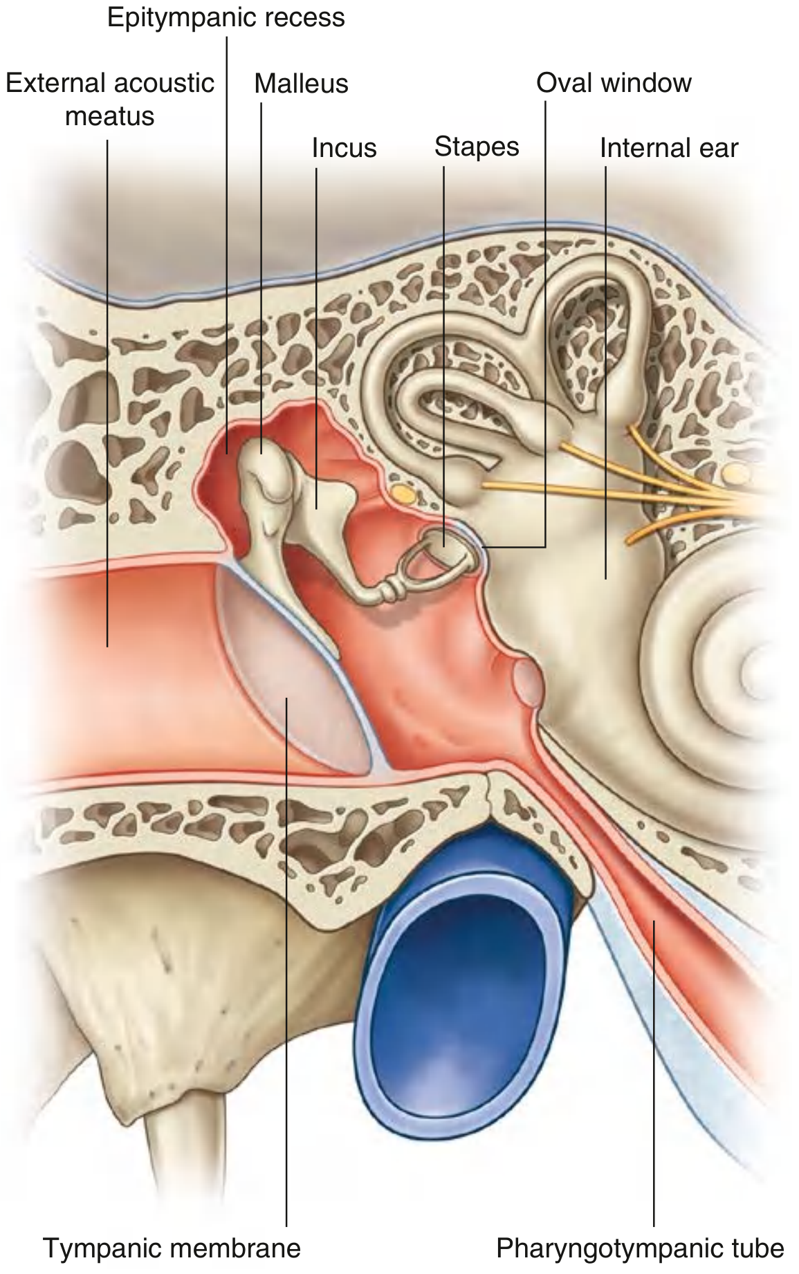
3. The Middle Ear
3a. Spatial Divisions of the Middle Ear
| Division | Location |
|---|---|
| Epitympanum (attic) | Superior, above the tympanic annulus |
| Mesotympanum | Immediately medial to the tympanic membrane |
| Hypotympanum | Inferior portion |
3b. Walls of the Middle Ear
| Wall | Name | Key Contents |
|---|---|---|
| Roof | Tegmental wall | Tegmen tympani (thin bone separating from middle cranial fossa) |
| Floor | Jugular wall | Thin bone over internal jugular vein |
| Lateral | Membranous wall | Almost entirely formed by the tympanic membrane |
| Medial | Labyrinthine wall | Promontory, oval window, round window, tympanic plexus |
| Posterior | Mastoid wall | Aditus to mastoid antrum, pyramidal eminence (stapedius tendon), chorda tympani entry |
| Anterior | Anterior wall | Internal carotid artery, pharyngotympanic tube opening, tensor tympani canal |
- Promontory: rounded bulge caused by the basal coil of the cochlea
- Oval window (fenestra vestibuli): posterosuperior to promontory; sealed by the footplate of the stapes
- Round window (fenestra cochleae): inferior to oval window; sealed by the secondary tympanic membrane; allows fluid pressure waves in the cochlea to dissipate
- Tympanic plexus: on the promontory, formed primarily by the tympanic branch of CN IX (Jacobson's nerve)
3c. The Tympanic Membrane (TM)
- Separates the EAC from the middle ear
- Divided into the pars flaccida (superiorly - slack, lacking fibrous layer) and pars tensa (the main body - has a fibrous layer)
- The umbo is the lowest point, attached to the tip of the malleus handle
- The TM vibrates in response to sound pressure waves and transmits motion to the ossicular chain
3d. The Ossicular Chain
| Ossicle | Connection | Notable Feature |
|---|---|---|
| Malleus ("hammer") | Handle embedded in TM | Largest ossicle |
| Incus ("anvil") | Between malleus & stapes | Long process has single nutrient vessel - most vulnerable to injury |
| Stapes ("stirrup") | Footplate in oval window | Smallest bone in the body |
- Malleus-incus: synovial joint
- Incus-stapes: synovial joint
- The ossicles amplify sound by ~25-30 dB through the surface area ratio of the TM to the oval window plus the lever arm of the ossicular chain
- Tensor tympani: attached to the malleus handle; innervated by CN V3 (medial pterygoid nerve); dampens loud sounds
- Stapedius: attached to the neck of the stapes; innervated by CN VII (facial nerve); the acoustic reflex - protective against intense noise
3e. The Eustachian (Pharyngotympanic) Tube
- Connects the middle ear to the nasopharynx; angled ~45° from middle ear to nasopharyngeal opening at the torus tubarius
- Proximal 1/3: bony (in petrous bone) - widest segment
- Distal 2/3: fibrocartilaginous, normally collapsed at rest; shaped like an inverted J in cross-section
- Bony-cartilaginous junction: narrowest point
- Opens during swallowing and yawning via contraction of tensor veli palatini - equalizes pressure between middle ear and atmosphere
- Internal carotid artery is closely associated with the medial wall at its tympanic opening (may be dehiscent)
3f. The Mastoid
- Air cells develop from outgrowth of the middle ear space; degree of pneumatization varies greatly between individuals
- Mastoid antrum: directly posterior to the epitympanum via the aditus
- Important surgical landmark: posterior to the pyramid of the mastoid
4. The Inner Ear
- Bony labyrinth: outer shell of dense bone filled with perilymph
- Membranous labyrinth: inner fluid-filled sacs and ducts filled with endolymph (K+-rich, Na+-poor - similar in composition to intracellular fluid)
| Fluid | Location | Ionic composition |
|---|---|---|
| Endolymph | Inside membranous labyrinth | High K⁺, low Na⁺ |
| Perilymph | Outside membranous labyrinth, within bony labyrinth | High Na⁺, low K⁺ (similar to CSF) |
4a. The Cochlea (Hearing)
| Compartment | Fluid | Separated by |
|---|---|---|
| Scala vestibuli | Perilymph | Reissner's membrane (above) |
| Scala media (cochlear duct) | Endolymph | Basilar membrane (below) |
| Scala tympani | Perilymph | Basilar membrane |
- Scala vestibuli and scala tympani communicate at the helicotrema (apex)
- Scala tympani ends at the round window
4b. The Organ of Corti (Auditory Transducer)
- Inner hair cells (IHCs): ~3,500; single row; flask-shaped; primary sensory receptors - 95% of afferent innervation
- Outer hair cells (OHCs): ~12,000; three rows; cylindrical; mainly efferent innervation; provide cochlear amplification (active electromotility)
- Tectorial membrane: acellular; overlies the hair cell bundles; OHC stereocilia are embedded in it
- Tunnel of Corti: delineated by inner and outer pillar cells
- Supporting cells: Deiters cells, Hensen cells, Claudius cells, Boettcher cells
- Base of cochlea → detects high frequencies (basilar membrane narrow, stiff)
- Apex of cochlea → detects low frequencies (basilar membrane wide, floppy)
- Stapes footplate movement displaces perilymph in scala vestibuli
- Pressure wave creates a traveling wave along the basilar membrane
- The wave peaks at the region tuned to that frequency
- Basilar membrane deflection moves hair cell stereocilia
- Stereocilia bending opens mechanically gated K⁺ channels
- K⁺ influx depolarizes the hair cell → neurotransmitter (glutamate) release
- Action potentials travel via CN VIII to the auditory cortex
4c. Vestibular Apparatus (Balance)
Semicircular Canals - Angular Acceleration
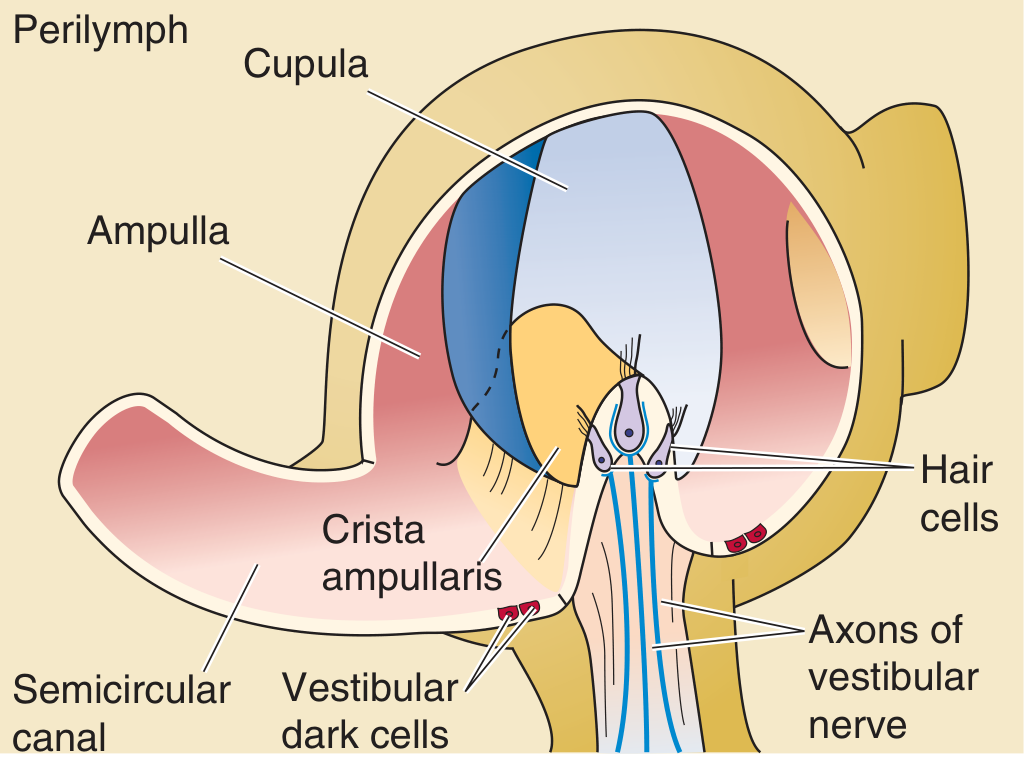
- Anterior (superior) semicircular canal
- Posterior semicircular canal
- Lateral (horizontal) semicircular canal
Otolith Organs - Linear Acceleration
| Organ | Macula Orientation | Senses |
|---|---|---|
| Utricle | Horizontal (ceiling) | Lateral tilt and translation; horizontal acceleration |
| Saccule | Vertical (wall) | Front-to-back tilt and translation; vertical acceleration; gravity |
4d. Vestibular Ganglia and Nerve Pathways
- Superior vestibular ganglion: receives input from utricle, anterior saccule, anterior and lateral semicircular canals
- Inferior vestibular ganglion: receives input from posterior saccule and posterior semicircular canal
- Both join the vestibulocochlear nerve (CN VIII), which enters the brainstem at the pontomedullary junction
- Central projections: thalamus, cortex, cerebellum, descending spinal cord, extraocular motor nuclei (for the vestibulo-ocular reflex, VOR)
5. Embryology of the Ear
| Timepoint | Development |
|---|---|
| 3 weeks gestation | Otic placode appears; invaginates to form otocyst (otic vesicle) → membranous labyrinth |
| 5 weeks | Endolymphatic duct forms first; then cochlear duct and vestibular precursor; Pars superior → semicircular canals; Pars inferior → saccule + cochlea |
| 6 weeks | Cochlear duct begins growing from saccule |
| 10 weeks | Cochlear duct completes 2.5 turns |
| 20 weeks | Full adult cochlear size reached |
| 21 weeks | Organ of Corti becomes functional |
| Postnatal | Endolymphatic sac/duct continues growing until age 3-4 years |
-
Middle and outer ear structures derived from 1st and 2nd branchial arches/grooves
-
K.J. Lee's Essential Otolaryngology
6. The Facial Nerve in the Ear
| Segment | Landmarks / Clinical Importance |
|---|---|
| Meatal (IAC) | Enters the temporal bone via the internal auditory canal |
| Labyrinthine | Narrowest segment; watershed vascular supply; most vulnerable to herpes zoster and trauma |
| Geniculate ganglion | Site of herpes zoster oticus (Ramsay Hunt syndrome); dehiscent in 15-20% |
| Tympanic | Runs above oval window; most frequently dehiscent here |
| Mastoid | Descends vertically; exits at stylomastoid foramen |
- Cummings Otolaryngology, Chapter 126
7. Clinical Correlations
| Finding | Likely Structure Involved |
|---|---|
| Conductive hearing loss | EAC occlusion, TM perforation, ossicular chain disruption, middle ear effusion |
| Sensorineural hearing loss | Cochlear hair cell damage (noise, ototoxic drugs, aging) or CN VIII lesion |
| Vertigo - rotational | Semicircular canal / crista ampullaris (e.g., BPPV - displaced otoconia) |
| Facial palsy + ear pain + vesicles | Ramsay Hunt syndrome (herpes zoster at geniculate ganglion) |
| Cholesteatoma in epitympanum | Pars flaccida retraction pocket; erosion of long process of incus (most vulnerable ossicle) |
| Tinnitus + unilateral SNHL + vertigo | Meniere's disease (endolymphatic hydrops) |
| Acute otitis media | Eustachian tube dysfunction → middle ear negative pressure → effusion → infection |
| Malignant otitis externa | Pseudomonas spreading through fissures of Santorini into temporal bone (typically in diabetics) |
8. Quick Review: Key Numbers to Remember
| Fact | Value |
|---|---|
| EAC length (adults) | ~2.5 cm |
| Cerumen pH | 6.0 - 6.5 |
| Cochlear turns | 2.5 |
| Inner hair cells | ~3,500 (single row) |
| Outer hair cells | ~12,000 (3 rows) |
| Endocochlear potential | +80 mV |
| Cochlear function at gestation | 21 weeks |
| Total vestibular sensory patches | 5 (3 cristae + 2 maculae) |
| Ossicle amplification | ~25-30 dB |
| Eustachian tube angle | ~45° |
| Hair cells per vestibular bundle | 50-100 stereocilia + 1 kinocilium |
Generate a presentation with ear anatomy slides
Detailed 3D anatomical cross-section of the human ear showing all three regions: outer ear (pinna/auricle, external auditory canal), middle ear (tympanic membrane, malleus, incus, stapes ossicles, Eustachian tube), and inner ear (cochlea with spiral shape, semicircular canals, vestibule, auditory nerve). Each structure labeled with clean white text. Medical illustration style, dark navy blue background, vibrant anatomical colors, photorealistic 3D rendering
3D anatomical illustration of the external human ear showing: pinna/auricle with labeled parts (helix, antihelix, tragus, antitragus, lobule, concha, fossa), external auditory canal cross-section showing cartilaginous lateral third and bony medial two-thirds, tympanic membrane visible at end. Labels with arrows, dark teal background, medical illustration style
Detailed 3D anatomical illustration of the human middle ear showing: tympanic membrane (eardrum) with pars flaccida and pars tensa labeled, malleus hammer bone attached to eardrum, incus anvil bone, stapes stirrup bone in oval window, Eustachian tube connecting to nasopharynx, round window, tensor tympani and stapedius muscles. Labels and arrows, dark blue background, vibrant educational medical illustration
3D cutaway anatomical illustration of the human inner ear (labyrinth) showing: bony cochlea in spiral 2.5 turns with scala vestibuli, scala media, scala tympani labeled, organ of Corti with hair cells, three semicircular canals (anterior, posterior, lateral), vestibule, utricle, saccule, oval window, round window, CN VIII vestibulocochlear nerve. Dark purple background, vivid colors, educational medical diagram with labels
Scientific diagram of the organ of Corti cross-section showing: basilar membrane, scala vestibuli above, scala media (cochlear duct) in middle with endolymph, scala tympani below with perilymph, Reissner's membrane, stria vascularis on lateral wall, inner hair cells (single row), outer hair cells (three rows), tectorial membrane, inner and outer pillar cells, tunnel of Corti, Deiters cells, Hensen cells. Labeled arrows, black background, vivid colors, high detail medical illustration
3D anatomical diagram of the vestibular system showing: three semicircular canals (superior/anterior, posterior, lateral/horizontal) in perpendicular planes, ampulla with crista ampullaris and cupula in each canal, utricle (horizontal macula with otoconia/calcium carbonate crystals), saccule (vertical macula), endolymph fluid, vestibular nerve branches going to brain. Labeled, dark background, educational medical illustration style
429 Too Many Requests. {'message': '{\n "error": {\n "code": 429,\n "message": "Resource exhausted. Please try again later. Please refer to https://cloud.google.com/vertex-ai/generative-ai/docs/error-code-429 for more details.",\n "status": "RESOURCE_EXHAUSTED"\n }\n}\n', 'status': 'Too Many Requests'}
We encountered an error while processing your request. Please try again or reach out to us for help.
Generate a presentation with ear anatomy slides
An error occurred (ValidationException) when calling the ConverseStream operation: Expected toolResult blocks at messages.40.content for the following Ids: tooluse_xOVv3LtEKkg7DXpXAWdexp, tooluse_SFNE074GLlHSHzwPsxmMOc, tooluse_s1YKPuoN3TodzqCxAJOD6o, tooluse_dYdPw2z3FckuvMTRmr3cFO
We encountered an error while processing your request. Please try again or reach out to us for help.
Can you please generate AI images of middle ear anatomy, at least seven, eight pictures to, uh, easily understand middle ear anatomy of the ear? Like, please generate in a easy way.
An error occurred (ValidationException) when calling the ConverseStream operation: Expected toolResult blocks at messages.40.content for the following Ids: tooluse_xOVv3LtEKkg7DXpXAWdexp, tooluse_SFNE074GLlHSHzwPsxmMOc, tooluse_s1YKPuoN3TodzqCxAJOD6o, tooluse_dYdPw2z3FckuvMTRmr3cFO
We encountered an error while processing your request. Please try again or reach out to us for help.