analyse_media
Reading File
Finding Sources
Finding Sources
Searching Images

intersphincteric perianal fistula MRI anatomy diagram
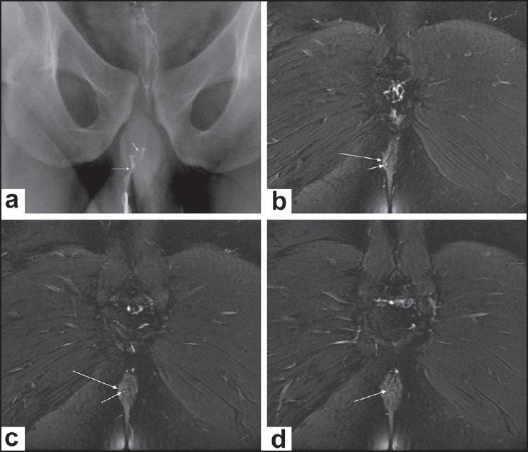
This composite figure illustrates a human intersphincteric perianal fistula using two imaging modalities. Image (a) is a conventional radiographic fistulogram showing a thin, contrast-filled tract near the midline with an internal communication point within the anal canal. Images (b), (c), and (d) are sequential posterior-to-anterior coronal fat-saturated T2-weighted MRI scans. These images demonstrate the anatomy of the anal sphincter complex where the external anal sphincter appears as a hypointense (dark) lateral structure and the internal sphincter/mucosa is relatively hyperintense. A small, high-signal intensity (bright) tract is clearly visible within the intersphincteric plane, located medially to the right external sphincter. The sequence captures the fistula's course as it originates in the intersphincteric space and eventually opens into the anal canal (arrow in d). The fat-saturated MRI technique provides excellent contrast between the fluid-filled inflammatory tract and the surrounding muscular structures and suppressed ischioanal fossa fat, which is essential for accurate surgical planning and classification of perianal fistulous disease.
This composite figure illustrates a human intersphincteric perianal fistula using two imaging modalities. Image (a) is a conventional radiographic fistulogram showing a thin, contrast-filled tract near the midline with an internal communication point within the anal canal. Images (b), (c), and (d) are sequential posterior-to-anterior coronal fat-saturated T2-weighted MRI scans. These images demonstrate the anatomy of the anal sphincter complex where the external anal sphincter appears as a hypointense (dark) lateral structure and the internal sphincter/mucosa is relatively hyperintense. A small, high-signal intensity (bright) tract is clearly visible within the intersphincteric plane, located medially to the right external sphincter. The sequence captures the fistula's course as it originates in the intersphincteric space and eventually opens into the anal canal (arrow in d). The fat-saturated MRI technique provides excellent contrast between the fluid-filled inflammatory tract and the surrounding muscular structures and suppressed ischioanal fossa fat, which is essential for accurate surgical planning and classification of perianal fistulous disease.

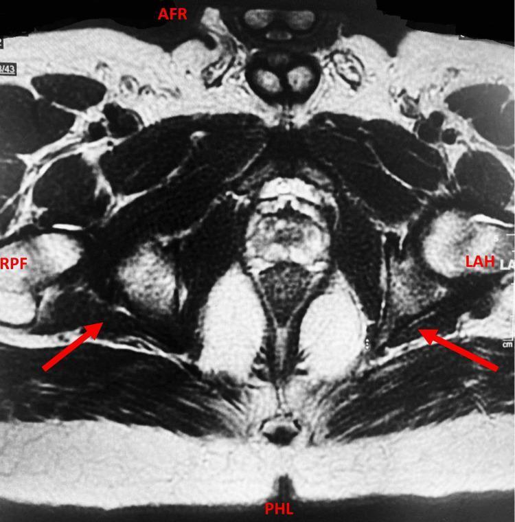
This diagnostic image is an axial MRI scan of the male pelvis, specifically a dedicated fistula protocol study with intravenous contrast. The image highlights the perianal region and pelvic floor anatomy. Anatomical landmarks include the gluteal muscles, pelvic bones, and the anal canal. Red arrows indicate bilateral, hyperintense linear tracts representing intersphincteric perianal fistulas. These tracts are situated within the intersphincteric space between the internal and external anal sphincters, with internal openings visualized at approximately the 5 o'clock and 7 o'clock positions. The rectum and sigmoid colon show evidence of mild circumferential wall thickening, suggesting an underlying inflammatory process. The surrounding soft tissues show subtle signal changes consistent with inflammation but lack evidence of complex trans-sphincteric extension, supralevator involvement, or localized abscess formation. This imaging is clinically significant for the classification of perianal Crohn's disease or chronic inflammatory conditions like Schistosomiasis-related granulomatous colitis, facilitating surgical planning and disease staging.
This diagnostic image is an axial MRI scan of the male pelvis, specifically a dedicated fistula protocol study with intravenous contrast. The image highlights the perianal region and pelvic floor anatomy. Anatomical landmarks include the gluteal muscles, pelvic bones, and the anal canal. Red arrows indicate bilateral, hyperintense linear tracts representing intersphincteric perianal fistulas. These tracts are situated within the intersphincteric space between the internal and external anal sphincters, with internal openings visualized at approximately the 5 o'clock and 7 o'clock positions. The rectum and sigmoid colon show evidence of mild circumferential wall thickening, suggesting an underlying inflammatory process. The surrounding soft tissues show subtle signal changes consistent with inflammation but lack evidence of complex trans-sphincteric extension, supralevator involvement, or localized abscess formation. This imaging is clinically significant for the classification of perianal Crohn's disease or chronic inflammatory conditions like Schistosomiasis-related granulomatous colitis, facilitating surgical planning and disease staging.

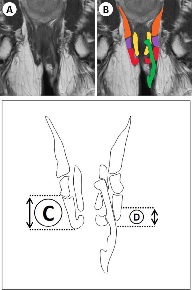
This composite educational image demonstrates the radiological assessment of perianal anatomy and fistula measurement using MRI. Panel A shows an unlabeled coronal T2-weighted MRI sequence of the perianal region. Panel B provides a color-coded anatomical overlay on the same MRI: the fistula tract is highlighted in green, the internal anal sphincter in yellow, the external anal sphincter (EAS) in red, the puborectal muscle in purple, and the levator ani in orange. The bottom schematic diagram (Panels C and D) illustrates the methodology for quantitative measurement. Label C indicates the vertical length or height of the external anal sphincter, while Label D represents the level of fistula penetration, defined as the vertical distance to the most cranial aspect of the tract penetrating the EAS or puborectal muscle. This resource is designed for radiologists and colorectal surgeons to standardize the classification and reporting of complex perianal fistulas.

This composite educational image demonstrates the radiological assessment of perianal anatomy and fistula measurement using MRI. Panel A shows an unlabeled coronal T2-weighted MRI sequence of the perianal region. Panel B provides a color-coded anatomical overlay on the same MRI: the fistula tract is highlighted in green, the internal anal sphincter in yellow, the external anal sphincter (EAS) in red, the puborectal muscle in purple, and the levator ani in orange. The bottom schematic diagram (Panels C and D) illustrates the methodology for quantitative measurement. Label C indicates the vertical length or height of the external anal sphincter, while Label D represents the level of fistula penetration, defined as the vertical distance to the most cranial aspect of the tract penetrating the EAS or puborectal muscle. This resource is designed for radiologists and colorectal surgeons to standardize the classification and reporting of complex perianal fistulas.

Reading File
Reading File
Reading File
Loading Image
Here is a full explanation of this MRI report for Mr. N. Rajesh Kannan.

MRI Report Explanation - Perianal Fistula

Patient: Mr. N. Rajesh Kannan, 51 years / Male Date: 29th July 2026 | Referring Doctor: Dr. D. Karthikeyan, MS, DMAS

What is the Diagnosis?

The MRI shows a Simple Intersphincteric Perianal Fistula - this is the most common type of anal fistula (accounting for ~45% of all cases). It is classified as Parks Type 1.
An anal fistula is an abnormal tunnel connecting the inside of the anal canal (the internal opening) to the skin around the anus (the external opening). It almost always results from a prior perianal abscess. - Sabiston Textbook of Surgery, p. 2163

Parks Classification - Where This Fistula Fits
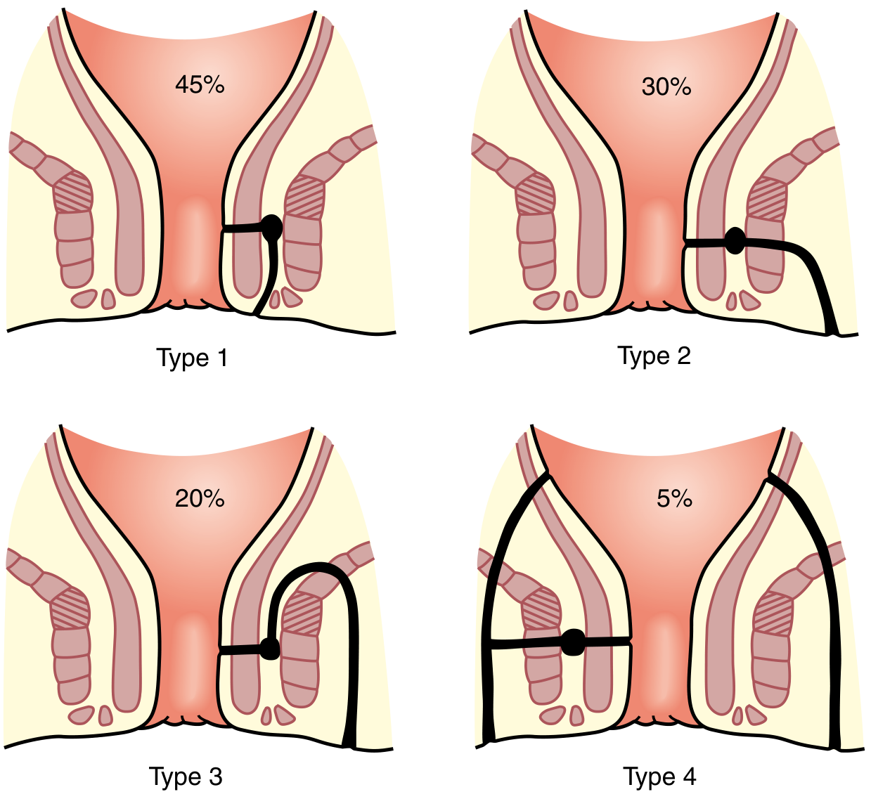
Parks Classification of Fistula-in-Ano: Type 1 (intersphincteric, 45%), Type 2 (transsphincteric, 30%), Type 3 (suprasphincteric, 20%), Type 4 (extrasphincteric, 5%)
This patient has a Type 1 (Intersphincteric) fistula. The tract lies within the intersphincteric space - the narrow gap between the internal and external anal sphincter muscles - and does NOT cross through the outer sphincter. - Mulholland & Greenfield's Surgery, p. 3564

Breaking Down the MRI Findings

FindingWhat It Means
External openingOn the skin, 4.5 cm in front of the anal opening (anterior perianal skin)
Tract length5 cm long, running from outside inward through the intersphincteric plane
Internal openingInside the anal canal at the 12-1 o'clock position, about 1.3 cm above the anal verge (near the dentate line)
No secondary tractsOnly one simple tunnel - no branching or horseshoe extension
No collectionsNo pus pockets or abscesses alongside the tract
No supralevator extensionThe fistula stays below the levator ani muscle - it has NOT spread upward into the pelvis
The fact that the external opening is anterior (in front) and the internal opening is at 12-1 o'clock is consistent with Goodsall's rule - anterior external openings typically track radially (directly) into the anal canal. - Sabiston Textbook of Surgery, p. 2163

Why These Findings Are Important (and Reassuring)

  • "Simple" fistula - only one tract, no branches, no abscess, no upward spread. This is the most favorable category.
  • No supralevator extension - rules out a complex fistula that involves the pelvic floor muscles. Surgery is therefore more straightforward.
  • Intersphincteric - the tract does NOT cross or damage the external anal sphincter, which is the key muscle for bowel control. This means surgical treatment carries a very low risk of incontinence.

What Happens Next? (Surgical Implications)

For a simple intersphincteric fistula like this, the standard treatment is fistulotomy (laying open the tract). Key points:
  • Involves less than one-third of the external sphincter (actually none in intersphincteric type)
  • Fecal incontinence after fistulotomy in low-lying fistulae occurs in less than 5% of patients with normal sphincter function
  • Recurrence rate with fistulotomy: 2-8% - Sabiston Textbook of Surgery, p. 2163-2164
Other sphincter-sparing options include:
  • LIFT procedure (Ligation of Intersphincteric Fistula Tract) - success rates 40-95%
  • Mucosal advancement flap
  • Fibrin glue or fistula plug (less commonly used for simple fistulas)
The operating surgeon (Dr. D. Karthikeyan) will decide the best approach based on clinical examination, patient's continence status, and this MRI roadmap.

Summary in Simple Terms

The MRI confirms a benign, straightforward anal fistula - a small tunnel running from the skin near the anus to the inside of the anal canal, staying between the two sphincter muscles. It has no complications (no pus collection, no branching, no spread upward). This is a surgically correctable condition with excellent outcomes and very low risk to bowel control.
This is a shared conversation. Sign in to Orris to start your own chat.