
Airway Management in Anaesthesia
airway management anaesthesia Difficult Airway Society guidelines 2025
airway management anesthesia
-
Airway assessment
History, examination, Mallampati class, mouth opening, thyromental distance, neck movement, predictors of difficult mask ventilation and intubation. -
Basic airway techniques
Positioning, chin lift, jaw thrust, suction, oxygen delivery, bag-mask ventilation, oral and nasal airways. -
Supraglottic airway devices
Laryngeal mask airways, indications, contraindications, insertion, complications, and use as a rescue device. -
Tracheal intubation
Direct and video laryngoscopy, endotracheal tube selection, stylets/bougies, confirmation with waveform capnography, securing the tube. -
Rapid sequence induction
Indications, preparation, pre-oxygenation, cricoid pressure considerations, induction and neuromuscular-blocking drugs, aspiration-risk management. -
Difficult airway management
Anticipated versus unanticipated difficulty, awake intubation, rescue oxygenation, and "cannot intubate, cannot oxygenate" emergency management. -
Extubation and postoperative care
Extubation planning, high-risk extubation, residual neuromuscular block, airway obstruction, laryngospasm, bronchospasm, and post-extubation stridor. -
Special situations
Obstetric, paediatric, obese, trauma, cervical-spine injury, full-stomach, burns, and upper-airway pathology.
Detailed explanation of assessment, routine intubation, RSI, Difficult Airway Algorithm, Extubation and special situations
- Pre-anaesthetic airway assessment: history, examination, prediction limits, and planning.
- Routine tracheal intubation: preparation, positioning, laryngoscopy, tube placement, and capnographic confirmation.
- Rapid sequence induction (RSI): aspiration risk, preparation, drug selection principles, and failed-intubation response.
- Difficult airway algorithm: anticipated difficult airway, awake techniques, unanticipated failure, rescue oxygenation, and CICO emergency.
- Extubation: risk assessment, awake versus deep extubation, staged extubation, and complications.
- Special situations: obstetric, paediatric, obesity, trauma/cervical spine injury, burns, and airway obstruction.
Which module should I explain first in detailed exam-note format?
-
Airway assessment
History, examination, Mallampati class, mouth opening, thyromental distance, neck movement, predictors of difficult mask ventilation and intubation. -
Basic airway techniques
Positioning, chin lift, jaw thrust, suction, oxygen delivery, bag-mask ventilation, oral and nasal airways. -
Supraglottic airway devices
Laryngeal mask airways, indications, contraindications, insertion, complications, and use as a rescue device. -
Tracheal intubation
Direct and video laryngoscopy, endotracheal tube selection, stylets/bougies, confirmation with waveform capnography, securing the tube. -
Rapid sequence induction
Indications, preparation, pre-oxygenation, cricoid pressure considerations, induction and neuromuscular-blocking drugs, aspiration-risk management. -
Difficult airway management
Anticipated versus unanticipated difficulty, awake intubation, rescue oxygenation, and "cannot intubate, cannot oxygenate" emergency management. -
Extubation and postoperative care
Extubation planning, high-risk extubation, residual neuromuscular block, airway obstruction, laryngospasm, bronchospasm, and post-extubation stridor. -
Special situations
Obstetric, paediatric, obese, trauma, cervical-spine injury, full-stomach, burns, and upper-airway pathology.
- Pre-anaesthetic airway assessment: history, examination, prediction limits, and planning.
- Routine tracheal intubation: preparation, positioning, laryngoscopy, tube placement, and capnographic confirmation.
- Rapid sequence induction (RSI): aspiration risk, preparation, drug selection principles, and failed-intubation response.
- Difficult airway algorithm: anticipated difficult airway, awake techniques, unanticipated failure, rescue oxygenation, and CICO emergency.
- Extubation: risk assessment, awake versus deep extubation, staged extubation, and complications.
- Special situations: obstetric, paediatric, obesity, trauma/cervical spine injury, burns, and airway obstruction.
- Airway assessment
- Routine tracheal intubation
- Rapid sequence induction
- Difficult airway algorithm
- Extubation
- Special situations
Give detailed notes on airway assessment
airway assessment anesthesia
ASA 2022 difficult airway guidelines airway assessment risk factors preoperative evaluation
airway assessment Mallampati classification diagram anesthesia
https://www.asahq.org/-/media/sites/asahq/files/public/resou…
Airway Assessment in Anaesthesia
1. Objectives
- Difficult face-mask ventilation
- Difficult placement or ventilation through a supraglottic airway device (SGA)
- Difficult laryngoscopy
- Difficult tracheal intubation
- Difficult emergency invasive airway access
- Difficult extubation or post-extubation obstruction
- Increased risk of pulmonary aspiration
2. General Principles
- Assess every patient, including those with a previously uncomplicated anaesthetic.
- Take a focused history, inspect the airway, perform bedside tests, and review prior records.
- Predict the separate problems of:
- ventilation
- laryngoscopy/intubation
- aspiration
- emergency rescue access
- A normal examination does not exclude difficulty.
- If risk is identified, make an explicit plan:
- Plan A: primary technique
- Plan B: rescue intubation/ventilation technique
- Plan C: rescue oxygenation
- Plan D: emergency front-of-neck airway
- Communicate the plan to the team and document important findings.
3. History
A. Previous anaesthetic and airway history
- Previous difficult mask ventilation, SGA insertion, laryngoscopy, intubation, or extubation
- Previous awake intubation, fiberoptic intubation, video laryngoscopy, bougie use, or surgical airway
- Difficult or prolonged intubation
- Multiple intubation attempts
- Dental trauma during anaesthesia
- Postoperative sore throat, hoarseness, stridor, dysphagia, or airway swelling
- Previous anaesthetic chart, difficult-airway alert card, or electronic record
B. Symptoms suggesting upper-airway pathology
- Snoring, witnessed apnoea, daytime sleepiness, diagnosed obstructive sleep apnoea
- Stridor, noisy breathing, dyspnoea when supine
- Voice change or hoarseness
- Dysphagia, odynophagia, aspiration, coughing while eating
- Reflux or regurgitation
- Neck swelling, rapidly enlarging mass, goitre
- Facial pain, reduced mouth opening, temporomandibular-joint symptoms
- Recent infection, epiglottitis-type symptoms, airway burns, inhalational injury
- Nasal obstruction if nasal intubation is contemplated
C. Relevant diseases and conditions
- Obesity, especially large neck circumference
- Obstructive sleep apnoea
- Diabetes with limited cervical mobility
- Rheumatoid arthritis or ankylosing spondylitis
- Cervical-spine instability or immobilisation
- Temporomandibular-joint disease
- Acromegaly
- Goitre, neck mass, mediastinal mass
- Head and neck tumour, previous surgery, or radiotherapy
- Maxillofacial trauma, facial burns, airway burns
- Congenital craniofacial syndromes, for example Pierre Robin sequence, Treacher Collins syndrome, Down syndrome, or Klippel-Feil syndrome.
D. Aspiration-risk assessment
- Inadequate fasting or recent meal
- Emergency surgery or trauma
- Bowel obstruction or acute intra-abdominal pathology
- Symptomatic gastro-oesophageal reflux
- Pregnancy
- Diabetic gastroparesis or other causes of delayed gastric emptying
- Opioid use, reduced consciousness, or critical illness
4. Physical Examination
A. Overall inspection: face and neck
- Facial asymmetry, deformity, trauma, scars, burns
- Receding chin or micrognathia
- Protruding upper incisors
- Large tongue or macroglossia
- Beard, which may prevent an effective mask seal
- Short, thick neck
- Neck mass, goitre, tracheal deviation, prior tracheostomy scar
- Obesity and large neck circumference
- Cervical collar or other restriction to access and movement
B. Mouth opening: inter-incisor gap
- Measure the distance between the upper and lower incisors.
- An opening at least 3 cm is generally desirable for adult laryngoscopy.
- Less than 3 cm, or roughly fewer than two fingerbreadths, suggests limited space for insertion and manipulation of a laryngoscope, SGA, or endotracheal tube.
C. Dentition
- Prominent maxillary incisors
- Loose, fractured, capped, or prosthetic teeth
- Edentulous mouth
- Dental crowding
- Overbite
D. Oropharyngeal anatomy and modified Mallampati classification
Technique
- Keep the head neutral.
- Open the mouth fully.
- Protrude the tongue fully.
- Do not phonate.
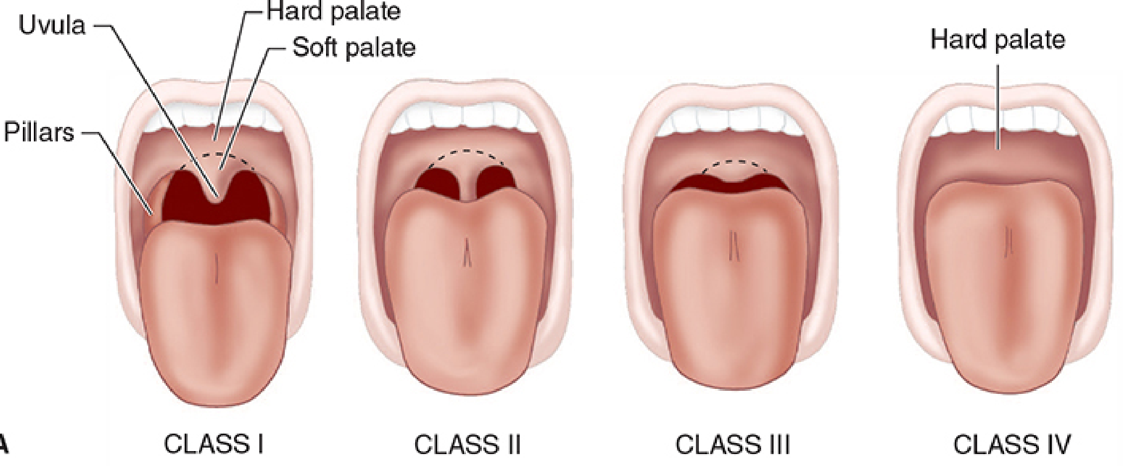
Modified Mallampati classes
| Class | Visible structures | Interpretation |
|---|---|---|
| I | Soft palate, fauces, uvula, and pillars | Usually favourable |
| II | Soft palate, fauces, and part of uvula | Usually favourable |
| III | Soft palate and base of uvula | Increased likelihood of difficult laryngoscopy |
| IV | Hard palate only | Greater concern for difficult laryngoscopy |
E. Thyromental distance
- A distance greater than about three fingerbreadths is desirable.
- A short distance suggests a reduced mandibular space and difficulty displacing the tongue during direct laryngoscopy.
F. Mandibular protrusion and upper-lip bite test
- Ability to bite beyond the vermilion border or lower edge of the upper lip suggests adequate mandibular protrusion.
- Inability to bite the upper lip suggests reduced mandibular mobility and increased likelihood of difficult laryngoscopy.
G. Neck movement
- Flexion and extension
- Ability to adopt a sniffing position
- Pain, stiffness, deformity, or neurological symptoms
H. Submandibular space and jaw anatomy
- Receding chin or micrognathia
- Length and compliance of the submandibular space
- Macroglossia
- High-arched palate
- Floor-of-mouth swelling
5. Prediction of Difficult Mask Ventilation
- Older age
- Male sex
- Obesity or increased BMI
- Large neck circumference
- Beard
- Snoring or obstructive sleep apnoea
- Mallampati III or IV
- Reduced thyromental distance
- Previous difficult intubation
- Limited mandibular protrusion
- History of neck radiation
Practical implication
- Ensure experienced help is immediately available.
- Use optimal positioning, usually head-elevated or ramped in obesity.
- Have oral/nasal airways available.
- Prepare two-person mask ventilation.
- Prepare an SGA early.
- Ensure a difficult-airway trolley and emergency front-of-neck kit are immediately accessible.
6. Prediction of Difficult Laryngoscopy and Intubation
- Previous difficult laryngoscopy or intubation
- Mallampati III or IV
- Inter-incisor gap less than 3 cm
- Thyromental distance less than three fingerbreadths
- Poor mandibular protrusion or failed upper-lip bite test
- Receding mandible or micrognathia
- Prominent upper incisors
- Restricted neck extension
- Large neck circumference
- Neck mass, airway tumour, goitre, or previous neck radiation
- Blood, secretions, airway swelling, or distorted anatomy
- Cervical-spine immobilisation
Multiple abnormal features are much more important than one isolated abnormal test.
7. Assessment of the Anticipated Difficult Airway
- Difficult laryngoscopy or intubation
- Difficult mask ventilation or SGA ventilation
- High aspiration risk
- Intolerance of even a brief period of apnoea, for example severe obesity, hypoxaemia, sepsis, pregnancy, or critical respiratory disease
- Difficulty with emergency invasive airway access
8. Advanced Assessment
- Flexible nasendoscopy or laryngoscopy
- Review of CT, MRI, neck radiographs, or previous endoscopy
- Bedside ultrasound
- Specialist ENT assessment
- Review of previous anaesthetic records and airway images
9. Documentation and Airway Plan
- Airway assessment findings
- Predicted problems: mask, SGA, laryngoscopy, intubation, aspiration, extubation
- Equipment selected
- Primary and rescue plans
- Need for senior assistance or ENT backup
- Whether awake intubation is considered
- Postoperative airway concerns and extubation strategy
10. Exam-Style Summary: “Airway Assessment Checklist”
History
- Previous difficult airway or anaesthetic record?
- Snoring/OSA, stridor, dysphagia, voice change?
- Head-neck surgery, radiation, trauma, infection, tumour?
- Cervical-spine or TMJ disease?
- Aspiration risk?
Examination
- Face and neck: obesity, beard, scars, mass, short neck, receding jaw
- Mouth opening: at least 3 cm desirable
- Dentition: loose/prominent teeth
- Mallampati class
- Thyromental distance: more than 3 fingerbreadths desirable
- Mandibular protrusion/upper-lip bite test
- Neck flexion and extension
- Identify/palpate cricothyroid membrane when difficulty is expected
Plan
- Predict difficulty with mask, SGA, laryngoscopy, intubation, and rescue access.
- Prepare oxygenation, primary technique, rescue equipment, skilled assistance, and extubation strategy.
- Use awake airway management when the consequences of failed ventilation after induction would be unacceptable.
Conditions when airway management is done
indications for airway management endotracheal intubation anaesthesia airway protection ventilation oxygenation guidelines
Conditions/Indications for Airway Management
1. During anaesthesia
- General anaesthesia, especially when unconsciousness and anaesthetic drugs reduce airway tone and protective reflexes.
- Procedures requiring controlled ventilation, muscle relaxation, or a motionless surgical field.
- Operations where the airway must be secured because access will be limited, for example:
- Prone position
- Lateral position
- Robotic surgery
- Head and neck surgery
- Long procedures
- Surgery involving a shared airway, such as ENT, maxillofacial, dental, and upper-airway procedures.
When is a tracheal tube preferred over an SGA in anaesthesia?
- Risk of aspiration
- Need for controlled positive-pressure ventilation at higher airway pressures
- Need for prolonged ventilation
- Surgical position or procedure making rescue airway access difficult
- Head, neck, thoracic, or abdominal surgery where an SGA may be unsuitable
- Airway contamination by blood, secretions, or gastric contents
- Significant airway obstruction or abnormal anatomy
2. Airway obstruction or threatened obstruction
- Foreign body in the upper airway
- Tongue fall-back in an unconscious patient
- Facial trauma or maxillofacial fractures
- Burns to the face or inhalational injury
- Angioedema or anaphylaxis
- Epiglottitis, croup, retropharyngeal abscess, Ludwig angina
- Laryngeal oedema after intubation, surgery, or allergic reaction
- Upper-airway tumour, goitre, neck mass, or expanding neck haematoma
- Bilateral vocal-cord paralysis
- Severe obstructive sleep apnoea with loss of consciousness
- Postoperative airway swelling or bleeding, especially after neck surgery
3. Failure of oxygenation
- Severe pneumonia
- Acute pulmonary oedema
- Acute respiratory distress syndrome
- Severe asthma or COPD exacerbation with respiratory failure
- Pulmonary embolism with respiratory compromise
- Severe chest trauma
- Near drowning
- Severe hypoxaemia in sepsis or shock
- Cardiac arrest
4. Failure of ventilation
- Apnoea or impending respiratory arrest
- Drug overdose, especially opioids or sedatives
- Depressed consciousness from traumatic brain injury, stroke, seizures, or metabolic encephalopathy
- Neuromuscular weakness, for example Guillain-Barré syndrome, myasthenic crisis, or high spinal cord injury
- Exhaustion from severe asthma or COPD
- Severe hypercapnic respiratory acidosis
- Respiratory-muscle fatigue in critical illness
5. Inability to protect the airway
- Reduced consciousness, including coma
- Severe head injury
- Acute stroke with impaired swallowing or airway reflexes
- Status epilepticus
- Intoxication with alcohol, sedatives, or drugs
- Post-cardiac arrest state
- Severe delirium or agitation requiring deep sedation
- Neuromuscular disease with bulbar weakness
- Recurrent vomiting in a patient with impaired consciousness
6. High aspiration risk
- Non-fasted patient requiring emergency surgery
- Intestinal obstruction
- Full stomach after trauma
- Active vomiting or gastrointestinal bleeding
- Severe gastro-oesophageal reflux or hiatus hernia
- Pregnancy, particularly during urgent operative delivery
- Delayed gastric emptying, for example diabetic gastroparesis
- Reduced consciousness
- Acute abdomen
- Critical illness
7. Need for airway access during procedures
- Bronchoscopy
- Upper gastrointestinal endoscopy in selected high-risk patients
- Interventional radiology or MRI in patients requiring deep sedation
- Cardioversion
- Electroconvulsive therapy
- Dental and maxillofacial procedures
- ENT surgery
- Tracheal, laryngeal, or thoracic procedures
8. Cardiac arrest and peri-arrest states
- Bag-mask ventilation
- Supraglottic airway
- Tracheal intubation by an experienced operator
Quick Classification
| Clinical problem | Airway goal | Usual level of intervention |
|---|---|---|
| Mild obstruction in unconscious patient | Open airway | Head tilt-chin lift or jaw thrust, oral/nasal airway |
| Mild hypoxaemia with intact airway | Improve oxygenation | Supplemental oxygen |
| Sedation with partial loss of airway tone | Maintain patency and ventilation | Positioning, airway adjunct, mask support |
| Inadequate mask ventilation | Rescue oxygenation | Two-person mask technique, airway adjunct, SGA |
| Aspiration risk or need for definitive control | Protect airway | Cuffed tracheal tube |
| Apnoea, severe respiratory failure | Ventilate and oxygenate | Tracheal intubation and mechanical ventilation |
| Cannot intubate, cannot oxygenate | Restore oxygenation urgently | Emergency front-of-neck access |
Key exam statement
Airway management is indicated when there is actual or potential failure of airway patency, oxygenation, ventilation, airway protection, or when anaesthesia and surgery require a controlled airway.
Explain indications for intubation versus an SGA
supraglottic airway tracheal intubation anesthesia
supraglottic airway versus tracheal intubation indications anaesthesia aspiration positive pressure ventilation guidance
Tracheal Intubation Versus Supraglottic Airway Device
Core distinction
| Feature | SGA | Cuffed tracheal tube |
|---|---|---|
| Position | Above glottis | In trachea, below vocal cords |
| Aspiration protection | Limited. Not a definitive protection | Greatest available protection, though not absolute |
| Airway pressure tolerated | Lower, due to leak risk | Higher airway pressures possible |
| Speed/ease of placement | Often quick and less stimulating | Usually needs laryngoscopy or an intubating technique |
| Muscle relaxant requirement | Often not required | Often, but not always, used |
| Postoperative sore throat/cough | Often less frequent | May be more frequent |
| Rescue use | Important rescue ventilation device in failed intubation | Definitive airway once inserted and confirmed by capnography |
1. Indications favouring tracheal intubation
A. Risk of pulmonary aspiration
- Non-fasted patient
- Emergency surgery
- Active vomiting
- Bowel obstruction or acute abdomen
- Upper gastrointestinal bleeding
- Full stomach after trauma
- Significant symptomatic reflux or regurgitation
- Delayed gastric emptying, such as diabetic gastroparesis
- Depressed consciousness or impaired protective reflexes
- Pregnancy when emergency surgery or operative delivery is required
- Airway surgery with blood/debris contaminating the pharynx
B. Need for high-pressure or tightly controlled positive-pressure ventilation
- Severe obesity with poor respiratory compliance
- Acute respiratory failure or critical illness
- Significant lung disease, including low compliance lungs
- Bronchospasm requiring controlled ventilation
- Thoracic surgery
- Prolonged major surgery
- Laparoscopy with pneumoperitoneum, especially when airway pressures are high
- Steep Trendelenburg position
- Procedures requiring hyperventilation or tightly controlled carbon dioxide levels
C. Need for a secure airway because access will become limited
- Prone surgery
- Complex lateral position
- Robotic surgery
- Head and neck surgery with surgical drapes limiting access
- Long operations
- Procedures outside the operating theatre where access is constrained
- Transport of an unstable or ventilated patient
D. Shared-airway or airway-contaminating surgery
- ENT surgery
- Oral, dental, and maxillofacial surgery
- Nasal surgery with significant bleeding risk
- Laryngeal, tracheal, or bronchial surgery
- Surgery where blood, irrigation fluid, smoke, debris, or surgical instruments may compromise the airway
E. Need for prolonged ventilatory support or repeated suctioning
- Prolonged mechanical ventilation
- Frequent tracheobronchial suctioning
- Intensive-care ventilation
- Management of coma, severe trauma, or severe neurological disease
- Airway protection after major aspiration or with copious secretions
F. Inability to protect the airway
- Coma or severe reduction in consciousness
- Traumatic brain injury
- Stroke with impaired swallowing
- Status epilepticus
- Sedative, opioid, alcohol, or drug intoxication
- Post-cardiac-arrest care
- Severe neuromuscular or bulbar weakness
G. Specific operative requirements
- Complete immobility with neuromuscular blockade
- Controlled lung isolation or one-lung ventilation
- Accurate control of ventilation during intracranial procedures
- A protected airway in major abdominal or thoracic surgery
2. Indications favouring an SGA
A. Elective, fasted patient with low aspiration risk
- Appropriately fasted
- No significant reflux, obstruction, vomiting, or delayed gastric emptying
- No major airway bleeding risk
- No requirement for prolonged ventilation
- Predicted low airway pressures
- Procedure compatible with an SGA
B. Procedures expected to need low-to-moderate airway pressures
- Lungs are reasonably compliant
- Ventilation requirements are modest
- High PEEP or high peak inspiratory pressure is not expected
- The patient can be ventilated without a substantial leak
C. When less airway stimulation is desired
- Less haemodynamic stimulation
- Less coughing at emergence
- Less postoperative sore throat or hoarseness in some patients
- No need for laryngoscopy in routine cases
D. Rescue oxygenation in difficult airway management
- Buy time for reassessment and calling for help
- Act as a conduit for flexible-scope intubation in selected devices
- Serve as a bridge while preparing emergency invasive airway access
E. Airway management when intubation is not required
3. Contraindications or strong cautions for primary SGA use
- Known full stomach or significant aspiration risk
- Active vomiting, gastrointestinal obstruction, or major upper-GI bleeding
- Severe obesity with anticipated high ventilation pressures
- Markedly reduced pulmonary compliance or high airway resistance
- Need for prolonged ventilation
- Surgery in prone position, if displacement would be hard to address
- Major head, neck, airway, oral, or maxillofacial surgery
- Upper-airway obstruction distal to the device
- Severe restricted mouth opening that prevents device insertion
- High likelihood of difficult SGA ventilation
- Need for lung isolation
- Airway contamination with blood or secretions
4. Practical decision framework
1. Is aspiration risk elevated?
- Yes: usually choose a cuffed tracheal tube, often with RSI or an awake technique depending on airway difficulty.
- No: an SGA may be appropriate.
2. Will high-pressure ventilation, high PEEP, or prolonged ventilation be required?
- Yes: choose tracheal intubation.
- No: an SGA may be appropriate.
3. Will the airway be inaccessible or contaminated during surgery?
- Yes: choose tracheal intubation.
- No: consider SGA if other conditions are favourable.
4. Is there anticipated difficult intubation?
- Do not automatically induce anaesthesia and “try an SGA.”
- Assess whether difficult mask ventilation, difficult SGA ventilation, aspiration risk, or intolerance of apnoea is also likely.
- Consider awake tracheal intubation if failure after induction would be hazardous.
- Have an SGA prepared as a rescue device when appropriate.
5. Exam answer in one paragraph
A supraglottic airway is suitable for a selected fasted elective patient with low aspiration risk, anticipated low airway pressures, short or moderate-duration surgery, and no need for a protected or highly secure airway. Tracheal intubation is indicated when aspiration risk is high, when controlled high-pressure or prolonged ventilation is needed, when airway access will be limited, during shared-airway or contaminated-field surgery, when airway protection or repeated suctioning is required, and in patients with impaired consciousness or severe respiratory failure. An SGA is also an important rescue device following failed intubation, but it is not equivalent to a cuffed tracheal tube for aspiration protection.
If there is foreign body obstruction in upper airway what method is used for airway management
American Red Cross choking first aid adult child infant responsive unresponsive foreign body airway obstruction
foreign body airway obstruction guidelines
1. Partial obstruction: patient can cough or speak
- Do not give blind finger sweeps.
- Do not perform abdominal thrusts if the patient is coughing effectively.
- Observe closely because partial obstruction can worsen.
2. Complete obstruction in a conscious adult or child
- Call for emergency help.
- Give 5 back blows.
- If not relieved, give 5 abdominal thrusts.
- Repeat cycles of 5 back blows and 5 abdominal thrusts until the object is expelled or the patient becomes unresponsive.
3. Complete obstruction in a conscious infant under 1 year
- Give 5 back blows, with the infant supported face-down and head lower than the trunk.
- Then give 5 chest thrusts.
- Repeat until the object is expelled or the infant becomes unresponsive.
4. Patient becomes unresponsive
- Call emergency services and obtain an AED.
- After each set of chest compressions, open the mouth and remove the object only if it is clearly visible.
- Attempt rescue breaths.
- Never do a blind finger sweep, because it may push the foreign body deeper.
5. In hospital or anaesthesia setting
If the object is visible in the mouth or pharynx
- Direct laryngoscopy
- Removal with Magill forceps by a trained clinician
- Suction for blood/secretions, but suction alone rarely removes a solid impacted object
If the foreign body is below the glottis or cannot be safely removed directly
- Rigid bronchoscopy is usually the definitive method of removal, especially in children.
- Flexible bronchoscopy may be appropriate in selected stable adults or for more distal foreign bodies.
If oxygenation is failing
- Attempt tracheal intubation only by an experienced clinician, recognizing that the tube may dislodge or worsen the obstruction.
- If there is a cannot intubate, cannot oxygenate situation, perform emergency front-of-neck airway access, usually cricothyrotomy in adults.
Exam-style answer
Upper-airway foreign body obstruction is initially managed according to consciousness and severity. An effective cough is encouraged in partial obstruction. Complete obstruction in a conscious adult or child is treated with alternating five back blows and five abdominal thrusts; in infants, five back blows and five chest thrusts are used. If the patient becomes unconscious, CPR is started and only visible foreign bodies are removed. In hospital, direct laryngoscopy with Magill forceps is used for a visible laryngeal foreign body, while rigid bronchoscopy is the definitive treatment for a tracheobronchial foreign body. Emergency surgical airway access is required if oxygenation cannot be achieved.