

Diagnosis and treatment
asthenozoospermia treatment male infertility
Semen Analysis Interpretation
Results Summary
| Parameter | Patient's Result | WHO 5th Edition Reference Range |
|---|---|---|
| Ejaculate volume | 1 mL | ≥ 1.5 mL (lower reference limit) |
| Colour | Milky white | Normal |
| Liquefaction time | Normal | Normal (within 60 min) |
| Viscosity | Thin | Normal |
| Motile spermatozoa | Only 2-3 motile seen; rest all non-motile | Progressive motility ≥ 32%; total motility ≥ 40% |
Diagnoses
1. Severe Asthenozoospermia (Asthenospermia)
2. Seminal Hypovolemia (Low Ejaculate Volume)
- Retrograde ejaculation (partial)
- Ejaculatory duct obstruction
- Hypoplasia of prostate or seminal vesicles (may reflect age-related androgen decline)
- Alpha-blocker medications (e.g., tamsulosin for BPH - common at age 60)
- Neurological conditions (diabetes, neuropathy)
3. Possible Necrospermia (to exclude)
Causes to Consider in a 60-Year-Old Male
- Age-related decline - Sperm motility declines significantly with advancing age; at 60, this is a major contributing factor
- Oxidative stress - Accumulation of reactive oxygen species damages sperm flagella over time
- Varicocele - Common cause of progressive motility decline
- Infection / accessory gland inflammation - Prostatitis, epididymitis
- Medications - Alpha-blockers (tamsulosin for BPH), sulfasalazine, calcium channel blockers, chemotherapy
- Systemic disease - Diabetes mellitus (causes neuropathy and oxidative damage), hypothyroidism
- Ejaculatory dysfunction / partial retrograde ejaculation - Explains both low volume and poor motility
- Mitochondrial dysfunction - Mitochondria power the sperm flagellum; dysfunction leads to immotility
Workup Recommended
Repeat Semen Analysis (as flagged by lab)
- Repeat after 2-3 months abstaining 2-5 days before collection
- Include sperm vitality (eosin-nigrosin stain) and morphology (Kruger strict criteria)
Additional Investigations
- Post-ejaculatory urinalysis - To rule out retrograde ejaculation (given low volume)
- Hormonal profile: FSH, LH, total testosterone, prolactin, TSH
- Transrectal ultrasound (TRUS) if ejaculatory duct obstruction suspected
- Scrotal Doppler ultrasound - To look for varicocele
- Urine culture / semen culture - If infection suspected
- Fasting glucose / HbA1c - Rule out diabetes
Treatment
1. Treat the Underlying Cause
- Retrograde ejaculation: Sympathomimetic agents (pseudoephedrine, imipramine) or sperm retrieval from post-ejaculatory urine
- Varicocele: Surgical varicocelectomy can improve motility
- Infection: Appropriate antibiotics
- Hormone deficiency: Testosterone/FSH supplementation under specialist guidance
- Medication review: Discontinue offending drugs if possible (e.g., tamsulosin - switch to alternative BPH therapy)
2. Antioxidant Nutraceuticals (Evidence-Based)
- Coenzyme Q10 (CoQ10) 200-300 mg/day - directly improves mitochondrial function in sperm flagella; shown in placebo-controlled trials to improve sperm density and motility
- Vitamin E 400 IU/day + Vitamin C 1000 mg/day
- Zinc 66 mg/day
- L-carnitine / L-acetyl-carnitine - Improves sperm energy metabolism
- Selenium 200 mcg/day
3. Empirical Hormonal Therapy (Endocrinologist Guidance)
- Clomiphene citrate or gonadotropins may be tried if hypogonadism is confirmed
- A 2025 network meta-analysis (Santi et al., Eur J Endocrinol, PMID 41347881) evaluated antiestrogens vs. FSH for idiopathic male infertility - both showed modest benefit
4. Assisted Reproductive Technologies (ART)
- Intrauterine insemination (IUI) - If some motile sperm are present after sperm washing
- Intracytoplasmic Sperm Injection (ICSI) - Even a single live sperm can be used to fertilize an egg; this is the preferred method when only a few motile sperm exist
- Sperm cryopreservation prior to ART
Key Clinical Note
At age 60, the primary goal of evaluation depends critically on why this semen analysis was done. If fertility/fatherhood is not the concern, treatment is not required. However, severe asthenozoospermia at this age may also reflect underlying systemic disease (diabetes, infection, vascular disease) that warrants investigation regardless of fertility goals.
Search for recent clinical guidelines on male infertility diagnosis and management
male infertility diagnosis management guidelines
male infertility clinical guidelines 2024 2025 EAU AUA ASRM
PMID: 40118737
PMID: 41312724
PMID: 41208521
PMID: 41678706
Recent Clinical Guidelines on Male Infertility (2023-2026)
1. EAU Guidelines on Male Infertility - 2025 Update
- All infertile men should have a full urological assessment
- Female partner must be evaluated simultaneously
- New evidence base for diagnostic workup, including updated semen analysis thresholds
- Two new sections added: exome sequencing (genetic workup) and probiotic treatment
- Counsel all infertile men on associated health risks (cardiovascular, metabolic, oncological)
- Shared decision-making between partners regarding timing and treatment strategies
- Significant updates to recommendations for genetic investigations
2. AUA/ASRM Guideline Amendment - 2024
| Area | Update |
|---|---|
| Y-chromosome microdeletion testing | Revised indications and thresholds |
| Azoospermia evaluation | Updated workup algorithm |
| Karyotype testing | Updated recommendations |
| Imaging | Clarified which modalities should (and should not) be used; new indications for pelvic MRI in infertile males |
| Nonazoospermic patients | New recommendation on testicular sperm use |
| Semen analysis reference ranges | Updated to align with WHO 6th Edition (2021) reference values |
| Terminology | New table defining common semen analysis terms |
3. WHO Guideline on Prevention, Diagnosis, and Treatment of Infertility - 2026
- Recommendations on when to repeat semen analysis (2 recommendations)
- Recommendations on varicocele treatment (4 recommendations)
- Notably, the GDG did not make a recommendation for or against antioxidant supplements in males - citing absence of, or low/very low certainty of, evidence
- Focuses strongly on equity, universal health coverage, and access in low-resource settings
4. First Australian Evidence-Based Guidelines on Male Infertility - 2025
- Reproductive and medical history
- Physical examination including scrotal exam
- Semen analysis
- Simultaneous female partner evaluation
- Hormonal profile + testicular volume estimation
- Sperm DNA fragmentation testing (selected cases)
- Somatic genetic testing and imaging (as indicated)
5. Brazilian Joint Position Statement on Male Hypogonadism - 2026
- Diagnosis requires morning total testosterone confirmation + gonadotropin assessment
- Functional hypogonadism (obesity-related) is increasingly common and should be recognized
- Fertility-preserving strategies when testosterone therapy is not appropriate: SERMs (clomiphene), hCG, aromatase inhibitors
- Testosterone replacement therapy is NOT used when fertility preservation is desired
Consensus Across All 2024-2026 Guidelines
| Domain | Consensus Recommendation |
|---|---|
| Initial workup | Semen analysis (×2, WHO 6th Ed. thresholds) + hormonal profile + history + exam |
| Partner evaluation | Simultaneous female assessment in all cases |
| Genetic testing | Karyotype + Y-chromosome microdeletion for severe oligospermia/azoospermia |
| Varicocele | Treat if clinical varicocele + abnormal semen parameters |
| Azoospermia | Distinguish obstructive vs. non-obstructive; micro-TESE preferred for NOA |
| Antioxidants | Evidence remains uncertain; WHO 2026 withholds formal recommendation |
| Health counselling | Counsel all infertile men about cardiovascular and metabolic health risks |
| ART | ICSI preferred when severe motility defects are present |
Key Reference Documents
- EAU 2025 Male Infertility Guidelines - Eur Urol (most comprehensive current international guideline)
- AUA/ASRM 2024 Amendment - J Urol (North America)
- WHO 2026 Infertility Guideline - Hum Reprod (global equity focus)
- Australian Guidelines 2025 - MJA (first country-specific evidence-based guideline)

Lips swelling after teeth extraction it is since 2 month what could be cause and treatment
persistent lip swelling after dental extraction orofacial granulomatosis
orofacial granulomatosis lip swelling treatment
cheilitis granulomatosa lip swelling orofacial granulomatosis
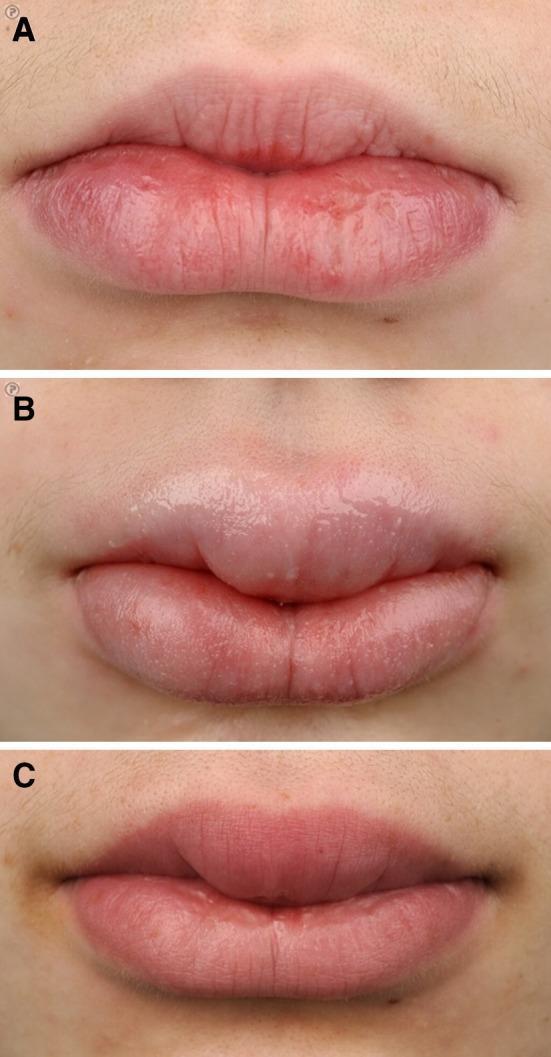
A series of three clinical photographs (a, b, and c) documenting the progression and treatment response of orofacial granulomatosis (cheilitis granulomatosa) in a pediatric patient. Photograph (a) shows the initial presentation with mild erythema and localized edema of the lower lip, displaying a dry texture and fine surface fissuring. Photograph (b) demonstrates a significant progression to macrocheilia, characterized by marked, diffuse swelling of both the upper and lower lips. The lip surfaces appear smooth, tense, and glossy, indicating severe interstitial edema. Photograph (c) depicts the clinical status after one year of combination therapy with infliximab and azathioprine. There is a visible reduction in swelling compared to (b), though mild persistent enlargement remains compared to the baseline in (a). The surface texture has regained normal anatomical creases and a matte appearance. This sequence serves as a clinical reference for the extraintestinal manifestations of Crohn's disease and the efficacy of biological maintenance therapy in managing orofacial granulomatosis.
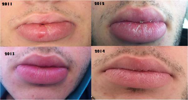
A series of four clinical photographs demonstrating the progression of cheilitis granulomatosa of the lower lip in a patient with Crohn's disease over a four-year period (2011–2014) during infliximab therapy. The 2011 image shows severe, macrocheilia-type swelling of the lower lip with an indistinct vermillion border, distorted shape, and a visible surface erosion or ulceration. The 2012 image reveals persistent but slightly reduced edema with significant xerosis and fine white desquamation. By 2013, there is a further decrease in lip volume and a progressive return of the natural anatomical contour. The final 2014 image shows significant clinical improvement with minimal residual swelling, a clearly defined vermillion border, and a relatively smooth surface texture. This longitudinal comparison illustrates the therapeutic response of orofacial granulomatosis to anti-TNF-alpha biological therapy, highlighting the resolution of granulomatous inflammation and secondary cutaneous manifestations like edema and surface erosions.
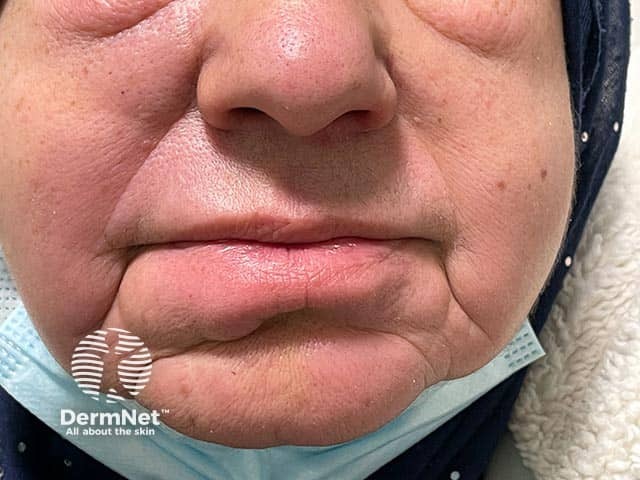
Clinical photography of the perioral region shows diffuse lower-lip edema attributable to granulomatous cheilitis, a manifestation of orofacial granulomatosis subsequently associated with Crohn disease. The frontal facial view emphasizes swelling of the lower lip with mucosal erythema, a thickened vermilion border, and subtle tightening of perioral folds. No acute mucosal ulceration is evident; soft-tissue expansion dominates the phenotype. The case notes progression to gastrointestinal Crohn disease, underscoring the well-established link between orofacial granulomatosis and intestinal inflammatory bowel disease. This image exemplifies lip involvement as a prominent extraintestinal sign that may precede or accompany GI symptoms. Differential considerations include cheilitis granulomatosa, orofacial granulomatosis, sarcoidosis, Melkersson-Rosenthal syndrome, and angioedema or contact cheilitis. Imaging modality is clinical photography with a color-balanced digital capture, using a standardized frontal orientation and no contrast or staining. Pathophysiology involves granulomatous inflammation causing localized edema of the lips and perioral tissues. Clinically, recognition of orofacial granulomatosis as a potential early marker of Crohn disease supports prompt GI evaluation, histologic confirmation when indicated, and multidisciplinary management with dermatology and gastroenterology. Educational and research use includes dermato-gastroenterology correlation, case-based teaching, and dataset enrichment for orofacial granulomatous disease. This image supports education, research annotation, and algorithmic retrieval of granulomatous lip disease.
Clinical Assessment: Persistent Lip Swelling After Tooth Extraction (2 Months)
What I Can See in the Image
The Key Clinical Point
Differential Diagnosis (Most Likely to Least Likely)
1. Orofacial Granulomatosis / Cheilitis Granulomatosa (TOP SUSPECT)
- Sudden onset with progressive course terminating in chronic enlargement of the lips
- Usually upper lip first, then lower lip - both are affected here
- Swelling is smooth, non-pitting, without ulceration, fissuring, or scaling
- Swelling remains permanently if untreated
"Cheilitis granulomatosa is characterized by a sudden onset and progressive course, terminating in chronic enlargement of the lips... Usually, only enlargement is present, without ulceration, fissuring, or scaling." - Andrews' Diseases of the Skin
- Cheilitis granulomatosa can be the first sign of Crohn's disease (before gut symptoms appear)
- Sarcoidosis
- Melkersson-Rosenthal syndrome (if also has facial palsy or scrotal/fissured tongue)
2. Melkersson-Rosenthal Syndrome
- Triad: recurrent lip/facial swelling + facial nerve palsy + fissured (scrotal) tongue
- Dental infection can precipitate it
- Incomplete form (only lip swelling) is common
3. Chronic Dental Infection / Periapical Abscess
- Residual infection at the extraction socket can cause persistent swelling
- Usually accompanied by pain, tenderness, pus discharge, or fever - if these are present, this moves up the differential
- Should be ruled out first with dental X-ray (periapical/OPG)
4. Angioedema (Hereditary or Drug-Induced)
- Usually episodic and recurrent, not constant for 2 months
- Drug-induced (ACE inhibitors are a common cause - e.g., ramipril, enalapril)
- Ask about medications
5. Sarcoidosis
- Can cause lip granulomas
- Check for systemic features: lymphadenopathy, respiratory symptoms, eye involvement
6. Crohn's Disease (Oral/Extraintestinal Manifestation)
- Lip swelling can precede gut symptoms by months-years
- Check for abdominal pain, diarrhea, weight loss
7. Lymphangioma / Hemangioma / Neurofibroma
- Structural lesion causing lip enlargement - usually present from childhood
Investigations Required
| Investigation | Purpose |
|---|---|
| Dental OPG X-ray | Rule out residual infection, dry socket, retained root |
| Lip biopsy (incisional) | Confirm granulomatous inflammation - the most important test |
| Full blood count + ESR/CRP | Infection or inflammation |
| Serum ACE + chest X-ray | Screen for sarcoidosis |
| Serum calcium | Elevated in sarcoidosis |
| Colonoscopy / GI referral | If Crohn's disease suspected |
| Patch testing (contact allergy panel) | Rule out allergic contact cheilitis |
| Review all medications | Rule out ACE inhibitor-induced angioedema |
Treatment
Step 1 - Treat Any Residual Dental Infection First
- Antibiotic therapy for odontogenic infection has been reported to lead to remission of orofacial granulomatosis.
Step 2 - Specific Treatment for Cheilitis Granulomatosa/OFG
- Intralesional corticosteroid injections (triamcinolone acetonide) - usually successful but temporary effect; repeated injections needed
- Intralesional corticosteroids + one or more oral agents:
- Doxycycline (anti-inflammatory)
- Hydroxychloroquine
- Dapsone
- Colchicine
- Sulfasalazine
- Topical tacrolimus ointment (applied to lips)
- Anti-TNF agents (infliximab, adalimumab) - shown effective, especially if Crohn's-associated (see clinical images above showing good response)
- Surgical lip reduction (cheiloplasty) - for permanently deformed lips; best combined with intralesional corticosteroids
- Compression therapy - improves outcome without side effects
Urgent Action Needed
This patient must see a dental surgeon + oral medicine specialist / dermatologist urgently. Two months of persistent post-extraction lip swelling requires:
- Dental X-ray to exclude residual infection
- Lip biopsy for definitive diagnosis
- Gastroenterology review to exclude Crohn's disease
Do not delay - the longer granulomatous swelling goes untreated, the more likely it becomes permanent due to connective tissue fibrosis.