
Lateral skull base approach, indications and complications in detail according to standard ENT textbook in simplified manner
lateral skull base surgical approaches translabyrinthine middle fossa retrosigmoid diagram
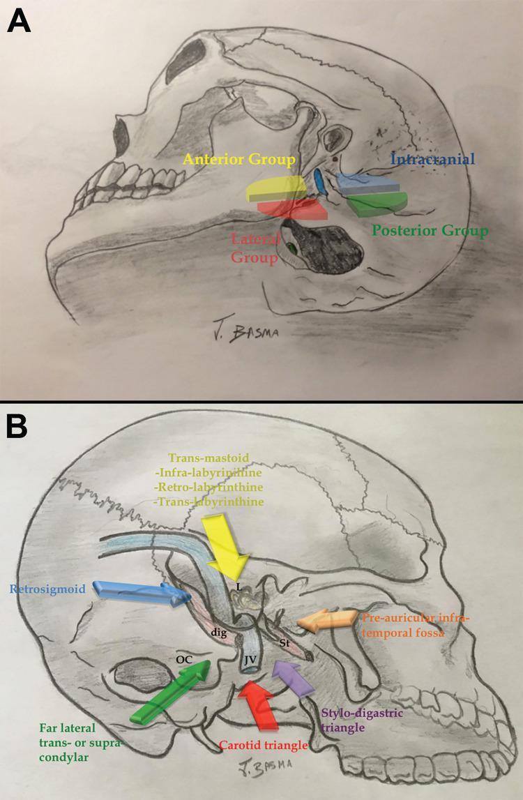
Anatomical illustrations outlining surgical corridors to the jugular foramen. Panel A depicts a lateral view of a human skull with color-coded regions classifying Rhoton’s surgical approaches into four groups: Anterior (yellow/orange), Lateral (red), Posterior (green), and Intracranial (blue). Panel B provides a detailed schematic of specific surgical trajectories and anatomical landmarks. Pathways include the Trans-mastoid approach (yellow arrow) with infra-, retro-, and trans-labyrinthine components; the Retrosigmoid approach (blue arrow); the Far lateral trans- or supra-condylar approach (green arrow) targeting the occipital condyle (OC); and the Pre-auricular infra-temporal fossa approach (orange arrow). Inferior trajectories through the Carotid triangle (red) and Stylo-digastric triangle (purple) are also shown. Key labels identify the digastric muscle (dig), styloid process (St), jugular vein (JV), and labyrinth (L). This educational diagram illustrates the complex three-dimensional 'angles of attack' required for skull base surgery, specifically for treating tumors such as glomus jugulare or schwannomas.
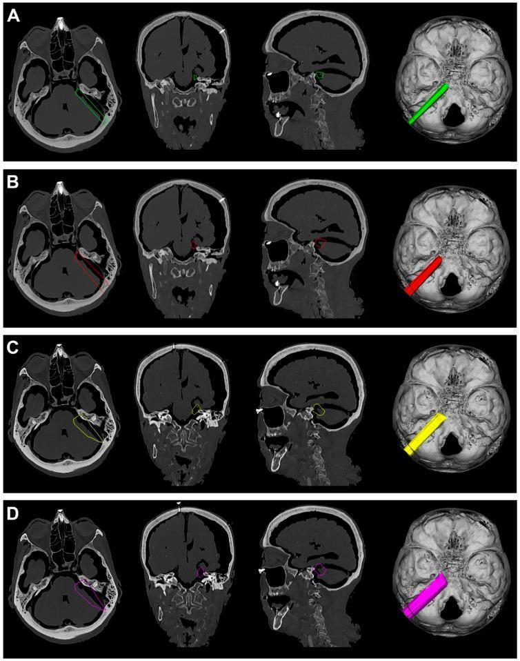
A comparative diagnostic and surgical simulation display consisting of four rows (A-D) showcasing surgical approaches to the skull base. Each row presents four views: axial CT, coronal CT, sagittal CT, and a 3D-rendered intracranial superior view. The images illustrate variations of the Retrosigmoid Approach (RSA) and Retrosigmoid Al-Mefty’s Approach (RSAS) with different brain retraction levels (10 mm and 15 mm). Colored overlays (Green in A, Red in B, Yellow in C, and Magenta in D) define the surgical corridors and work volumes directed toward Meckel’s cave and the petroclival region. The axial and 3D views demonstrate the linear trajectory and entry angle from the posterior fossa toward the middle fossa transition. Coronal and sagittal views highlight the cross-sectional area of the simulated surgical corridor relative to critical neurovascular structures and the skull base. This educational resource is designed for neurosurgical planning and anatomical training, focusing on quantifying surgical exposure and working volumes for various microsurgical transcranial approaches (MTAs).
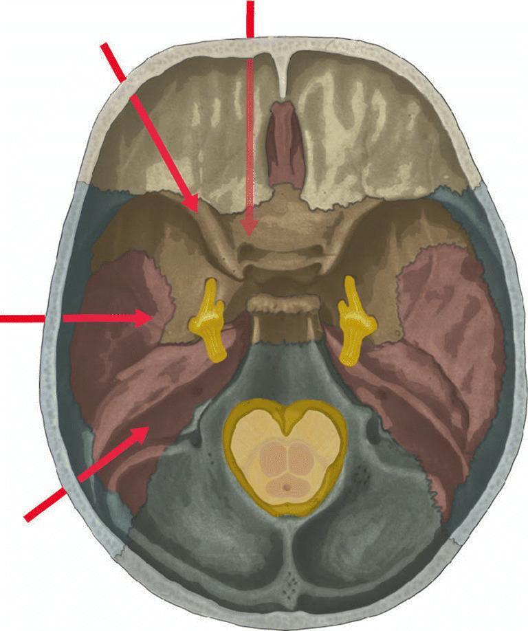
This medical illustration presents a superior view of the internal skull base, color-coded to delineate major anatomical regions and neurosurgical pathways. The anterior cranial fossa is shaded in light beige, the middle cranial fossa in rust/brown, and the posterior cranial fossa in grey. Key neuroanatomical structures are highlighted in yellow, including the trigeminal ganglia situated within Meckel's cave bilaterally and the brainstem cross-section positioned over the foramen magnum. Four red arrows originate from the periphery to illustrate different surgical approaches to Meckel's cave: antero-medial (endonasal/transpterygoid route), antero-lateral (superior orbital fissure/cavernous sinus route), lateral (subtemporal/transtentorial route), and posterior (suboccipital route). This diagram serves as a pedagogical tool for neurosurgical planning and skull base anatomy, specifically focusing on lesions such as trigeminal schwannomas or lymphoma involving the trigeminal cave.
Lateral Skull Base Approaches - A Complete Simplified Guide
What is the Lateral Skull Base?
- Bounded anteriorly by the greater wing of sphenoid and zygomatic arch
- Bounded posteriorly by the occipital bone
- Laterally formed by the squamous and petrous temporal bone
Overview of Lateral Skull Base Approaches
| Approach | Hearing | Main Use |
|---|---|---|
| Translabyrinthine | Sacrificed | Large acoustic neuromas, CPA tumors |
| Middle Cranial Fossa | Preserved | Small intracanalicular tumors |
| Retrosigmoid / Retrolabyrinthine | Preserved (possible) | Small-medium CPA tumors |
| Transcochlear / Transotic | Sacrificed | Petrous apex, clivus lesions |
| Lateral Temporal Bone Resection | Sacrificed | EAC/temporal bone malignancies |
| Lateral (Fisch) Infratemporal Approach | Sacrificed | Large infratemporal/parapharyngeal lesions |
1. Translabyrinthine Approach
What it is
Steps (Key Stages)
- Extended post-auricular incision
- Complete cortical mastoidectomy
- Bony labyrinthectomy (removal of all semicircular canals)
- Skeletonization of sigmoid sinus, jugular bulb, and vertical facial nerve
- Skeletonization of the IAC
- Identification of facial nerve at the lateral end of the IAC (fundus)
- Opening of posterior fossa dura
- Tumor removal with microsurgical technique
- Closure - obliteration with abdominal fat, closure of middle ear as blind sac
Indications
- Medium to large acoustic neuromas (vestibular schwannomas) - the principal indication
- Any CPA or IAC lesion where hearing is not serviceable (non-functional hearing)
- Small acoustic neuromas in patients with no useful hearing
- Meningiomas, non-acoustic schwannomas, gliomas, chordomas, skull base chondrosarcomas of the CPA
- Complete facial nerve decompression (when hearing is absent)
- Vestibular neurectomy (when hearing is absent)
- Can be extended to access: petrous apex + clivus (transcochlear), jugular foramen, hypoglossal canal, foramen magnum
Advantages
- No cerebellar retraction needed
- Early facial nerve identification at the lateral IAC fundus - the safest point
- Wide, direct access to the entire CPA
- Can tackle tumors of any size
- Highest rate of facial nerve preservation compared to other approaches
- Low rate of postoperative headache
Disadvantages
- Permanent hearing loss (hearing must be sacrificed)
- Higher incidence of CSF fistula compared to other approaches (10-15% of CPA surgeries)
- Limited access to IAC fundus in some variants - not applicable here
2. Middle Cranial Fossa (MCF) Approach
What it is
Steps
- Incision begins near midline of calvarium, 2 cm behind hairline, carried inferiorly in front of the ear
- Temporal lobe retraction upward (extradural)
- Identification of the IAC through the tegmen/petrous ridge
- Drilling of the IAC from above
- Facial nerve identified and preserved
- Tumor removal
- The temporal branch of the facial nerve is protected by elevating temporal fascia during skin elevation
Indications
- Small, mainly intracanalicular tumors (confined within the IAC)
- Young patients where hearing preservation is the priority
- Small acoustic neuromas with good/serviceable hearing
- When the tumor does not extend significantly into the CPA
- Facial nerve decompression (IAC and geniculate ganglion)
- Superior semicircular canal dehiscence repair
Advantages
- Hearing preservation possible
- Direct access to the IAC fundus
- No need to traverse the labyrinth
Disadvantages
- Requires temporal lobe retraction - risk of temporal lobe injury, postoperative epilepsy, dysphasia (especially if dominant lobe)
- Higher risk of facial nerve injury (~16%) - anatomical course of the facial nerve may compromise tumor exposure
- Limited for large tumors
- Needs osmotic agents/diuretics for brain relaxation
- Technically demanding in elderly with more adherent temporal dura
3. Retrosigmoid (Suboccipital) Approach
What it is
Indications
- Small to medium acoustic neuromas with serviceable hearing where the tumor does NOT extend to the fundus of the IAC
- CPA meningiomas
- Other posterior fossa tumors with preserved hearing
- Extensive posterior fossa lesions
- When translabyrinthine or MCF approach is not suitable
Advantages
- Hearing preservation is possible (when tumor does not reach the fundus)
- Lower facial nerve palsy rate (~6%) compared to MCF
- Lower CSF leak rate (~9%) compared to translabyrinthine
- Wider posterior fossa access
Disadvantages
- Requires cerebellar retraction - associated with its own complications
- Cannot reach the IAC fundus - if tumor extends to the fundus, hearing preservation is compromised or impossible
- Higher incidence of postoperative headache (due to bone removal near occipital muscles)
- Needs osmotic/diuretic agents for brain relaxation
4. Transcochlear / Transotic Approach
What it is
Indications
- Lesions of the petrous apex
- Extensive clivus lesions
- Cholesterol granuloma of petrous apex
- Chordomas
- When maximum anterior medial exposure is needed and hearing is absent
5. Lateral (Partial) Temporal Bone Resection (LTBR)
What it is
Indications
- Malignant neoplasms localized to the osseous EAC without transgression through the tympanic membrane
- Squamous cell carcinoma of the EAC (most common)
- Other EAC malignancies
Subtypes (Cummings)
- Sleeve resection - for superficial malignancies of the cartilaginous canal only (no bone removal)
- Lateral/Partial temporal bone resection - for osseous canal involvement
- Subtotal temporal bone resection - for more extensive disease
- Total temporal bone resection - for most advanced disease
6. Lateral (Fisch) Infratemporal Fossa Approach
What it is
Steps
- Extended post-auricular incision, EAC closed as blind sac, auricle retracted anteriorly
- Extended mastoidectomy + total parotidectomy
- Facial nerve rerouting (anterior transposition)
- Zygomatic arch removed
- Temporalis muscle divided - middle cranial fossa base accessed
- Condylar process of mandible may be removed for wider exposure
- Mandibular branch of trigeminal nerve divided + pterygoid muscles removed
- Internal carotid artery identified and retracted
- Parapharyngeal space + cartilaginous Eustachian tube resected en bloc with tumor
Indications
- Recurrent nasopharyngeal carcinoma (NPC)
- Large glomus jugulare / glomus vagale tumors
- Extensive parapharyngeal space tumors
- Tumors of the infratemporal fossa
- Lesions requiring en bloc resection of parapharyngeal structures
Advantages
- Early identification and protection of internal carotid artery
- Direct access to deep neck and parapharyngeal space
Disadvantages
- Extensive mobilization of normal tissues required - high morbidity
- Poor visualization of nasopharyngeal midline and anterior extension
- Limited access to nasal cavity/paranasal sinuses
- Permanent conductive hearing loss (EAC closed)
- Facial nerve morbidity from rerouting
Complications of Lateral Skull Base Surgery
1. Hearing Loss
- Sensorineural hearing loss from drill-generated acoustic trauma
- Direct drill contact with ossicles or endosteal membrane of the cochlea
- Prevention: avoid contacting ossicles; use systemic + local corticosteroids
- If inner ear is inadvertently opened: recognize immediately, repair, avoid suction of perilymph/endolymph
2. CSF Leak (Most Common Major Complication)
- 10-15% of CPA surgery procedures overall
- Highest risk with translabyrinthine approach
- Retrosigmoid: ~9% CSF leak rate
- CSF rhinorrhea: from mastoid air cells communicating with Eustachian tube
- Management:
- Replace compression mastoid dressing
- Eustachian tube obliteration (routinely performed - reduces rhinorrhea to <5%)
- Lumbar CSF drainage for 3 days (persistent leaks)
- Hydroxyapatite cement/titanium mesh cranioplasty (highly effective)
- Surgical closure of EAC + direct obliteration of Eustachian tube (for refractory cases)
3. Facial Nerve Injury
- Most feared complication of lateral skull base surgery
- Translabyrinthine approach: 57% HB Grade I-II when total resection attempted; 77% HB Grade I-II with near-total (functional) resection
- Retrosigmoid approach: ~6% facial nerve palsy rate
- Middle cranial fossa approach: ~16% facial nerve damage (highest among the three)
- Management:
- Immediate repair or interposition nerve graft if transected
- Facial nerve monitoring (EMG) intraoperatively - routine in all cases
- Facial-hypoglossal anastomosis (delayed reanimation)
- Temporalis muscle transfer
- Nerve substitution with V3 or combined V3+hypoglossal anastomosis
- Botox for synkinesis management
- Physical therapy (Diels technique)
4. Meningitis
- Any procedure opening the subarachnoid space risks postoperative meningitis
- Mean onset: 8 days postoperatively for acoustic neuroma surgery
- Presents with: toxic appearance, stiff neck, fever, headache
- Diagnosis: CSF studies
- Management: aggressive antibiotic therapy - avoids serious complications
- Prevention: perioperative prophylactic antibiotics (routine)
5. Anterior Inferior Cerebellar Artery (AICA) Injury
- Tumor manipulation at the brainstem can cause ischemia in the AICA distribution
- Presents as intraoperative vital sign changes
- Management: stop manipulation temporarily; if persistent, terminate procedure
- Complete AICA interruption can cause Atkinson syndrome - lateral tegmental pontine infarction, often fatal
- Non-fatal partial forms also occur
6. Ophthalmologic Complications
- Combination of facial paralysis + corneal insensitivity (V1 involvement) = severe corneal risk
- More common with large tumors
- Management:
- Lubricating drops/ointments, moisture chambers, nightly eye taping
- Gold weight implantation in upper lid (preferred in many centers)
- Brow lift + eyelid springs for prolonged paralysis (>6 months)
- Close ophthalmologic monitoring
7. Cerebral / Cerebellar Complications
- Temporal lobe retraction (MCF approach): risk of postoperative epilepsy, temporary dysphasia if dominant lobe retracted
- Cerebellar retraction (retrosigmoid approach): cerebellar contusion, edema
- Prevention: use of osmotic agents + diuretics for brain relaxation in approaches requiring retraction (NOT needed in translabyrinthine)
8. Vascular Complications
- Sigmoid sinus injury - can cause massive hemorrhage, air embolism
- Internal carotid artery injury (infratemporal approaches)
- Jugular bulb injury
- Prevention: preoperative embolization for vascular tumors (glomus); intraoperative EMG monitoring; careful skeletonization technique
9. Lower Cranial Nerve Injury
- Relevant in jugular foramen surgery and extended approaches
- CN IX, X, XI injury: dysphagia, aspiration, hoarseness, shoulder drop
- CN XII injury: tongue deviation/atrophy
10. Postoperative Headache
- Most common after retrosigmoid approach (due to disruption of occipital musculature)
- Less common with translabyrinthine approach
Choosing the Right Approach - Summary
| Situation | Best Approach |
|---|---|
| Large acoustic neuroma, no useful hearing | Translabyrinthine |
| Small intracanalicular tumor, good hearing, young patient | Middle fossa |
| Small-medium CPA tumor, good hearing, tumor not reaching fundus | Retrosigmoid |
| Petrous apex/clivus lesion | Transcochlear |
| EAC/temporal bone malignancy | Temporal bone resection |
| Large infratemporal/parapharyngeal tumor | Fisch infratemporal |
As stated in Cummings: "The notion that a single approach should be used with all lesions of the posterior fossa is counterintuitive and subjects patients to unnecessary morbidity. The best hearing and functional outcomes result from individualizing the surgical approach to patient and tumor characteristics."