
Im 1st year mbbs student at Hngu university [ Bhagyoday medical college Kadi ] in exam imp questions list i saw one question is 8 mark question is About DNA [ structure, salient features, Types]give a answer of this question easy to understand English language,when I read this question answer I understand easily, give answer this type when I read this question answer first time so much answer I understand easily in first time and when i write this in exam my sir will give me full out of full marks and easy to read and understand,answer give as 1 st year mbbs [ Biochemistry ]and easy to understand and write Thank you
DNA - Structure, Salient Features, and Types
INTRODUCTION
PART 1: CHEMICAL STRUCTURE OF DNA (Building Blocks)
Each Nucleotide Has 3 Parts:
| Component | Details |
|---|---|
| Nitrogenous Base | Purines: Adenine (A), Guanine (G) / Pyrimidines: Cytosine (C), Thymine (T) |
| Sugar | Deoxyribose (5-carbon sugar, no -OH at carbon 2) |
| Phosphate group | One inorganic phosphate |
How Nucleotides Are Joined:
PART 2: WATSON-CRICK DOUBLE HELIX MODEL (Secondary Structure)
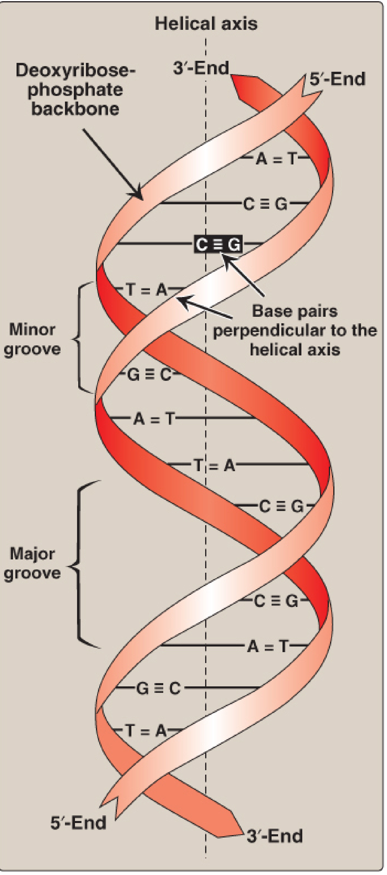
Key Points of the Double Helix:
- Two strands coil around a common central axis (the helical axis) - like a twisted ladder.
- Antiparallel orientation - one strand runs 5'→3' and the other runs 3'→5' (in opposite directions).
- Backbone is on the outside - the sugar-phosphate (deoxyribose-phosphate) backbone is hydrophilic and faces outward.
- Bases are on the inside - the hydrophobic nitrogenous bases are stacked inward (like steps of a ladder).
- Base pairing (Chargaff's Rule):
- A pairs with T (Adenine - Thymine) → 2 hydrogen bonds
- G pairs with C (Guanine - Cytosine) → 3 hydrogen bonds
- So A = T and G = C in any double-stranded DNA. The total purines always equal total pyrimidines.
- Major and minor grooves - Because of the helical twist, two unequal grooves form along the surface:
- Major groove (wide) - where most regulatory proteins and transcription factors bind.
- Minor groove (narrow) - where some drugs (e.g., dactinomycin/actinomycin D) intercalate.
- Right-handed helix - the helix turns in a clockwise direction (called right-handed).
- 10.4 base pairs per turn of the helix, with a rise of 3.4 Å (angstroms) per base pair, and a full turn every 34 Å.
PART 3: SALIENT FEATURES OF DNA
| Feature | Details |
|---|---|
| Double-stranded | Two complementary strands held by H-bonds |
| Antiparallel strands | 5'→3' direction is opposite in the two strands |
| Complementary base pairing | A=T (2 H-bonds), G≡C (3 H-bonds) |
| Right-handed helix | Turns clockwise (B-form) |
| Major & minor grooves | Sites for protein/drug binding |
| Stable structure | Stabilized by H-bonds + base stacking interactions |
| Chargaff's rule | A=T, G=C; total purines = total pyrimidines |
| Semiconservative replication | Each new DNA has one old strand + one new strand |
| Genetic code stored as base sequence | Sequence of bases = information for protein synthesis |
| Location | Nucleus (chromatin), mitochondria; prokaryotes: nucleoid |
PART 4: TYPES OF DNA
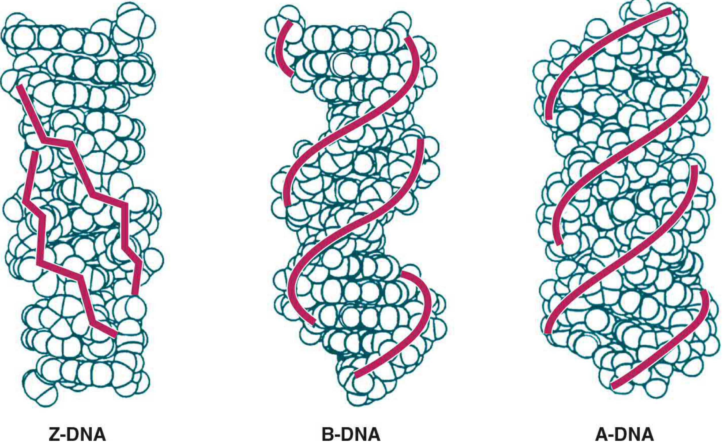
1. B-DNA (Most Important)
- The normal form found in cells under physiological (water-rich) conditions.
- Right-handed double helix.
- 10.4 bp per turn (approximately 10 bp per turn).
- Rise per base pair: 3.4 Å.
- Has clear major and minor grooves.
- This is the Watson-Crick model.
2. A-DNA
- Found when DNA is in low humidity (dehydrated conditions) or in DNA-RNA hybrid molecules.
- Also right-handed, but more compact and wider.
- 11 bp per turn.
- Rise per base pair: 2.3 Å (shorter/more compact than B-form).
- Has a deeper major groove.
3. Z-DNA
- Left-handed helix (turns counter-clockwise) - the opposite of A and B forms.
- Named "Z" because the phosphate backbone zigzags (zigs and zags in a Z-shaped pattern).
- 12 bp per turn (approximately 11-12 bp).
- Rise per base pair: 3.8 Å.
- Narrower and more elongated.
- Found transiently in cells during transcription (RNA synthesis).
- Physiological role is still being studied.
Comparison Table: Types of DNA
| Feature | B-DNA | A-DNA | Z-DNA |
|---|---|---|---|
| Handedness | Right | Right | Left |
| bp per turn | 10.4 | 11 | 12 |
| Rise per bp | 3.4 Å | 2.3 Å | 3.8 Å |
| Groove | Major+Minor | Deep major | Shallow minor |
| Found in | Normal cells | DNA-RNA hybrid | Transcription sites |
ADDITIONAL NOTE: Linear vs Circular DNA
- Eukaryotes - linear dsDNA in nucleus (organized into chromosomes with histone proteins); circular dsDNA in mitochondria.
- Prokaryotes - single, circular dsDNA molecule in the nucleoid.
- Plasmids - small, circular, extrachromosomal DNA in bacteria (used in genetic engineering).
QUICK MEMORY TRICK
"All Bankers Get Tired" = A bonds T, G bonds C (base pairing rule) B-DNA = Body (normal), A-DNA = compact (like A for Altered), Z-DNA = Zigzag (left-handed)
Im 1st year mbbs student at Hngu university [ Bhagyoday medical college Kadi ] in exam imp questions list i saw one question is 5 mark question is Structure And Function of T-RNA give a answer of this question easy to understand English language,when I read this question answer I understand easily, give answer this type when I read this question answer first time so much answer I understand easily in first time and when i write this in exam my sir will give me full out of full marks and easy to read and understand,answer give as 1 st year mbbs [ Biochemistry ]and easy to understand and write Thank you
tRNA (Transfer RNA) - Structure and Function
INTRODUCTION
PART 1: STRUCTURE OF tRNA
Basic Facts:
- tRNA is a single-stranded RNA molecule.
- Length: 74 to 95 nucleotides long.
- Sedimentation coefficient: 4S (smallest RNA).
- Contains many unusual (modified) bases - e.g., Dihydrouracil (D), Pseudouracil (ψ), Inosine, Ribothymidine.
- Makes up about 15-20% of total cellular RNA.
- There is at least one tRNA for each of the 20 amino acids. In humans, there are at least 50 different tRNA species.
Secondary Structure: THE CLOVERLEAF
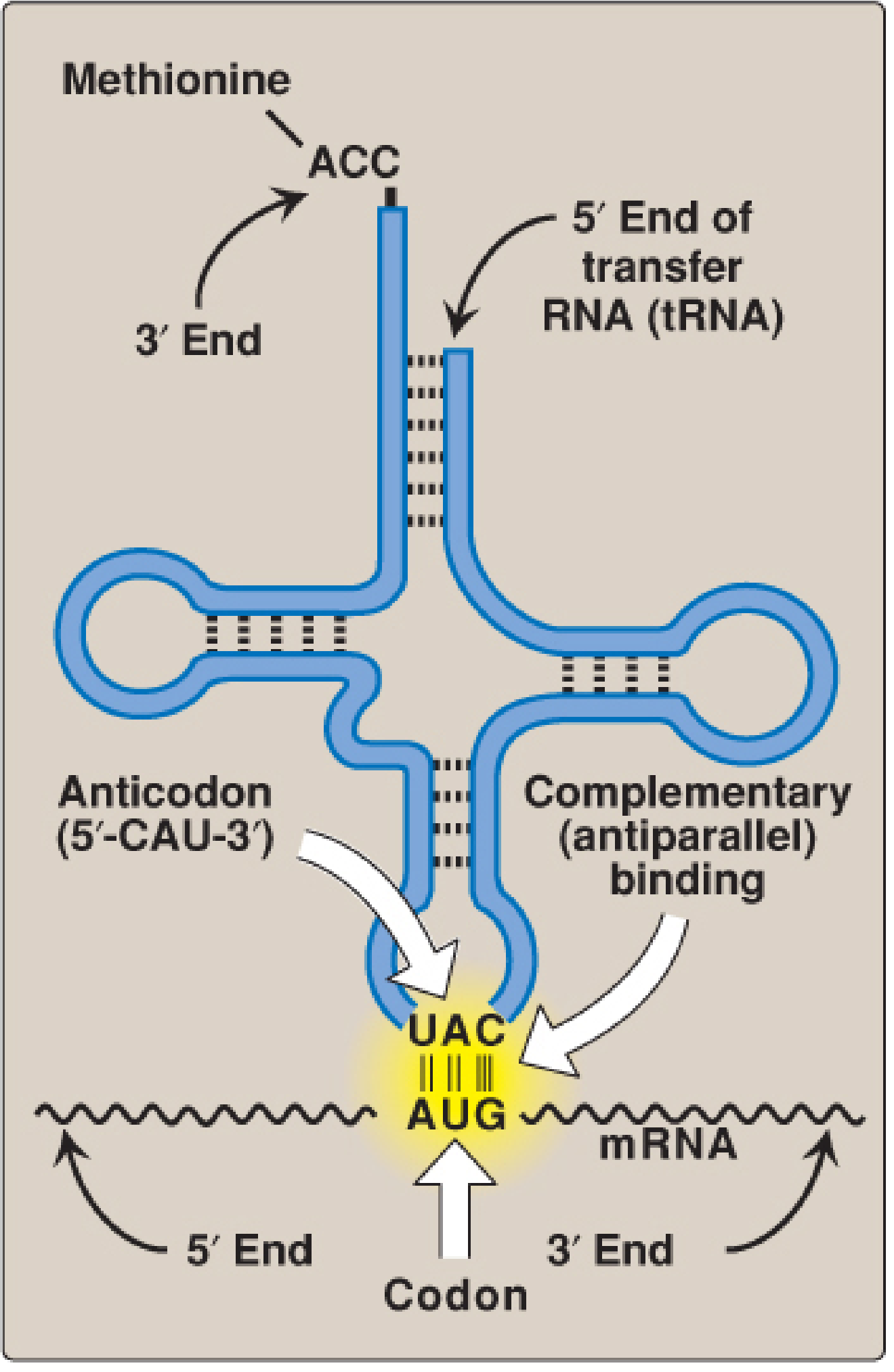
| Part | Name | Function |
|---|---|---|
| 1. Acceptor Arm (Stem) | Amino acid acceptor arm | 3' end always ends in -CCA-OH. The amino acid attaches here. |
| 2. D-Loop (D arm) | Dihydrouracil loop | Contains modified base Dihydrouracil (D). Helps tRNA bind to aminoacyl-tRNA synthetase enzyme. |
| 3. Anticodon Loop (Anticodon arm) | Anticodon arm | Contains the anticodon - 3 bases that pair with the mRNA codon. Most important part for reading mRNA. |
| 4. TψC Loop (T arm) | Ribothymidine-Pseudouracil-Cytosine loop | Contains T, ψ (pseudouracil), C. Helps tRNA bind to the ribosome. |
| 5. Variable Loop | Extra arm | Variable in size. Helps classify tRNA types (class I = small, class II = large). |
3D (Tertiary) Structure: L-SHAPE
- The acceptor arm is at one end of the L.
- The anticodon loop is at the other end of the L.
- These two important functional ends are kept far apart - one binds the amino acid, the other reads the mRNA.
Key Structural Features to Remember:
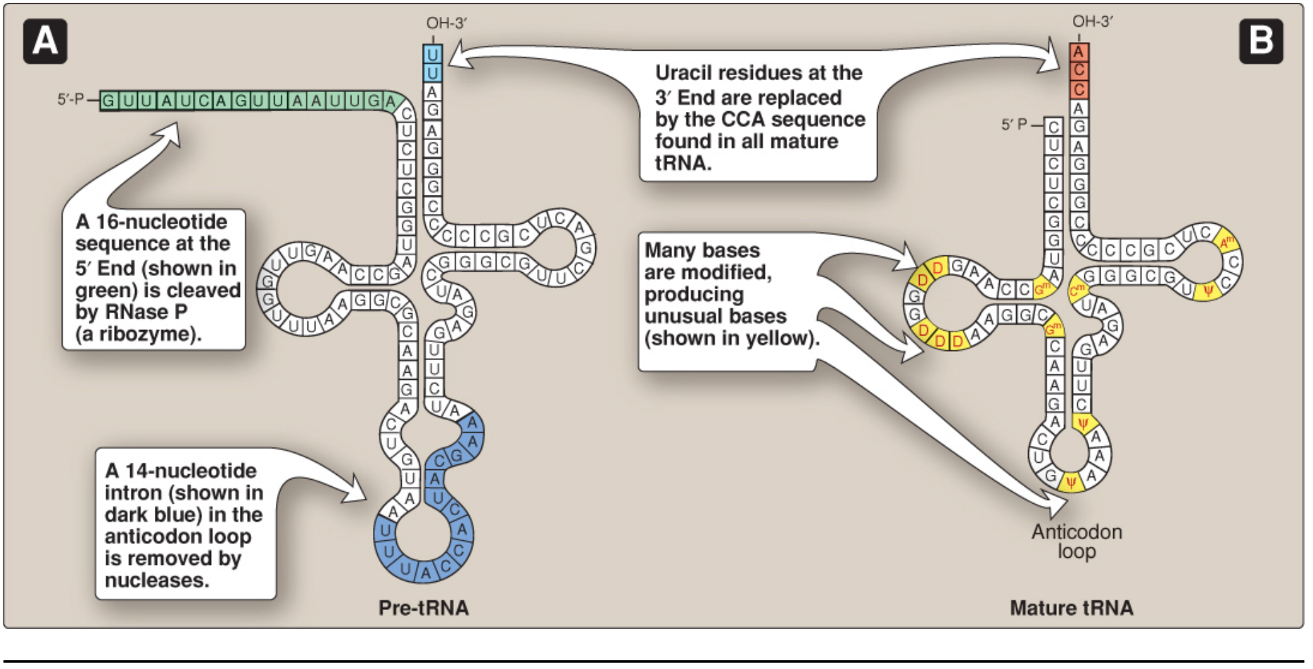
- 3' end always = -CCA-OH (universal, added post-transcriptionally by enzyme nucleotidyltransferase)
- 5' end always = phosphate group
- Contains modified/unusual bases (not found in other RNA types)
- Extensive intrachain base pairing - holds the cloverleaf shape
PART 2: FUNCTIONS OF tRNA
1. Adaptor Molecule (Most Important Function)
2. Carries Amino Acids ("Charging" of tRNA)
- Each tRNA is charged (loaded) with its specific amino acid by an enzyme called Aminoacyl-tRNA Synthetase.
- There are 20 different synthetase enzymes - one for each amino acid.
- The amino acid attaches by ester bond to the 3'-OH of the CCA end (acceptor arm).
- This process requires ATP.
- A tRNA with an amino acid attached = Charged tRNA (aminoacyl-tRNA).
- A tRNA without an amino acid = Uncharged tRNA.
3. Reads the mRNA Code (Anticodon-Codon Pairing)
- The anticodon in the anticodon loop of tRNA is a 3-base sequence.
- It pairs with the complementary codon on mRNA in an antiparallel direction.
- Example: If mRNA codon = AUG (codes for Methionine), then tRNA anticodon = UAC.
4. Delivers Amino Acid to Ribosome
- The charged tRNA carries the amino acid to the ribosome.
- At the ribosome, the amino acid is added to the growing polypeptide chain.
- After releasing the amino acid, the tRNA leaves the ribosome as uncharged tRNA.
5. Proofreading
- Aminoacyl-tRNA synthetases also have proofreading (editing) activity - they remove a wrongly attached amino acid, ensuring accuracy of protein synthesis.
SUMMARY TABLE
| Feature | Details |
|---|---|
| Full name | Transfer RNA |
| Size | 74-95 nucleotides; 4S |
| Secondary structure | Cloverleaf (4 arms + 4 loops) |
| 3D structure | L-shaped |
| 3' end | Always -CCA-OH (amino acid attaches here) |
| Anticodon | 3-base sequence; pairs with mRNA codon |
| Unusual bases | Dihydrouracil, Pseudouracil, Inosine, etc. |
| Key enzyme | Aminoacyl-tRNA synthetase (charges tRNA with amino acid) |
| Main function | Carries amino acid to ribosome; reads mRNA code |
| % of cell RNA | ~15-20% |
QUICK MEMORY TRICK
"tRNA = Taxi RNA"
- Picks up passenger (amino acid) at the CCA-3' end
- Reads the address (anticodon pairs with mRNA codon)
- Drops off passenger at ribosome for protein building
Im 1st year mbbs student at Hngu university [ Bhagyoday medical college Kadi ] in exam imp questions list i saw one question is 5 mark question is genetic code give a answer of this question easy to understand English language,when I read this question answer I understand easily, give answer this type when I read this question answer first time so much answer I understand easily in first time and when i write this in exam my sir will give me full out of full marks and easy to read and understand,answer give as 1 st year mbbs [ Biochemistry ]and easy to understand and write Thank you
Genetic Code
INTRODUCTION
WHAT IS A CODON?
- The genetic code is a triplet code - every 3 consecutive nucleotide bases on mRNA = 1 codon.
- Each codon codes for one specific amino acid (or a stop signal).
- Total possible codons: 4³ = 64 codons (4 bases taken 3 at a time).
- These 64 codons code for only 20 amino acids + stop signals.
THE CODON TABLE
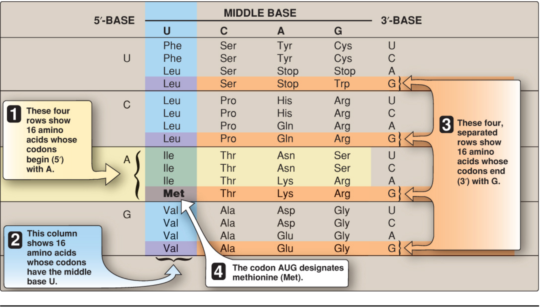
Start Codon:
- AUG = codes for Methionine (Met) = the START/Initiation codon for all proteins.
- Every protein begins with Methionine (though it may be removed later).
Stop Codons (Termination Codons):
- UAA - "Ochre"
- UAG - "Amber"
- UGA - "Opal" (also called "Umber")
- These 3 stop codons do NOT code for any amino acid. They signal the ribosome to stop making the protein.
- So: 61 codons code for 20 amino acids, and 3 codons are stop signals.
PROPERTIES / CHARACTERISTICS OF THE GENETIC CODE
1. TRIPLET CODE (Three-letter code)
- Each codon consists of 3 nucleotide bases (a triplet).
- Example: AUG, GCU, UAA
- A sequence of 3 bases = 1 amino acid.
2. DEGENERATE (Redundant)
- Most amino acids are coded by more than one codon.
- Example: Leucine is coded by 6 codons (UUA, UUG, CUU, CUC, CUA, CUG).
- Only Methionine (AUG) and Tryptophan (UGG) have just 1 codon each.
- The third base of the codon (3' end) is the one that usually varies - this is called "wobble position".
- This degeneracy protects the organism - a mutation in the third base often still codes for the same amino acid.
3. UNAMBIGUOUS (Specific)
- Each codon codes for only ONE specific amino acid - never two.
- The same codon never means two different amino acids.
- AUG always = Methionine. Never anything else.
- Remember: Degenerate = one amino acid can have many codons. Unambiguous = one codon has only one meaning.
4. NON-OVERLAPPING
- Codons do NOT share bases with each other.
- The mRNA is read 3 bases at a time, one after the other, without overlap.
- Example: AUGCCUGAA is read as: AUG | CCU | GAA (not AUG, UGC, GCC...)
5. COMMALESS (No punctuation)
- There are no gaps or "commas" between codons on the mRNA.
- Reading starts at a fixed point (AUG) and continues continuously without skipping, until a stop codon is reached.
- This fixed reading order is called the Reading Frame.
6. UNIVERSAL
- The genetic code is the same in ALL living organisms - bacteria, plants, animals, humans.
- AUG means Methionine in a bacterium and in a human cell.
- This is why genetic engineering works - human insulin gene placed in E. coli bacteria still produces human insulin!
- Exception: Mitochondria use a slightly different code. For example, UGA codes for Tryptophan in mitochondria (instead of being a stop codon).
7. POLAR (Read in one direction only)
- mRNA is always read 5' → 3' direction.
- Codons are always written from 5' end to 3' end.
MUTATIONS AND THE GENETIC CODE
| Type | What happens | Effect |
|---|---|---|
| Silent mutation | Changed codon still codes for SAME amino acid | No effect on protein |
| Missense mutation | Changed codon codes for DIFFERENT amino acid | Protein may be altered (e.g., Sickle cell anemia: GAG → GUG, Glu → Val) |
| Nonsense mutation | Changed codon becomes a STOP codon | Protein is shortened (truncated) |
SUMMARY TABLE: Properties of Genetic Code
| Property | Meaning | Simple way to remember |
|---|---|---|
| Triplet | 3 bases = 1 codon | 3 letters = 1 word |
| Degenerate | 1 amino acid, many codons | Many roads to same city |
| Unambiguous | 1 codon = only 1 amino acid | 1 address = 1 house |
| Non-overlapping | Bases not shared between codons | Read 3 at a time, move forward |
| Commaless | No gaps between codons | Continuous reading |
| Universal | Same in all organisms | One language for all life |
QUICK MEMORY TRICK
"The Genetic Code is DUNDUC"
- Degenerate
- Unambiguous
- Non-overlapping
- Direction: 5'→3'
- Universal
- Commaless
Im 1st year mbbs student at Hngu university [ Bhagyoday medical college Kadi ] in exam imp questions list i saw one question is 8 mark question is About DNA [ structure, salient features, Types]give a answer of this question easy to understand English language,when I read this question answer I understand easily, give answer this type when I read this question answer first time so much answer I understand easily in first time and when i write this in exam my sir will give me full out of full marks and easy to read and understand,answer give as 1 st year mbbs [ Biochemistry ]and easy to understand and write Thank you Give a answer from Satyanarayana Biochemistry book
- Clear numbered points
- Simple language
- Neat tables
- Diagrams described simply
- Exactly the style Indian MBBS exams expect
DNA - Structure, Salient Features and Types
(As per Satyanarayana Biochemistry - 1st Year MBBS)
INTRODUCTION
- DNA is the genetic material of the cell - it stores and transmits hereditary information.
- It is present in the nucleus (chromosomes), mitochondria, and in prokaryotes (nucleoid region).
- DNA was first isolated by Friedrich Miescher in 1869 from pus cells.
- Its double helical structure was proposed by Watson and Crick in 1953 - one of the greatest discoveries in biology.
PART I: CHEMICAL COMPOSITION (Building Blocks of DNA)
Each Nucleotide Has 3 Components:
| Type | Bases |
|---|---|
| Purines (double ring) | Adenine (A), Guanine (G) |
| Pyrimidines (single ring) | Cytosine (C), Thymine (T) |
Memory tip: "Pure As Gold" = Purines = Adenine + Guanine
How Nucleotides Join:
- Nucleotides are joined by 3'→5' Phosphodiester bonds
- The phosphate of one nucleotide links to the 3'-OH of the next
- This forms a long unbranched chain with a free 5' end and a free 3' end
PART II: STRUCTURE OF DNA - WATSON AND CRICK MODEL (B-DNA)
- Chargaff's rules of base pairing
- X-ray crystallography data from Rosalind Franklin and Maurice Wilkins
The Double Helix - Key Points:
- DNA has two polynucleotide strands coiled around a common central axis (helical axis)
- The overall shape resembles a twisted ladder (spiral staircase)
- The two strands run in opposite directions
- One strand goes 5'→3' and the other goes 3'→5'
- This is called antiparallel arrangement
- The deoxyribose-phosphate backbone (hydrophilic) is on the outside of the helix
- Like the two side rails of a ladder
- The nitrogenous bases (hydrophobic) are stacked on the inside
- Like the steps of a ladder
- Bases are perpendicular to the helical axis
- Bases on one strand pair with bases on the opposite strand:
- A pairs with T → connected by 2 Hydrogen bonds
- G pairs with C → connected by 3 Hydrogen bonds
- This means: A = T and G = C in any double-stranded DNA
- Total Purines = Total Pyrimidines
Memory tip: "AT - Two bonds (2)", "GC - Three bonds (3)" → AT home, Get Cozy with Three
- Due to the helical twisting, two grooves are formed on the surface:
- Major groove (wide) - where regulatory proteins and enzymes bind
- Minor groove (narrow) - where some drugs intercalate (e.g., Actinomycin D)
- The helix turns clockwise when viewed from above
- This is called a right-handed helix
PART III: SALIENT FEATURES OF DNA (Watson-Crick B-DNA)
| S.No | Feature | Details |
|---|---|---|
| 1 | Diameter | 20 Å (2 nm) |
| 2 | Rise per base pair | 3.4 Å per base pair |
| 3 | Base pairs per turn | 10 base pairs per complete turn |
| 4 | Length of one full turn | 34 Å (10 × 3.4 Å) |
| 5 | Direction | Right-handed helix |
| 6 | Backbone | Sugar-phosphate on outside |
| 7 | Bases | Stacked on inside |
| 8 | Base pairing | A=T (2H-bonds), G≡C (3H-bonds) |
| 9 | Strand orientation | Antiparallel |
| 10 | Stabilizing forces | H-bonds + base stacking interactions |
| 11 | Chargaff's rule | A=T, G=C; Purines = Pyrimidines |
PART IV: TYPES OF DNA
1. B-DNA
- The most common form found in cells under normal (physiological) conditions
- Right-handed helix
- 10 bp per turn
- Rise per bp = 3.4 Å
- Has clear major and minor grooves
- This is the Watson-Crick model - the one you always study
2. A-DNA
- Found in dehydrated (low water) conditions
- Also found in DNA-RNA hybrid molecules
- Right-handed helix (same as B, but different dimensions)
- 11 bp per turn
- Rise per bp = 2.3 Å (shorter, more compact)
- Wider and shorter than B-DNA
- Deeper major groove
3. Z-DNA
- Left-handed helix (turns counter-clockwise - opposite to A and B)
- Named "Z" because the phosphate backbone zigzags in a Z-pattern
- 12 bp per turn
- Rise per bp = 3.8 Å (longer, more stretched)
- Narrow and elongated
- Found transiently during active transcription (RNA synthesis)
- Less common; stabilized by specific Z-DNA binding proteins
Comparison Table - Types of DNA
| Feature | B-DNA | A-DNA | Z-DNA |
|---|---|---|---|
| Handedness | Right | Right | Left |
| bp per turn | 10 | 11 | 12 |
| Rise per bp | 3.4 Å | 2.3 Å | 3.8 Å |
| Diameter | 20 Å | 23 Å | 18 Å |
| Grooves | Major + Minor | Deep major | Shallow minor |
| Occurrence | Normal cells | DNA-RNA hybrid | Transcription sites |
Memory tip for handedness: "B is for Body (normal/right)", "Z is for Zigzag (left)"
ADDITIONAL IMPORTANT POINTS
Linear vs. Circular DNA:
- Eukaryotes: Linear DNA in nucleus (bound to histone proteins → chromatin → chromosomes); Circular DNA in mitochondria
- Prokaryotes: Single circular DNA in nucleoid (no membrane)
- Plasmids: Small circular DNA in bacteria (used in genetic engineering)
Stabilization of DNA Double Helix:
- Hydrogen bonds between complementary base pairs (A-T and G-C)
- Base stacking interactions (hydrophobic interactions between stacked bases inside the helix)
- Ionic interactions between negatively charged phosphate groups and positive ions (Mg²⁺, histones)
EXAM DIAGRAM - WATSON-CRICK DOUBLE HELIX
- 5' end and 3' end of both strands (antiparallel)
- Deoxyribose-phosphate backbone (outside)
- A=T and G≡C base pairs (inside)
- Major groove and minor groove
- Helical axis
- 3.4 Å between base pairs, 34 Å per turn
QUICK SUMMARY FOR RAPID REVISION
DNA = Double helix, Antiparallel, Complementary base pairing (A=T, G≡C), Right-handed (B-form), 10 bp/turn, 3.4 Å rise, Major + Minor grooves
3 Types: B (normal, right-handed) → A (compact, right-handed) → Z (Zigzag, LEFT-handed)
Ask about dna
- Watson-Crick Double Helix Model
- Types of DNA (A, B, Z forms)
- Salient features of DNA
- Nucleotides, nucleosides
- Purines and pyrimidines
- Phosphodiester bonds
- DNA Replication (semi-conservative)
- DNA Repair mechanisms
- Transcription (DNA → RNA)
- Chargaff's rules
- Denaturation and renaturation of DNA
- Supercoiling of DNA
- Chromatin structure and histones
- Mitochondrial DNA
- DNA vs RNA
- Prokaryotic vs Eukaryotic DNA
Purin and pyrimidines
Purines and Pyrimidines
(1st Year MBBS - Biochemistry)
INTRODUCTION
- They contain Nitrogen atoms in their rings
- They are basic (slightly alkaline) in nature
PART I: PURINES
What are Purines?
- Purines have a DOUBLE RING structure
- One ring is a pyrimidine ring (6-membered) fused with an imidazole ring (5-membered)
- They are larger molecules
Purine Bases:
| Base | Symbol | Found in |
|---|---|---|
| Adenine | A | DNA + RNA |
| Guanine | G | DNA + RNA |
Memory tip: "Pure As Gold" = Purines = Adenine + Guanine
PART II: PYRIMIDINES
What are Pyrimidines?
- Pyrimidines have a SINGLE RING structure (6-membered ring only)
- They are smaller molecules
Pyrimidine Bases:
| Base | Symbol | Found in |
|---|---|---|
| Cytosine | C | DNA + RNA |
| Thymine | T | DNA only |
| Uracil | U | RNA only |
Memory tip: "CUT the PY" = Cytosine, Uracil, Thymine = PYrimidines
STRUCTURES OF BASES (Must know for exam)
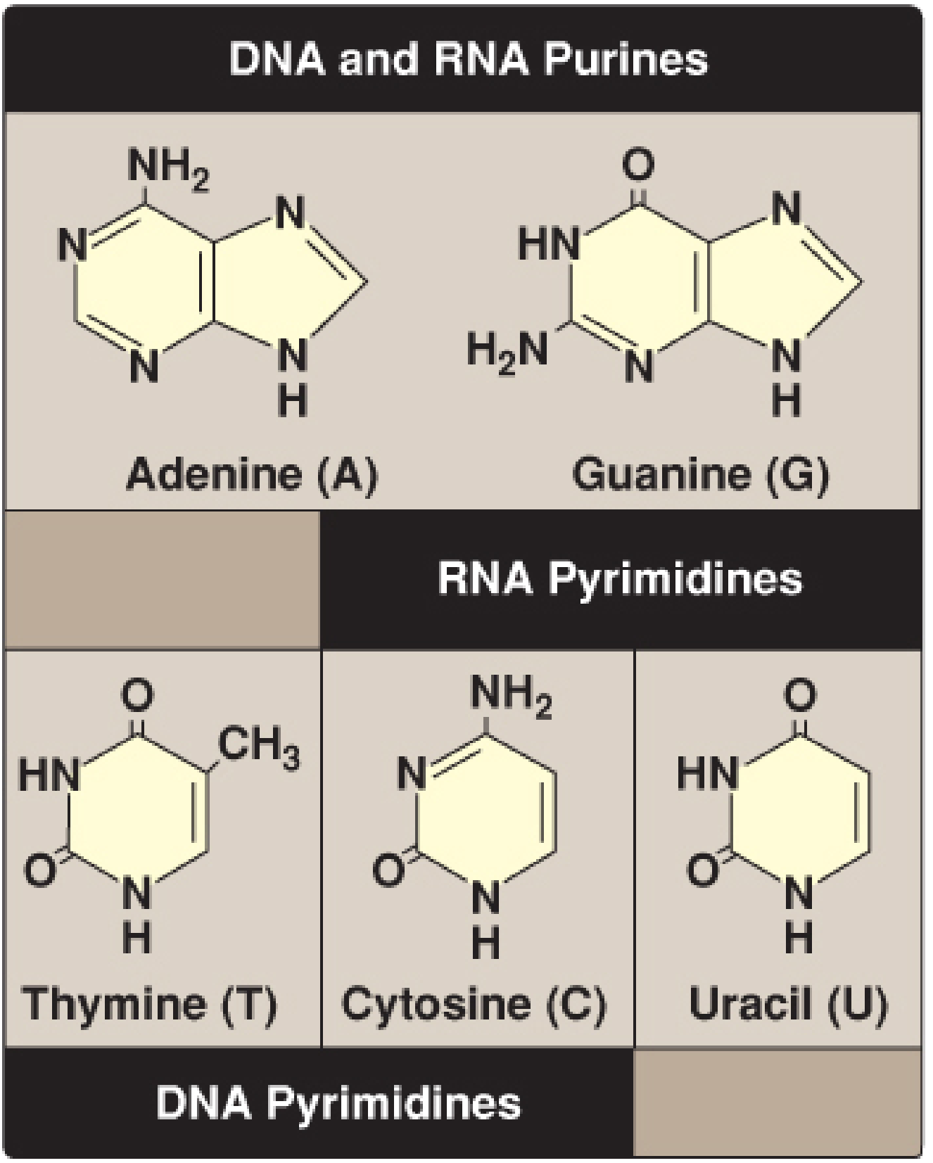
Key Structural Points:
- Adenine = Purine with an amino group (-NH₂) at position 6
- Guanine = Purine with a keto group (=O) at position 6 and amino group at position 2
- Thymine = Pyrimidine with a methyl group (-CH₃) at position 5 (this is what makes it different from Uracil)
- Cytosine = Pyrimidine with an amino group (-NH₂) at position 4
- Uracil = Pyrimidine like Thymine but without the methyl group - only found in RNA
Important: Thymine = Uracil + one methyl group (-CH₃)
PART III: NUCLEOSIDES
Nucleoside = Base + Sugar (NO phosphate)
| Base | Sugar | Nucleoside Name |
|---|---|---|
| Adenine + Ribose | → | Adenosine |
| Guanine + Ribose | → | Guanosine |
| Cytosine + Ribose | → | Cytidine |
| Uracil + Ribose | → | Uridine |
| Adenine + Deoxyribose | → | Deoxyadenosine |
| Guanine + Deoxyribose | → | Deoxyguanosine |
| Cytosine + Deoxyribose | → | Deoxycytidine |
| Thymine + Deoxyribose | → | Thymidine (or Deoxythymidine) |
Two types of sugar:
- Ribose (has -OH at 2' carbon) → used in RNA
- Deoxyribose (has only -H at 2' carbon, no -OH) → used in DNA
PART IV: NUCLEOTIDES
Nucleotide = Base + Sugar + Phosphate
| Number of Phosphates | Name |
|---|---|
| 1 phosphate | Nucleoside Monophosphate (NMP) - e.g., AMP |
| 2 phosphates | Nucleoside Diphosphate (NDP) - e.g., ADP |
| 3 phosphates | Nucleoside Triphosphate (NTP) - e.g., ATP |
ATP (Adenosine Triphosphate) = the energy currency of the cell - a nucleotide!
PART V: COMPARISON TABLE - PURINES vs PYRIMIDINES
| Feature | Purines | Pyrimidines |
|---|---|---|
| Ring structure | Double ring (bicyclic) | Single ring (monocyclic) |
| Size | Larger | Smaller |
| Bases | Adenine (A), Guanine (G) | Cytosine (C), Thymine (T), Uracil (U) |
| Found in DNA | A, G | C, T |
| Found in RNA | A, G | C, U |
| Molecular weight | Higher | Lower |
PART VI: BASE PAIRING IN DNA (Very Important!)
- Adenine (Purine) pairs with Thymine (Pyrimidine) → 2 Hydrogen bonds
- Guanine (Purine) pairs with Cytosine (Pyrimidine) → 3 Hydrogen bonds
Note: G-C pair is stronger than A-T pair because 3 H-bonds > 2 H-bonds. DNA with more G-C content has a higher melting temperature (Tm)
PART VII: UNUSUAL (MODIFIED) BASES
- Dihydrouracil (D) - in tRNA
- Pseudouracil (ψ) - in tRNA
- Inosine - in tRNA (anticodon)
- 5-Methylcytosine - in DNA (gene silencing/methylation)
- 7-Methylguanosine - the cap of eukaryotic mRNA
QUICK SUMMARY FOR EXAM
| Purines | Pyrimidines | |
|---|---|---|
| Ring | Double | Single |
| Bases | A, G | C, T (DNA), U (RNA) |
| Base pairing | A=T (2H), G≡C (3H) | Same |
Memory Tricks:
- "Pure Gold" = PURines = Adenine + Guanine
- "CUT the PY" = PYrimidines = Cytosine + Uracil + Thymine
- "AT 2, GC 3" = A-T has 2 H-bonds, G-C has 3 H-bonds
- Thymine = only in DNA | Uracil = only in RNA