
Lung mucormycosis treatment and diagnostic algorithm
I'll start by reading the medical library skill, then search for lung mucormycosis content in parallel.
Searching sources...
Reading File
Now I'll run parallel searches across the medical library, PubMed, and web simultaneously.
Finding Sources
Finding Sources
Searching PubMed
pulmonary mucormycosis diagnosis treatment
Searching Images
lung mucormycosis CT scan chest imaging reverse halo sign
I found key references. Now I'll read the most relevant textbook sections in parallel - Murray & Nadel's Respiratory Medicine, Goldman-Cecil, and Harrison's.
I have excellent content. Let me read the key treatment and diagnosis sections from Murray & Nadel's block_29 and Harrison's for the treatment details, and also get the radiology section.
Reading File
Reading File
Reading File
Excellent content gathered. Let me get the antifungal therapy details from Harrison's and the full diagnosis section from Goldman-Cecil.
Reading File
Reading File
Searching the Web
pulmonary mucormycosis diagnostic algorithm 2024 2025 ECMM guidelines
I now have comprehensive data from multiple authoritative sources. Let me fetch the CT scan image from Goldman-Cecil and the histopathology image for visual illustration.
Loading Image
Loading Image
I now have all the information needed to provide a comprehensive, well-illustrated answer. Here is the complete reference guide:
Lung Mucormycosis: Diagnostic Algorithm and Treatment
Overview
Pulmonary mucormycosis is the second most common form of mucormycosis (after rhinocerebral), caused by molds of the order Mucorales - primarily Rhizopus, Mucor, Lichtheimia (formerly Absidia), and Cunninghamella species. It is characterized by rapid angioinvasion, vessel thrombosis, and tissue necrosis. Mortality remains extremely high, particularly in disseminated disease.
Risk Factors
| High-Risk Category | Specific Conditions |
|---|---|
| Hematologic | Prolonged neutropenia, AML, allogeneic HSCT, aplastic anemia |
| Metabolic | Diabetic ketoacidosis (DKA), uncontrolled diabetes, metabolic acidosis |
| Immunosuppressive therapy | High-dose corticosteroids, calcineurin inhibitors, ibrutinib |
| Iron overload | Deferoxamine therapy, hemochromatosis, multiple transfusions |
| Antifungal prophylaxis gap | Prior voriconazole or echinocandin use (no Mucorales coverage) |
| Other | Solid organ transplant, COVID-19-associated, trauma/burns |
Key mechanistic insight: Mucorales exploit elevated iron (free serum iron in acidotic states) and impaired phagocyte function (neutropenia, DKA). Acidosis upregulates the GRP78 receptor on endothelial cells, which the Mucorales use for cell invasion. - Goldman-Cecil Medicine, p. 3360
Diagnostic Algorithm
CLINICAL SUSPICION
(immunocompromised + fever refractory to antibiotics + pulmonary infiltrate
 OR prior azole/echinocandin prophylaxis + new pulmonary lesion)
         |
         v
STEP 1: IMMEDIATE IMAGING
  CT Chest (high-resolution)
  - Halo sign (nodule + surrounding GGO) → angioinvasion
  - Reversed halo / atoll sign → strongly suggestive of Mucorales
  - Multi-lobar consolidation, cavitation (26-40%), air-crescent sign (5-12.5%)
  - Extension across fissures, chest wall, or diaphragm = hallmark
         |
         v
STEP 2: MICROBIOLOGIC SAMPLING
  Preferred: Bronchoscopy + BAL + TRANSBRONCHIAL BIOPSY
  - BAL culture: LOW sensitivity - positive culture is significant but negative
    does NOT rule out mucormycosis
  - Tissue biopsy is the diagnostic gold standard
  - Laboratory: Do NOT homogenize tissue - cut into sections and place on media
  - Grow at both room temperature AND 37°C (variable optimal temperature)
  - Cultures positive in <50% of cases despite active infection
         |
         v
STEP 3: HISTOPATHOLOGY (GOLD STANDARD)
  H&E and GMS staining:
  - Broad, ribbon-like, aseptate/sparsely septate hyphae, 3-25 µm diameter
  - Random branching at right angles (vs. 45° branching in Aspergillus)
  - Angioinvasion with vessel wall invasion and thrombosis
  - Surrounding tissue infarction and necrosis
         |
         v
STEP 4: ANCILLARY TESTS
  - Serum galactomannan: typically NEGATIVE (distinguishes from Aspergillus)
  - Beta-D-glucan: typically NEGATIVE (not a cell wall component of Mucorales)
  - Mucorales PCR (BAL or serum): sensitivity ~85%, specificity ~90%
    (not widely available; important emerging tool)
  - MALDI-TOF or ITS sequencing for species identification on culture
         |
         v
STEP 5: STAGING / EXTENT OF DISEASE
  CT sinuses + CT/MRI brain (if rhinocerebral spread suspected)
  CT abdomen (dissemination to liver, spleen in profoundly neutropenic)
Key clue distinguishing pulmonary mucormycosis from invasive aspergillosis: (1) pansinusitis, (2) breakthrough on voriconazole/echinocandin prophylaxis, (3) negative serum galactomannan, (4) reversed halo sign on CT. - Goldman-Cecil Medicine, p. 3359
CT findings - Goldman-Cecil Fig. 312-1:
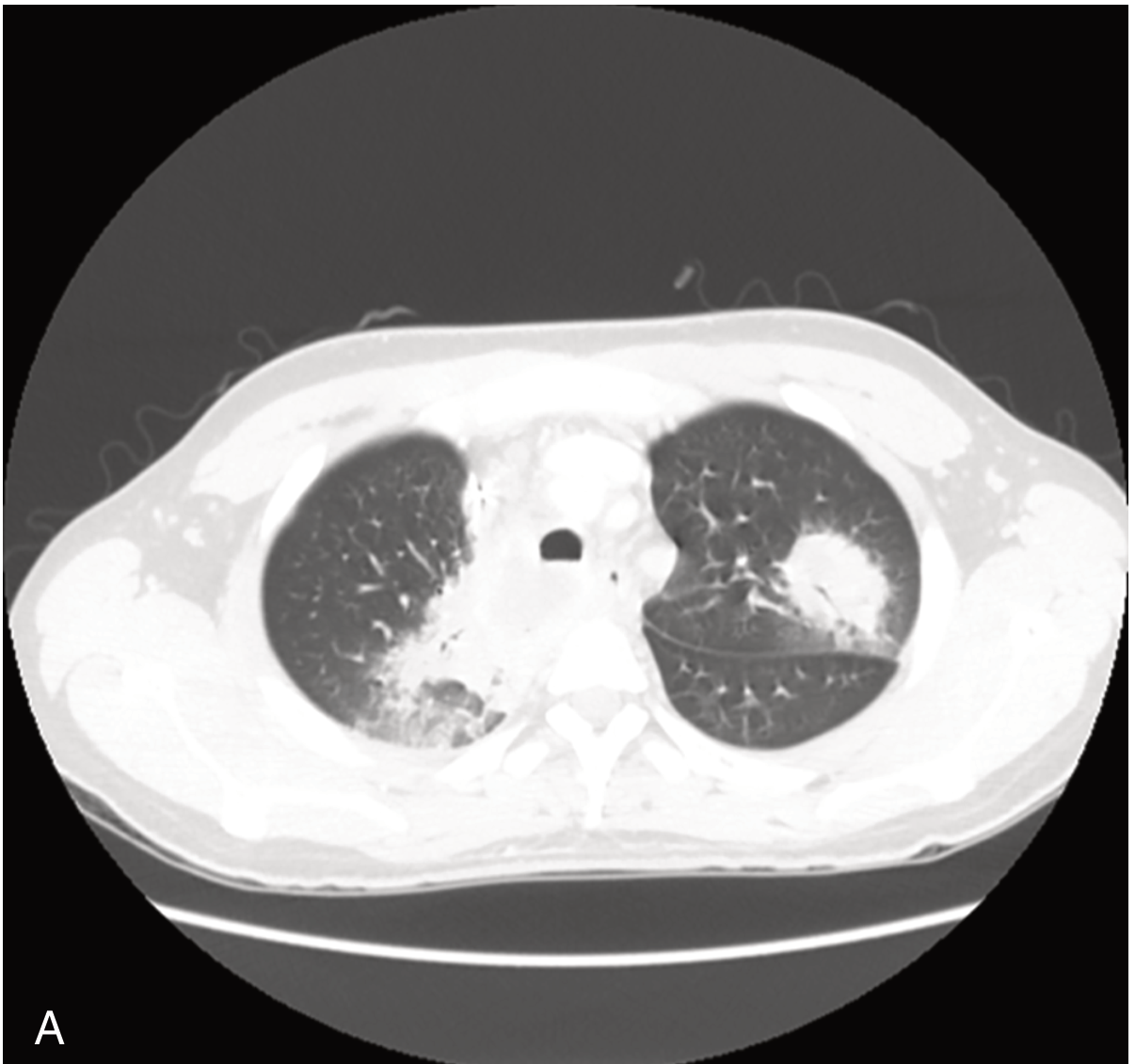
CT chest: Large right upper lobe consolidation with left lower lobe nodule in pulmonary mucormycosis - Goldman-Cecil Medicine
Histopathology - invasive Mucorales with angioinvasion:
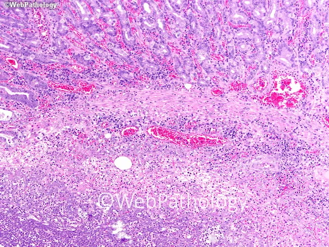
H&E: Broad zone of submucosal necrosis with wide, ribbon-like, non-septate hyphae and vascular invasion characteristic of Mucorales infection
Treatment Algorithm
Successful treatment requires ALL four simultaneous pillars - Harrison's Principles, 22e, p. 1763:
Pillar 1: Early Antifungal Therapy (START IMMEDIATELY - do NOT wait for confirmation)
First-Line Therapy
| Drug | Dose | Notes |
|---|---|---|
| Liposomal Amphotericin B (LAmB) | 5-10 mg/kg/day IV | Preferred for most cases; superior CNS penetration; FDA-approved |
| ABLC (Amphotericin B lipid complex) | 5 mg/kg/day IV | Alternative lipid formulation; may be less nephrotoxic than AmB-d |
| AmB deoxycholate | 1.0-1.5 mg/kg/day IV | Only if lipid formulations unavailable; high nephrotoxicity |
Delaying LAmB by 6+ days after diagnosis is associated with a twofold increase in mortality. - Murray & Nadel's Respiratory Medicine, p. 2925
Second-Line / Salvage Options
| Drug | Dose | Notes |
|---|---|---|
| Isavuconazole | 200 mg q8h x 6 doses (loading), then 200 mg once daily | FDA-approved for mucormycosis; useful when mold species uncertain (covers both Mucorales AND Aspergillus); oral bioavailability allows step-down |
| Posaconazole | 200 mg QID (suspension) or 300 mg daily (tablet) | Salvage only; do not use as monotherapy initial treatment; retrospective data only |
Voriconazole has no activity against Mucorales. Echinocandins alone are not active, though combination with LAmB may have additive benefit in select cases. - Harrison's Principles 22e
Combination Therapy (Emerging)
- LAmB + Isavuconazole or LAmB + Echinocandin: considered in salvage/refractory cases; no definitive RCT evidence; discussed in expert consensus (ECMM 2019 global guidelines, PMID: 31699664)
Pillar 2: Surgical Debridement
- Recommended wherever anatomically feasible (independent predictor of favorable outcome in multivariate analyses)
- Rationale: vessel thrombosis causes tissue necrosis with poor antifungal penetration - surgical removal is necessary to achieve adequate drug delivery
- Use intraoperative frozen sections to delineate infection margins
- For pulmonary disease: lobectomy, pneumonectomy, or wedge resection depending on extent
- Serial debridement may be needed
- Endoscopic debridement for tracheobronchial lesions
Pillar 3: Reverse Underlying Predisposing Factors
| Factor | Intervention |
|---|---|
| DKA / hyperglycemia | Insulin drip, correction of acidosis (sodium bicarbonate in mice models improved survival) |
| Corticosteroids | Taper to lowest immunosuppressive dose compatible with underlying disease treatment |
| Neutropenia | G-CSF; consider granulocyte infusions in selected cases |
| Iron overload / deferoxamine | Stop deferoxamine immediately - it provides iron to the fungus; iron administration must be avoided |
| Immunosuppression in transplant | Reduction where safe |
Pillar 4: Treat the Underlying Disease Concurrently
A common and fatal error is halting chemotherapy or transplant protocols to "first cure" the mucormycosis. This is incorrect because:
- Mucormycosis will not be definitively eradicated until near-normal immunity is restored
- Modern antifungals can temporize, allowing chemotherapy/transplant to proceed
- Death is most often from the underlying malignancy, not the infection alone
- Harrison's Principles 22e, p. 1764
Duration of Therapy and Step-Down
- Induction: LAmB IV until clinical stabilization (typically weeks to months)
- Step-down to oral: Isavuconazole or posaconazole extended-release once stable
- Total duration: No fixed endpoint - continue until: (1) resolution/stability of lesions on imaging, (2) restoration of immune function, and (3) clearance of predisposing factors
- Maintenance/secondary prophylaxis with isavuconazole or posaconazole is indicated while patient remains immunocompromised
Adjunctive Strategies (Limited Evidence)
| Strategy | Evidence |
|---|---|
| Deferasirox (iron chelator) | Early case reports showed benefit; subsequent RCT showed no benefit and possible harm in HSCT recipients - NOT recommended routinely |
| Hyperbaric oxygen | Case reports in rhinocerebral disease; not validated for pulmonary mucormycosis |
| Cytokine therapy (IFN-γ) | Case series only |
| Granulocyte transfusions | Limited data; considered in profound neutropenia |
Prognosis
| Clinical Form | Approximate Mortality |
|---|---|
| Isolated sinusitis (surgically resected) | ~20-30% |
| Pulmonary mucormycosis | ~50-65% |
| Disseminated mucormycosis | ~80-100% |
| HSCT-associated mucormycosis | ~70-80% |
Prognosis is improving with earlier diagnosis and better antifungal agents. - Goldman-Cecil Medicine, p. 3361
Key Differential: Pulmonary Mucormycosis vs. Invasive Aspergillosis
| Feature | Mucormycosis | Aspergillosis |
|---|---|---|
| Hyphae | Broad, aseptate, random branching | Narrow, septate, 45° branching |
| Serum galactomannan | Negative | Positive (sensitive) |
| Beta-D-glucan | Negative | Positive |
| Reversed halo sign on CT | More specific | Less common |
| Voriconazole coverage | No | Yes |
| Echinocandin coverage | No (monotherapy) | Adjunct use |
| Iron chelator predisposition | Yes (deferoxamine) | No |
Summary: Management in Brief
- High suspicion in at-risk host + new pulmonary infiltrate not responding to antibiotics
- CT chest immediately - look for halo, reversed halo, multi-lobar consolidation, cavitation
- Bronchoscopy + transbronchial biopsy (do not homogenize tissue for culture)
- Start LAmB 5-10 mg/kg/day empirically - do not wait for microbiologic confirmation
- Surgery if lesion is resectable
- Correct DKA, stop deferoxamine, reduce steroids, restore immune function
- Do not stop treatment of underlying malignancy while awaiting mucormycosis cure
- Step-down to isavuconazole orally once stable
Sources:
- Goldman-Cecil Medicine International Edition, Vol. 2, Chapter 312, pp. 3358-3362
- Harrison's Principles of Internal Medicine 22e (2025), Chapter 224, pp. 1763-1765
- Murray & Nadel's Textbook of Respiratory Medicine, Chapter on Opportunistic Mycoses
- Grainger & Allison's Diagnostic Radiology, Chapter 5 (Chest Radiology), p. 3704
- ECMM Global Guideline for Mucormycosis (Cornely et al., 2019) - PMID 31699664
- Muthu V et al. "Treatment of pulmonary mucormycosis: current concepts." Expert Rev Anti Infect Ther 2026 - PMID 41575183
This is a shared conversation. Sign in to Orris to start your own chat.