
Create a notes on WBC disorders based on dacie and lewis, McKenzie textbook of hematology for pathology residents
neutrophilia toxic granulation Dohle bodies peripheral blood smear
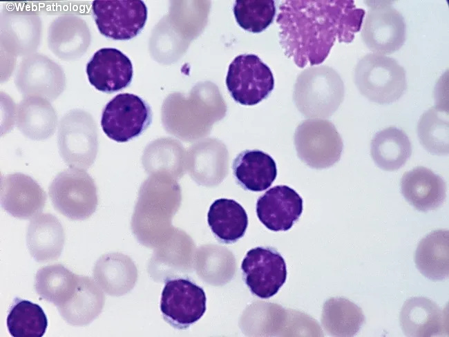
Peripheral blood smear from a patient with suspected chronic lymphocytic leukemia (CLL) shows a predominance of small, mature-appearing lymphocytes with scant cytoplasm and clumped nuclear chromatin, accompanied by fragile cells that readily disrupt during smear preparation, producing numerous smudge or basket cells visible on the slide. The image depicts a single smudge cell in the upper right, with numerous small darkly staining lymphocytes occupying the field. The smear was prepared with Wright-Giemsa stain and examined under light microscopy at high magnification (approximately 1000x total magnification with oil immersion). Clinically, CLL is defined by absolute mature lymphocytosis of ≥5.0 × 10^9/L sustained for ≥3 months, often asymptomatic and detected incidentally on complete blood count. Morphology alone is insufficient for diagnosis; immunophenotyping showing co-expression of CD5, CD19, CD23 with weak surface immunoglobulin and light chain restriction supports CLL diagnosis. Smudge cells result from fragile leukemic cells and reflect underlying cytoskeletal fragility, though they are not specific. The image illustrates typical features: monomorphic lymphocytes, scant cytoplasm, condensed chromatin, and absent prominent nucleoli. This finding has diagnostic significance, guiding further workup, including flow cytometry, cytogenetics, and molecular studies, to distinguish CLL from other chronic lymphoproliferative disorders such as small lymphocytic lymphoma (SLL), mantle cell lymphoma, prolymphocytic leukemia, and reactive lymphocytosis.
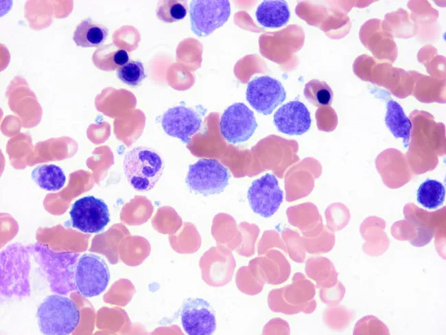
This image depicts a peripheral blood smear prepared with Wright-Giemsa stain and examined under light microscopy at high magnification. The predominant features are small, mature-appearing lymphocytes with scant cytoplasm and discrete basophilic nuclei interspersed among erythrocytes. A characteristic subset of circulating lymphocytes displays short, polarity-restricted cytoplasmic villi (finger-like projections) consistent with splenic marginal zone lymphoma (SMZL) involvement of peripheral blood. The villi are typically slender and localized to one pole of the cell, unlike the longer, circumferential villous extensions seen in hairy cell leukemia, which aids in differential diagnosis. Some lymphocytes appear slightly irregular or irregular nuclear contours; occasional larger atypical cells may be present but are less common. The background shows normocytic red cells with normal distribution; platelets are not prominent. This cytomorphology supports SMZL in the context of known splenomegaly or lymphoproliferative disease and is often corroborated by immunophenotyping and molecular studies. Clinically, peripheral blood involvement occurs in roughly half to two-thirds of SMZL cases and helps establish disease burden. The image illustrates the diagnostic utility of meticulous peripheral smear review for small-vessel lymphocytosis and villous lymphocytes, informing differential diagnoses, guiding flow cytometry panels, and contributing to monitoring and prognosis.
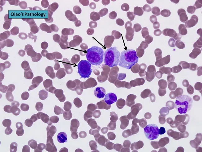
Peripheral blood smear prepared from a patient with suspected plasma cell leukemia (PCL) was examined by bright‑field light microscopy after Wright‑Giemsa staining. The smear shows several circulating plasma‑cell–like elements, including four conspicuous proplasmacytes with dispersed nuclear chromatin and prominent nucleoli, as indicated by arrows in the original figure. In addition, background erythrocytes exhibit rouleaux formation, a common feature in paraproteinemic states. The cells display eccentrically placed nuclei with pronounced basophilic cytoplasm and a coarse chromatin pattern; occasional cells show a perinuclear clearing (Golgi rim) suggestive of plasmacytic differentiation. Immunoglobulin light‑chain restriction is not determined on this smear alone but flow cytometry or immunohistochemistry would typically confirm clonal plasma cells. Relative proportions indicate that neoplastic plasma cells constitute a substantial fraction of leukocytes, consistent with plasma cell leukemia rather than reactive plasmacytosis. PCL is an aggressive myelomatous process often presenting with cytopenias, organomegaly, and sometimes lymphadenopathy; prognosis is poor, and treatment responsiveness is limited. This image illustrates characteristic morphologic features used for differential diagnosis against multiple myeloma, Waldenström macroglobulinemia, and other causes of plasmacytosis. Clinically relevant keywords include plasma cells, rouleaux, Wright‑Giemsa stain, 100X oil, peripheral blood, leukemia, myeloma, and hematologic malignancy.
eosinophilia peripheral blood eosinophil granules
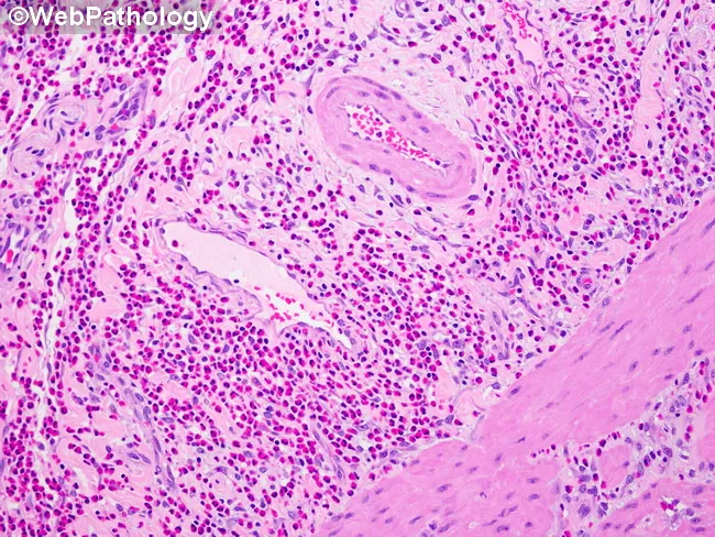
Histopathology; Hematoxylin and Eosin stained gallbladder tissue demonstrating pure eosinophilic infiltrate within the lamina propria. The mucosal architecture is preserved without stromal necrosis or significant neutrophilic infiltration. Eosinophils appear as round to irregular cells with bilobed or multilobed nuclei and abundant bright pink-orange cytoplasmic granules, forming dense networks in the lamina propria surrounding ductal structures and capillaries. The submucosa is largely unremarkable, and no mural granulomas or ulceration are evident. Vascular congestion may be present secondary to inflammatory edema. Scattered lymphocytes and plasma cells are minimal, with eosinophils constituting the predominant inflammatory cell type. A scattered macrophage population is observed adjacent to small vessels. No gallstones are visible in this field, and there is no evidence of acute neutrophilic cholangitis. This pattern is characteristic of eosinophilic cholecystitis, a rare inflammatory variant often linked to allergic or parasitic processes, drug reactions, or hypereosinophilic syndrome. Clinically, eosinophilic cholecystitis may present with right upper quadrant pain and cholecystitis-like symptoms, but peripheral eosinophilia or serologic allergy markers may be absent. The diagnostic significance lies in recognizing eosinophil-predominant inflammation, guiding differential diagnosis, investigative workup for eosinophilia, and management focusing on underlying triggers and symptomatic gallbladder relief.
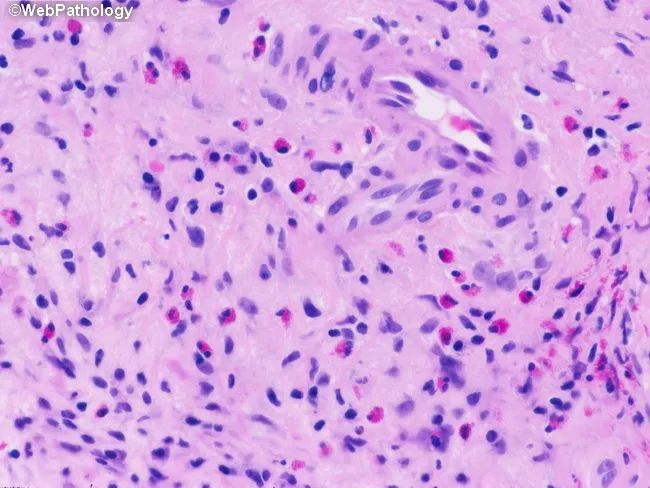
Histopathology: Light microscopy of a gastrointestinal mucosal biopsy stained with Hematoxylin and Eosin reveals a dense inflammatory infiltrate within the lamina propria. The predominant cellular population comprises eosinophils, characterized by bilobed or irregular nuclei and abundant bright pink cytoplasmic granules, intermixed with a smaller number of mononuclear lymphocytes. Epithelial architecture within this field shows intact surface epithelium without definite crypt distortion or ulceration; no granulomas or conspicuous vasculitis are evident in the sampled area. The surrounding stroma appears mildly edematous with scattered plasma cells and occasional neutrophils. In this image, the eosinophilic infiltrate is conspicuous and may extend around mucosal glands and vessels. Such a pattern is compatible with eosinophilic gastroenteritis or hypersensitivity reactions and could reflect parasitic infection, drug-induced hypersensitivity, or food allergy, depending on clinical context. Normal variant eosinophil counts in the lamina propria can be elevated in the GI tract; therefore correlation with peripheral eosinophilia and clinical symptoms is essential. Diagnostic significance: the presence of tissue eosinophilia in the lamina propria supports a non-neoplastic inflammatory process; differential diagnoses include eosinophilic colitis/enteritis, inflammatory bowel disease with eosinophilia, or secondary eosinophilia due to parasites. Clinical correlation with symptoms (abdominal pain, diarrhea), stool studies, and serologies is recommended.
leukemoid reaction left shift band neutrophils myelocytes blood smear
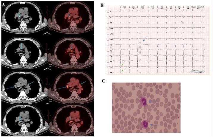
A multi-panel figure containing diagnostic imaging, an electrocardiogram (ECG), and a blood smear. Panel A presents axial PET/CT fusion and CT images of the thoracic and upper abdominal regions, showing a soft tissue mass near the esophageal hiatus and retroperitoneal abdominal trunk with hypermetabolic fluorodeoxyglucose (FDG) uptake, indicating tumor recurrence or metastasis. Panel B displays a 12-lead ECG strip demonstrating arrhythmias including premature atrial contractions (PACs) and premature ventricular contractions (PVCs). Green arrows highlight mild ST-segment depression in leads V5 and V6, while the blue arrow points to significant T-wave morphology changes, suggesting immunotherapy-related myocardial injury. Panel C is a peripheral blood smear micrograph showing two neutrophils with a left shift in nuclei (indicated by blue arrows), characterized by immature band forms, which is suggestive of an acute inflammatory response or infection. This composite illustrates the clinical monitoring of a patient with esophageal squamous cell carcinoma experiencing immune-related adverse events.
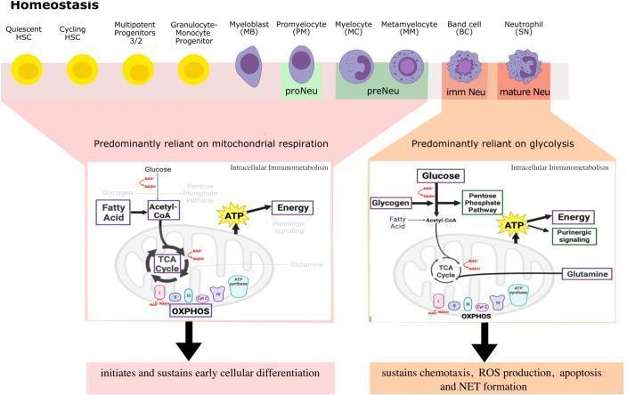
This hematology infographic illustrates the metabolic reprogramming of neutrophils during homeostasis. The top panel depicts neutrophil development (granulopoiesis) from hematopoietic stem cells (HSCs) through various stages: Myeloblast (MB), Promyelocyte (PM), Myelocyte (MC), Metamyelocyte (MM), Band cell (BC), and segmented Neutrophil (SN). Morphological changes show nuclear progression from round/ovoid to indented and ultimately multi-lobed. The bottom panel highlights a significant immunometabolic shift. Immature stages (HSCs to Metamyelocytes) are predominantly reliant on mitochondrial respiration, utilizing fatty acids and glucose to fuel the TCA cycle and oxidative phosphorylation (OXPHOS) for ATP generation; this metabolic state sustains early cellular differentiation. In contrast, mature neutrophils (Band cells and SNs) transition to being predominantly reliant on glycolysis. In these mature cells, glucose and glycogen fuel the pentose phosphate pathway and glycolysis to produce ATP, which sustains effector functions including chemotaxis, ROS production, apoptosis, and NET formation. The diagram emphasizes the shift from high mitochondrial activity in progenitors to glycolytic dominance in terminally differentiated innate immune cells.
WBC (Leukocyte) Disorders
Comprehensive Notes for Pathology Residents
SECTION 1 - NORMAL REFERENCE RANGES
| Cell Type | Adult Reference Range |
|---|---|
| Total WBC | 4.8-10.8 × 10³/µL |
| Neutrophils | 1.8-7.0 × 10³/µL (40-70%) |
| Lymphocytes | 1.5-4.0 × 10⁹/L (20-40%) |
| Monocytes | 0.1-0.8 × 10⁹/L (~4%) |
| Eosinophils | 0-0.5 × 10³/µL |
| Basophils | 0-0.2 × 10³/µL |
SECTION 2 - LEUKOCYTOSIS: OVERVIEW
- Size of myeloid/lymphoid precursor and storage pools in BM, thymus, and tissues
- Rate of release from storage pools into circulation
- Proportion adherent to vessel walls (the marginal granulocyte pool, MGP)
- Rate of extravasation into tissues
SECTION 3 - NEUTROPHILIC DISORDERS
3.1 Neutrophilia (Neutrophilic Leukocytosis)
Mechanisms (Henry's):
| Mechanism | Examples |
|---|---|
| Redistribution from MGP to CGP | Exercise, epinephrine, stress, glucocorticoids |
| Increased BM release | Acute infection, inflammation (TNF, IL-1 mediated) |
| Increased BM production | Chronic infection, G-CSF, myeloproliferative neoplasms (MPN) |
| Decreased egress to tissues | Glucocorticoids (block ICAM/selectin interactions) |
Causes (Robbins Table 13.2/13.3):
- Acute bacterial infections (especially pyogenic organisms) - commonest cause
- Sterile tissue necrosis (myocardial infarction, burns, surgery)
- Metabolic disorders (uremia, diabetic ketoacidosis, eclampsia, gout)
- Drugs (glucocorticoids, lithium, G-CSF)
- Physiologic (neonates, pregnancy, exercise)
- Chronic inflammation (growth factor-dependent)
- Paraneoplastic (e.g., Hodgkin lymphoma - growth factor-dependent)
- Myeloproliferative neoplasms (growth factor-independent)
Pearl: Physiologic leukocytosis from exercise/epinephrine is transient and normalizes within 30-60 minutes. No band/left shift is expected.
3.2 Qualitative Neutrophil Abnormalities
Toxic Changes (Henry's) - Acquired
| Finding | Description |
|---|---|
| Toxic granulation | Dark blue-purple cytoplasmic granules (peroxidase-positive); represent primary granules that failed to lose their staining properties |
| Döhle inclusion bodies | Small pale-blue oval inclusions in peripheral cytoplasm (Wright's stain); remnants of free ribosomes/rough ER from earlier developmental stage |
| Cytoplasmic vacuolation | Phagocytic vacuoles; strong indicator of bacterial sepsis when combined with toxic granulation |
| Hypersegmentation | ≥5 lobes in >5% of neutrophils; classic for B12/folate deficiency; also uremia |
Hereditary/Constitutional Anomalies (Henry's)
| Anomaly | Genetics | Morphology | Function |
|---|---|---|---|
| May-Hegglin anomaly | MYH9 gene, chr 22q12-13, autosomal dominant | Large Döhle-like pale blue RNA inclusions in ALL granulocytes + giant platelets ± thrombocytopenia | Normal |
| Alder-Reilly anomaly | Mucopolysaccharidoses (Hurler, Hunter, etc.) | Dense, large azurophilic granules in ALL WBCs (resembles toxic granulation but NOT transient); metachromatic lymphocyte inclusions (Gasser cells) | Normal granulocyte function |
| Chediak-Higashi syndrome | LYST gene, autosomal recessive | Giant fused lysosomes (giant granules) in granulocytes, lymphocytes, NK cells; partial oculocutaneous albinism | Severely impaired (recurrent pyogenic infections, NK cell dysfunction) |
| Pelger-Huet anomaly | LBR gene, autosomal dominant | Hyposegmented neutrophils (bilobed "pince-nez"/dumbbell nuclei, or round/ovoid); coarse chromatin | Normal - clinically benign |
| Pseudo-Pelger-Huet | Acquired (MDS, drugs, infection) | Identical morphology but acquired; important to distinguish from true Pelger-Huet | Normal |
Exam pearl: In May-Hegglin, Sebastian, Fechtner, and Epstein syndromes - all are MYH9-related, all have Döhle-like inclusions and giant platelets. Fechtner and Epstein additionally have nephritis and sensorineural hearing loss.
3.3 Neutropenia
Causes:
- Aplastic anemia, chemotherapy, radiation
- B12/folate deficiency (ineffective granulopoiesis)
- Myelophthisis (marrow replacement by tumor, granuloma, fibrosis)
- Congenital: Kostmann syndrome (severe congenital neutropenia - HAX1 mutation), cyclic neutropenia (ELANE gene, 21-day cycles)
- Myelodysplastic syndrome
- Immune: autoimmune neutropenia, drug-induced (quinidine, propylthiouracil, clozapine, sulfonamides), isoimmune neonatal neutropenia
- Hypersplenism
- Overwhelming sepsis (in neonates and elderly - important exam pearl)
- Pseudoneutropenia (benign ethnic neutropenia in people of African descent - MGP enlarged at expense of CGP, ANC can be as low as 0.2; normal bone marrow; no increased infection risk)
- Typhoid fever, Brucellosis, Tularemia, Rickettsial infection (cause relative neutropenia)
- Felty syndrome (RA + splenomegaly + neutropenia)
SECTION 4 - EOSINOPHILIC DISORDERS
4.1 Eosinophilia
- Mild: 0.5-1.5 × 10³/µL
- Moderate: 1.5-5.0 × 10³/µL
- Severe: >5.0 × 10³/µL (hypereosinophilia)
Causes (Robbins Table 13.3):
- Allergic disorders - asthma, allergic rhinitis, atopic dermatitis (IL-5 driven)
- Parasitic infestations - tissue-invasive parasites (Toxocara, Trichinella, Strongyloides); NOT intestinal protozoa
- Drug reactions - penicillin, aspirin, sulfonamides
- Malignancies - Hodgkin lymphoma (most important), some NHL; paraneoplastic
- Autoimmune/skin - pemphigus, dermatitis herpetiformis, eosinophilic fasciitis
- Vasculitides - eosinophilic granulomatosis with polyangiitis (Churg-Strauss)
- Atherosclerotic embolism (transient)
- Adrenal insufficiency (Addison's)
- Hypereosinophilic syndrome (HES): persistent eosinophilia >1.5 × 10³/µL for >6 months with end-organ damage (heart, lung, CNS, skin); must exclude reactive and clonal causes
- Clonal eosinophilia: FIP1L1-PDGFRA fusion (imatinib-responsive), ETV6-PDGFRB; classified as myeloid/lymphoid neoplasm with eosinophilia
Morphology clue (Henry's): In hypereosinophilic syndrome, eosinophils may show cytoplasmic vacuolation, reduced or absent granules (degranulation), and nuclear hyperlobulation - indicating activated/degranulating eosinophils.
SECTION 5 - BASOPHILIC DISORDERS
5.1 Basophilia
- Myeloproliferative neoplasms - MOST IMPORTANT (especially CML; basophilia >20% suggests blast phase transformation)
- Allergic reactions (food, drugs, foreign proteins)
- Inflammatory bowel disease
- Infections: variola (smallpox), varicella
- Hypothyroidism
Critical pearl: Basophilia almost always signals a myeloproliferative neoplasm (especially CML) when absolute basophilia is sustained. Always check BCR-ABL1.
SECTION 6 - MONOCYTIC DISORDERS
6.1 Morphology (Henry's)
6.2 Monocytosis
-
50% of absolute monocytosis cases were associated with hematologic neoplasms: AML (especially monocytic), lymphoma (Hodgkin predominant), multiple myeloma, MPN
- Infectious: tuberculosis, brucellosis, bacterial endocarditis, syphilis (but uncommon)
- Inflammatory: SLE, RA, IBD
- Post-splenectomy/recovery from agranulocytosis
- Myelodysplastic syndrome (chronic myelomonocytic leukemia - CMML)
Important: The classic finding in CMML is persistent monocytosis >1.0 × 10⁹/L for >3 months with clonal cytogenetic/molecular features.
6.3 Monocytopenia
- Hairy cell leukemia (classic association - virtually pathognomonic)
- Prednisone (falls within first hours of first dose, returns by 12h)
- MonoMac syndrome (GATA2 mutation) - monocytopenia + susceptibility to mycobacterial and fungal infections + NK cell deficiency
- B-lymphoblastic leukemia
- Chemotherapy (monocytopenia heralds onset of neutropenia - Quick Compendium)
SECTION 7 - LYMPHOCYTIC DISORDERS
7.1 Normal Lymphocytes (Henry's)
- Small lymphocytes: 6-10 µm, single sharply defined nucleus, heavy chromatin blocks, scant pale cytoplasm
- Large lymphocytes (LGLs): 12-16 µm, indented/folded nucleus, pale gray-blue cytoplasm with azurophilic granules
- Adults: lymphocytes = 20-40% of WBCs (1.5-4.0 × 10⁹/L)
- Children up to 5 years: lymphocytes up to 50% (highest in 1st year)
- Normal CD4:CD8 ratio = 1.0-3.4
7.2 Lymphocytosis
Reactive (Non-neoplastic) Lymphocytosis (Henry's):
| Cause | Lymphocyte Type | Key Features |
|---|---|---|
| EBV (infectious mononucleosis) | Reactive T cells (CD8+) | Atypical lymphocytes (Downey cells); heterophile antibody positive; splenomegaly; pharyngitis; lymphadenopathy |
| CMV mononucleosis | Reactive T cells | Similar to EBV but heterophile NEGATIVE; milder pharyngitis |
| Hepatitis | Reactive lymphocytes | Elevated transaminases |
| Acute infectious lymphocytosis | Small mature T cells | Children; WBC 20-50 × 10⁹/L; normal morphology; no adenopathy/splenomegaly; lasts 3-5 weeks |
| Pertussis (whooping cough) | Small mature T cells, normal CD4:CD8 | WBC can exceed 30 × 10⁹/L; HIGHEST in week 1-3; caused by pertussis toxin (PT) blocking chemokine G-coupled receptors, preventing lymphocyte retention in BM/spleen - redistribution, NOT increased lymphopoiesis |
| Adenovirus, varicella, measles, rubella | Reactive | Mild lymphocytosis |
| Stress lymphocytosis | Polyclonal | Acute (trauma, MI, surgery); transient |
- Larger than normal small lymphocytes
- Irregular, indented, or lobulated nuclei; dispersed chromatin
- Abundant pale-blue cytoplasm, often "scalloped" by adjacent RBCs
- NOT the same as neoplastic cells; they are activated T cells
- Three types: Type I (Rieder/band forms), Type II (monocytoid), Type III (blast-like)
7.3 Lymphopenia
- HIV infection (CD4+ T cell loss)
- SLE (anti-lymphocyte antibodies)
- Steroid therapy
- Anti-CD20 (rituximab) therapy
- Severe acute respiratory syndrome (SARS/COVID-19)
- Congenital immunodeficiency: Bruton's X-linked agammaglobulinemia, SCID, DiGeorge syndrome, common variable immunodeficiency (CVID)
- Radiation/chemotherapy
- Malnutrition (protein-calorie)
SECTION 8 - LEUKEMOID REACTION vs CML
8.1 Leukemoid Reaction
| Feature | Leukemoid Reaction | CML |
|---|---|---|
| LAP score (Leukocyte Alkaline Phosphatase) | High (>100) | Low (<10) |
| BCR-ABL1 (Philadelphia chromosome) | Absent | Present (>95%) |
| Basophilia | Absent/mild | Prominent |
| Splenomegaly | Absent/mild | Marked |
| Underlying cause | Identifiable (infection, drug) | None |
| Eosinophilia | Absent/mild | Often present |
| Toxic changes | Present | Absent |
Pearl: The LAP score has largely been replaced by BCR-ABL1 testing in clinical practice, but remains on exams as the classic differentiator.
SECTION 9 - NEOPLASTIC WBC DISORDERS: CLASSIFICATION FRAMEWORK
- Cell of origin (myeloid vs. lymphoid; B vs. T vs. NK)
- Maturation state (precursor/blast vs. mature)
- Cytogenetic and molecular alterations
- Clinical behavior (acute vs. chronic)
9.1 Major Categories
A. Myeloid Neoplasms
| Entity | Key Features |
|---|---|
| Acute Myeloid Leukemia (AML) | ≥20% blasts in BM/blood; myeloperoxidase positive; Auer rods pathognomonic; FAB M0-M7 classification |
| Myeloproliferative Neoplasms (MPN) | Clonal, effective maturation; increased output of one or more cell lines |
| - CML | BCR-ABL1 t(9;22); basophilia; splenomegaly; LAP low |
| - Polycythemia vera | JAK2 V617F (>95%); elevated RBC mass; low EPO |
| - Essential thrombocythemia | JAK2 (55%), CALR (25%), MPL mutations; platelets >450 |
| - Primary myelofibrosis | JAK2/CALR/MPL; leukoerythroblastosis; teardrop RBCs |
| Myelodysplastic Syndrome (MDS) | Dysplasia + ineffective hematopoiesis; cytopenias; ring sideroblasts; may transform to AML |
| CMML | Monocytes >1.0 × 10⁹/L; dysplasia; ASXL1, TET2 mutations |
B. Lymphoid Neoplasms
| Entity | Immunophenotype | Key Molecular | Clinical |
|---|---|---|---|
| B-ALL/LBL | TdT+, CD10+, CD19+, CD34+ | BCR-ABL1 (poor prognosis), ETV6-RUNX1 (good), hyperdiploidy | Childhood peak; BM/blood; CNS involvement |
| T-ALL/LBL | TdT+, CD3+, CD7+; mediastinal | NOTCH1 mutations | Adolescent males; anterior mediastinal mass |
| CLL/SLL | CD5+, CD19+, CD23+, CD20 (weak), FMC7-; smudge cells | 17p del (worst), 11q del, trisomy 12, 13q del (best) | Adults; indolent; Richter transformation |
| Follicular lymphoma | CD10+, BCL2+, CD20+ | t(14;18) BCL2-IGH | Adults; follicular growth; waxing/waning |
| Mantle cell lymphoma | CD5+, CD20+, cyclin D1+, BCL2+, FMC7+ | t(11;14) CCND1-IGH | Middle-aged males; mantle zone pattern; aggressive |
| DLBCL | CD20+, CD19+; variable BCL2, BCL6, MYC | MYC rearrangement (double/triple-hit = very aggressive) | Most common NHL; diffuse large B cells |
| Burkitt lymphoma | CD20+, CD10+, BCL6+, BCL2- | t(8;14) or t(2;8), t(8;22) MYC translocation; Ki67 ~100% | "Starry sky" pattern; jaw (endemic); ileocecal (sporadic) |
| Multiple myeloma | CD138+, CD38+, CD56+, CD19-, CD45- | t(4;14), t(14;16), del17p (high risk); FISH for staging | Bone pain, hypercalcemia, renal failure, anemia (CRAB) |
| Hodgkin lymphoma | RS cells: CD15+, CD30+, CD20-, LCA- | EBV association (esp. mixed cellularity, LDHL) | Bimodal age; mediastinal mass; B symptoms |
SECTION 10 - KEY IMMUNOPHENOTYPING MARKERS (Robbins, Table 13.5)
| Marker | Expression |
|---|---|
| CD3 | T cells (all mature) |
| CD4 | Helper T cells |
| CD5 | T cells + small subset B cells (also CLL, MCL) |
| CD8 | Cytotoxic T cells |
| CD10 | Pre-B cells, germinal center B cells (ALL, FL, Burkitt) |
| CD19 | Pre-B to mature B cells (not plasma cells) |
| CD20 | Pre-B (after CD19) to mature B cells (not plasma cells) - rituximab target |
| CD21 | Mature B cells, follicular dendritic cells (EBV receptor) |
| CD23 | Activated mature B cells (CLL+; MCL-) |
| CD30 | Activated B and T cells; Reed-Sternberg cells |
| CD34 | Hematopoietic stem/progenitor cells; blasts |
| CD45 (LCA) | All leukocytes |
| TdT | Precursor B and T cells (blasts) |
| MPO | Myeloid lineage |
| CD138 | Plasma cells |
| CD11c | Granulocytes, monocytes, macrophages; also hairy cell leukemia |
| CD14 | Monocytes |
| CD15 | Granulocytes; Reed-Sternberg cells |
| CD56 | NK cells; subset T cells; also myeloma cells |
SECTION 11 - MORPHOLOGY PEARLS FOR PATHOLOGY SIGN-OUT
| Morphology | Associated Condition |
|---|---|
| Smudge/basket cells | CLL (fragile leukemic lymphocytes) |
| Auer rods | AML (fused primary granules - pathognomonic for myeloid lineage) |
| "Starry sky" pattern | Burkitt lymphoma (tingible body macrophages amidst tumor cells) |
| "Hair-like" projections | Hairy cell leukemia (TRAP+, annexin A1+, CD11c+, CD25+, CD103+) |
| Bilobed "mirror-image" RS cells | Classical Hodgkin lymphoma |
| Lacunar cells | Nodular sclerosing Hodgkin lymphoma |
| Lymphocyte-predominant "popcorn" cells | NLPHL (CD20+, CD45+, CD15-, CD30-) |
| Cerebriform (Sézary) cells | Sézary syndrome/Mycosis fungoides (CD4+ T-cell) |
| Bilobed/dumbbell neutrophils | Pelger-Huet anomaly |
| Giant granules in granulocytes | Chediak-Higashi syndrome |
| Rouleaux + plasma cells in blood | Multiple myeloma / plasma cell leukemia |
| Teardrop poikilocytes + leukoerythroblastic picture | Primary myelofibrosis |
| Villous lymphocytes at one pole | Splenic marginal zone lymphoma (SMZL) |
| Circumferential villi | Hairy cell leukemia |
SECTION 12 - APPROACH TO LEUKOCYTOSIS/LEUKOPENIA (Algorithmic)
When encountering elevated WBC:
- Identify the predominant cell type (neutrophilia, lymphocytosis, monocytosis, eosinophilia)
- Check for left shift (bands, metamyelocytes, myelocytes in blood)
- Assess morphology (toxic changes? atypical cells? blasts?)
- Correlate with clinical context (infection, inflammation, drug history, lymphadenopathy)
- If WBC >50 × 10⁹/L or blasts identified → pursue BCR-ABL1, flow cytometry, BM biopsy
Red flags requiring immediate workup:
- Blasts on peripheral smear (any %)
- WBC >100 × 10⁹/L (leukostasis risk)
- Unexplained monocytosis >1.0 × 10⁹/L persisting >3 months
- Basophilia >20% in MPN (blast phase transformation)
- Lymphocytosis >5.0 × 10⁹/L with monoclonal B cells on flow
Sources
- Henry's Clinical Diagnosis and Management by Laboratory Methods - Ch. 31, 34 (Leukocytes, Non-neoplastic Disorders)
- Robbins, Cotran & Kumar Pathologic Basis of Disease - Ch. 13 (Diseases of White Blood Cells and Lymph Nodes)
- Goldman-Cecil Medicine - Leukocytosis and Leukopenia sections
- Quick Compendium of Clinical Pathology, 5th ed - Hematopathology section 4.2
- Goldman-Cecil Medicine - Table 13.2-13.3 (Mechanisms and Causes of Leukocytosis)