
What’s mph in mri brain
multifocal punctate hyperintensities MRI brain white matter meaning causes
multifocal punctate white matter hyperintensities MRI brain FLAIR T2
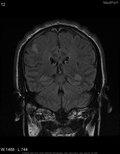
**Imaging Modality:** Magnetic Resonance Imaging (MRI), Coronal plane.
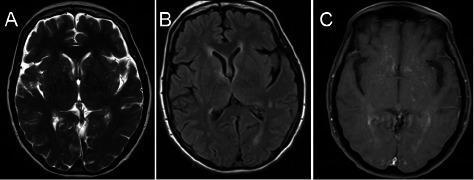
This diagnostic imaging panel presents three axial brain MRI sequences (T2-weighted, FLAIR, and post-contrast T1-weighted) illustrating periventricular and parenchymal abnormalities. Panel A (T2-weighted) shows areas of hyperintense signal prolongation in the periventricular white matter, particularly near the anterior and posterior horns of the lateral ventricles. Panel B (FLAIR) highlights these same hyperintensities by suppressing the cerebrospinal fluid (CSF) signal, revealing multifocal white matter involvement. Panel C (post-contrast T1-weighted) demonstrates mild, diffuse enhancement within the periventricular regions and subtle multifocal punctate enhancement throughout the brain parenchyma, indicative of blood-brain barrier disruption. These findings are clinically relevant for the diagnosis of inflammatory or vasculitic processes, such as lymphocytic vasculitis, presenting with progressive neurological symptoms like headaches and gait instability. The imaging is targeted at medical students and radiology residents as a representation of diffuse inflammatory central nervous system disease.
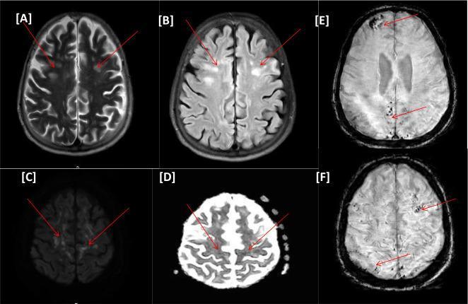
This diagnostic image set consists of six axial brain MRI sequences (A-F) from a clinical case of COVID-19-related encephalopathy. Images A (T2-weighted) and B (FLAIR) demonstrate bilateral, multifocal, hyperintense nodular and punctate lesions within the periventricular and subcortical white matter. Images C (DWI) and D (ADC map) reveal corresponding areas of diffusion restriction, highlighted by red arrows, indicating acute ischemic changes. Images E and F (Susceptibility-Weighted Imaging, SWI) show multiple punctate hypointense foci scattered across the frontal and posterior brain parenchyma, characteristic of susceptibility artifacts from microhemorrhagic changes or microbleeds. This combination of multifocal white matter hyperintensities, restricted diffusion, and microhemorrhages in a COVID-19 patient is highly suggestive of critical illness-associated cerebral microbleeds and leukoencephalopathy. The imaging highlights the neurological complications involving both vascular ischemia and microvascular hemorrhage within the brain parenchyma.
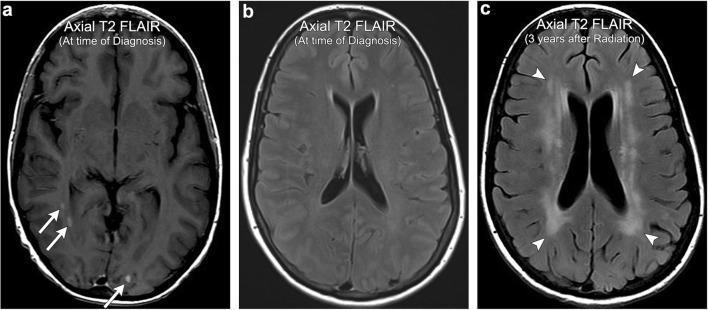
This diagnostic imaging sequence consists of three axial T2-weighted Fluid-Attenuated Inversion Recovery (FLAIR) MRI scans of the brain, illustrating the progression of radiation-induced leukoencephalopathy. Images (a) and (b), labeled 'At time of Diagnosis', show focal, punctate hyperintensities in the subcortical white matter (indicated by arrows), representing metastatic thyroid carcinoma. Image (c), labeled '3 years after Radiation', demonstrates a dramatic interval change following whole-brain radiation therapy (WBRT). There is now extensive, symmetric, and confluent T2 FLAIR hyperintensity within the periventricular white matter (indicated by arrowheads). A hallmark feature visible in image (c) is the relative sparing of the subcortical U-fibers, which distinguishes this condition from other leukoencephalopathies such as Progressive Multifocal Leukoencephalopathy (PML). The imaging findings are consistent with chronic, non-necrotic white matter injury secondary to radiation, characterized by progressive cognitive decline in a clinical context.
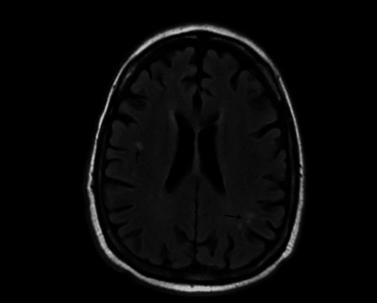
Diagnostic Image: This axial MRI slice of the brain, likely a Fluid-Attenuated Inversion Recovery (FLAIR) or T2-weighted sequence, demonstrates multifocal white matter changes. The primary pathology consists of small, punctate hyperintensities (indicated by black arrows) within the subcortical and deep white matter. Specifically, lesions are visible in the left frontal lobe region near the lateral ventricle's frontal horn and in the posterior left parietal/occipital region. The brain parenchyma shows preserved gray-white matter differentiation with normal sulcal and gyral patterns, and the ventricular system appears non-dilated and symmetric. In the clinical context provided, these non-specific white matter hyperintensities (WMH) are investigated for neuroinflammatory or infectious etiologies, such as neurosyphilis. The image serves as a clinical example of subcortical white matter disease and is suitable for educational modules on neuroradiology, neurology, and infectious disease manifestations in the central nervous system.
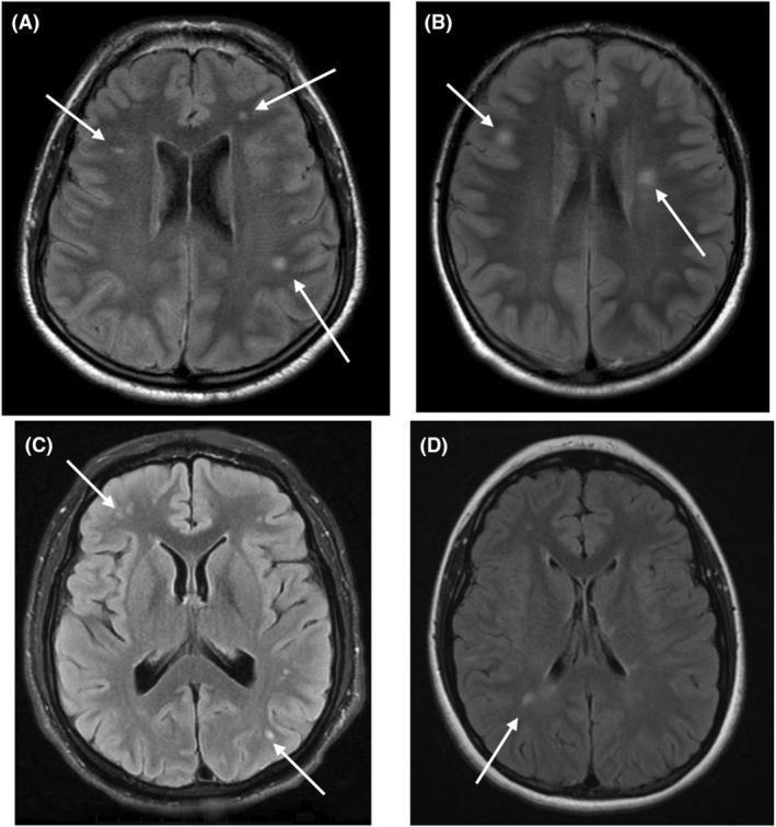
Diagnostic Image: A composite of four axial brain MRI slices using T2-weighted Fluid-Attenuated Inversion Recovery (FLAIR) sequences (A–D). The images demonstrate multifocal areas of signal prolongation (hyperintensity) within the cerebral white matter. White arrows identify specific lesions that vary in size, morphology, and distribution. (A) Multiple punctate and small patch-like lesions are visible in both subcortical and periventricular white matter. (B) Fewer, but larger and more confluent-appearing hyperintense foci are situated near the frontal and posterior horns of the lateral ventricles. (C) Shows a cluster of hyperintensities in the frontal subcortical white matter and a small punctate lesion in the posterior region. (D) Demonstrates a solitary, discrete hyperintense lesion in the deep white matter. These findings illustrate characteristic white matter signal abnormalities associated with Down syndrome-related regression disorder (DSRD). This material is intended for intermediate to advanced medical learners studying neuroradiology and neurodevelopmental pathology.
MPH in MRI Brain: Multifocal Punctate Hyperintensities
What Do They Look Like?
Where Are They Found?
- Periventricular white matter (around the lateral ventricles)
- Deep white matter (centrum semiovale, corona radiata)
- Subcortical white matter (just beneath cortex)
- Less commonly: deep gray matter (basal ganglia, thalamus), brainstem
Causes (Differential Diagnosis)
| Category | Examples |
|---|---|
| Small vessel / vascular disease (most common) | Hypertension, diabetes, smoking, age-related arteriosclerosis |
| Demyelinating disease | Multiple sclerosis (MS), neuromyelitis optica - lesions typically >3 mm, periventricular, ovoid, "Dawson fingers" |
| Migraine | Migraine-associated WMH in young patients |
| Vasculitis / inflammatory | Lupus (SLE), primary CNS vasculitis, sarcoidosis |
| Infectious | Neurosyphilis, Lyme disease, HIV encephalopathy |
| Metabolic / toxic | Vitamin B12 deficiency, radiation-induced leukoencephalopathy |
| Cerebral amyloid angiopathy (CAA) | Lobar distribution, elderly patients |
| CADASIL | Young patients with migraines + stroke + dementia history |
| Microinfarcts | Visible as punctate DWI hyperintensities in acute phase - per Harrison's Principles 22E |
| Incidental / age-related | Very common >60 years, prevalence increases with age |
Clinical Significance
-
Age-related / incidental: In older adults, a few punctate hyperintensities are common and often benign (found in ~1.9% of asymptomatic subjects as an incidental finding - Adams and Victor's Neurology, 12th Ed.).
-
Vascular risk marker: Higher lesion burden is linked to cognitive decline, gait disturbance, depression, and increased stroke risk. Detection should prompt assessment of blood pressure, diabetes, and lipids.
-
MS vs vascular: MS lesions tend to be ovoid, >3 mm, periventricular, juxtacortical, or infratentorial. Pure punctate lesions <3 mm are more often vascular. The McDonald criteria help distinguish.
-
Prognostic: Severity correlates with all-cause mortality, cognitive impairment, and functional decline - particularly in elderly patients.
Key MRI Sequences
| Sequence | Appearance |
|---|---|
| T2 / FLAIR | Bright (hyperintense) - best for detecting WMH |
| T1 | Dark (hypointense) |
| DWI | Acute microinfarcts restrict diffusion (bright DWI / dark ADC) |
| SWI | Microbleeds appear as dark punctate foci |
Clinical Approach When MPH Are Reported
- Correlate with age and clinical presentation
- Assess cardiovascular risk factors (HTN, DM, smoking, hyperlipidemia)
- Consider MS workup if young patient + atypical distribution + symptoms
- Look for associated findings: lacunar infarcts, microbleeds, brain atrophy
- Use Fazekas scale or Age-Related White Matter Changes (ARWMC) scale to grade severity