
How to write a long case after Trabeculectomy surgery done to present in MS opthalmology practical exam Use verified sources
how to write long case trabeculectomy MS ophthalmology practical exam glaucoma
trabeculectomy filtering bleb glaucoma surgery
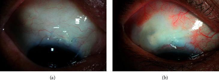
Two side-by-side slit-lamp clinical photographs showing filtering blebs in the superior bulbar conjunctiva following glaucoma surgery (trabeculectomy). Panel (a) displays a post-trabeculectomy bleb with amniotic membrane transplantation (AMT). It exhibits medium height, a horizontal extent of approximately 4 clock-hours, and mild vascularity, characterized by fine, dispersed conjunctival vessels over the elevated area. Panel (b) shows a post-trabeculectomy bleb without AMT. This bleb is also of medium height but has a slightly smaller horizontal extent of approximately 3 clock-hours. It appears cystic and translucent with notable avascularity over the main filtering area, contrasting with the surrounding peripheral conjunctival hypervascularity. These images are used to compare bleb morphology, specifically the Wuerzburg or Moorfields bleb grading parameters, in different surgical techniques for intraocular pressure (IOP) management.
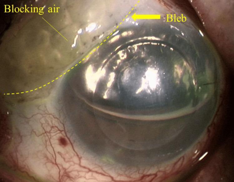
This slit-lamp clinical photograph demonstrates an ophthalmological complication following Descemet's stripping automated endothelial keratoplasty (DSAEK). The image focuses on a superior filtering bleb, previously created during a glaucoma-filtering surgery (trabeculectomy). Visible within the bleb is a phenomenon termed 'blocking air,' where air from the anterior chamber has migrated into the subconjunctival space. The bleb appears elevated and cystic, located at the superior limbal area. A dashed yellow line outlines the extent of the air bubble, which presents as a translucent, irregular area within the superior-left portion of the bleb. The cornea is central and appears slightly hazy, characteristic of early postoperative DSAEK. The underlying anterior chamber shows a visible fluid-air interface. The inferior conjunctiva and sclera exhibit moderate injection with prominent vascularization. This image illustrates the interaction between corneal transplant surgery and pre-existing glaucoma drainage structures, specifically highlighting how therapeutic air used to tamponade a corneal graft can unintentionally enter a functional filtering bleb, potentially impacting intraocular pressure and graft stability.
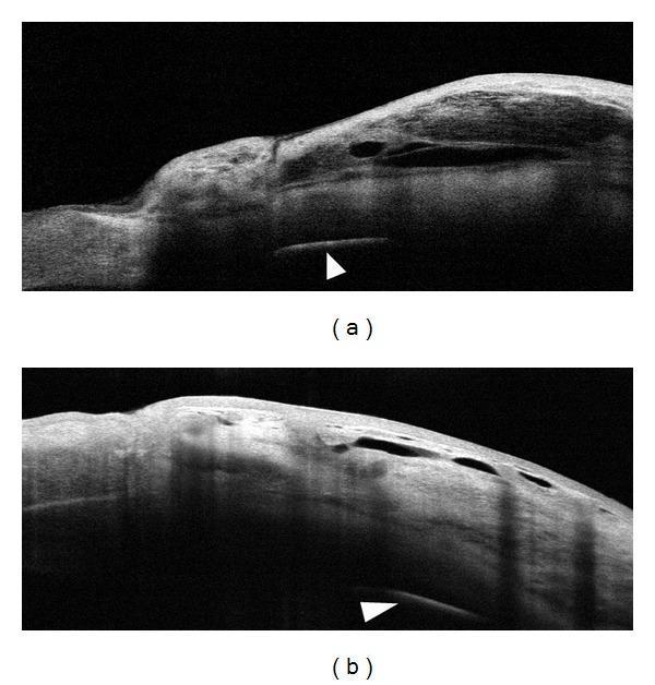
This Comparison Chart features two Anterior Segment Optical Coherence Tomography (AS-OCT) cross-sectional scans (labeled a and b) illustrating filtering bleb morphology following glaucoma surgery. The diagnostic images demonstrate the subconjunctival space and scleral layers three months post-trabeculectomy. Image (a) depicts a filtering bleb where a biodegradable collagen matrix (Ologen) was implanted, showing a slightly thicker bleb wall compared to image (b), which represents a standard trabeculectomy without an implant. Both scans reveal characteristic optically empty cystic spaces within the bleb wall, representing aqueous humor drainage pathways. White arrowheads in both images point to the scleral tissue and the interface between the conjunctival-episcleral layer and the deeper sclera, exhibiting relatively homogeneous reflectivity and similar tissue density across both cases. This clinical imaging is essential for ophthalmology students and clinicians to evaluate bleb functionality, wall thickness, and the presence of internal fluid-filled cavities, which are key indicators of surgical success in intraocular pressure management.
How to Write a Long Case After Trabeculectomy - MS Ophthalmology Practical Exam
Overview: What a Long Case Tests
- A thorough, structured history
- Systematic examination findings with clinical interpretation
- Understanding of the procedure done and why
- Recognition of the current status of the bleb and IOP control
- Knowledge of complications and their management
- A follow-up and management plan
SECTION 1 - HISTORY
1a. Presenting Complaint
- Gradual diminution of vision (usually peripheral field loss)
- Headache/halos (if angle closure component)
- Incidentally detected elevated IOP on routine examination
- Pain (acute angle closure)
1b. History of Present Illness
- Duration of known glaucoma
- Type of glaucoma (POAG, PACG, secondary, congenital)
- IOP readings before surgery (highest recorded IOP, target IOP)
- Medications tried before surgery - list each drop, duration used, reasons for stopping
- Beta-blockers (Timolol), Prostaglandin analogues (Latanoprost), CAIs (Dorzolamide), Alpha-2 agonists (Brimonidine)
- Laser procedures before surgery (ALT, SLT, LPI)
- Previous optic disc status and visual field stage
- Date of trabeculectomy
- Which eye was operated on
- Use of antimetabolites: Mitomycin C (MMC 0.2-0.4 mg/ml) or 5-FU (ask the patient or refer to records)
- Type of conjunctival flap: fornix-based or limbal-based
- Postoperative medications prescribed (steroid drops, atropine, antibiotics)
- Number of follow-up visits and any postoperative manipulations:
- Suture lysis (laser suture lysis)
- Releasable suture removal
- Bleb needling
- 5-FU subconjunctival injections
- Bleb massage
- Current IOP (controlled vs. not)
- Current medications (on drops or medication-free)
- Any drop in vision after surgery
- Symptoms: discharge, pain, redness, photophobia (risk of blebitis/endophthalmitis)
- Blurred vision (hypotony maculopathy, cataract, refractive shift)
1c. Past Ocular History
- Previous eye surgery (phacoemulsification, vitreoretinal surgery, corneal surgery)
- Ocular trauma
- Uveitis, diabetic retinopathy, retinal vein occlusion
1d. Past Medical History
- Diabetes mellitus, hypertension (relevant to glaucoma risk and healing)
- Thyroid disease (dysthyroid optic neuropathy DDx)
- Blood dyscrasias, anticoagulant use (bleeding risk during surgery)
- Autoimmune disease (ocular surface affects bleb health)
1e. Drug History
- Current eye drops and systemic medications
- Allergy to any eye drops (especially sulfonamides - Dorzolamide)
1f. Family History
- First-degree relatives with glaucoma (strong risk factor for POAG)
1g. Social History
- Monocularity (if opposite eye is blind, management decisions change)
- Occupation (legal driving visual field requirements)
SECTION 2 - EXAMINATION
2a. General Examination
- Visual acuity (distance and near, each eye, with and without correction)
- Pupils: RAPD (relative afferent pupillary defect) - present in advanced glaucoma with significant asymmetric optic nerve damage
- Method: Swinging flashlight test
- RAPD is a very important positive finding - document carefully
- Extraocular movements (rule out thyroid/orbital pathology)
- Periorbital examination
2b. Slit-Lamp Examination (Anterior Segment)
| Feature | What to Look For |
|---|---|
| Location | Superior (standard) or other |
| Height | Flat, low, moderate, high |
| Extent | Number of clock hours |
| Vascularity | Avascular (high risk - MMC bleb), normal, hyperaemic |
| Wall thickness | Thin-walled (risk of leak), moderate, thick |
| Microcysts | Present = good, functioning bleb |
| Encapsulation | Domed, firm, Tenon's cyst = failure |
| Leakage | Perform Seidel test |
- Instill fluorescein 2% and examine at slit lamp with cobalt blue filter
- Positive Seidel = fluorescein washed green/yellow from bleb = aqueous leak
- Negative = no leak
- Clarity, any epithelial defects adjacent to bleb (bleb dysaesthesia)
- Peripheral anterior synechiae (PAS) at the angle
- Surgical scar/paracentesis site (usually temporal)
- Depth: deep, shallow, flat (compare operated vs. fellow eye)
- Cells/flare (inflammation)
- Peripheral iridectomy: note whether patent (whitish, oval gap in peripheral iris, usually at 12 o'clock). Test patency - shine a slit beam and see if light transilluminates through it.
- Surgical iridectomy site
- Iris bombé (if PI not patent - pupillary block complication)
- Posterior synechiae
- Nuclear sclerosis/cortical cataract (common post-trabeculectomy complication)
- Posterior subcapsular cataract (from steroid use)
- Presence of IOL (pseudophakic)
2c. IOP Measurement (Goldman Applanation Tonometry)
- Record IOP of both eyes
- Mention the target IOP set for this patient (typically 12-15 mmHg in advanced disease)
- Compare with pre-operative IOP
- IOP < 5 mmHg = hypotony (risk of maculopathy, choroidal detachment)
- IOP 6-18 mmHg = well controlled (on or off drops)
- IOP > 21 mmHg = failure of filtration
2d. Gonioscopy
- Mention it even if not done in the exam - it is part of a complete glaucoma work-up
- Look for: angle grade (Shaffer grading), PAS, goniotomy changes, internal ostium of sclerostomy
2e. Fundus Examination (MOST IMPORTANT for grading severity)
- Cup-to-disc ratio (CDR): Vertical CDR (VCDR) is the most important - normal < 0.5, glaucomatous > 0.7
- Neuroretinal rim: ISNT rule - Inferior > Superior > Nasal > Temporal (normal). Glaucoma causes rim loss, especially inferior and superior
- Disc haemorrhages: Drance haemorrhages - splinter haemorrhages at disc margin = sign of progressive glaucoma
- RNFL defects: Arcuate nerve fibre layer defects
- Peripapillary atrophy (PPA): Zone alpha and beta PPA (beta zone = progressive glaucoma)
- Asymmetry: VCDR asymmetry > 0.2 between eyes is significant
2f. Visual Field Assessment
- Humphrey Visual Field (HVF) or Goldmann Perimetry
- Document pattern: arcuate scotoma, nasal step, paracentral scotoma, tunnel vision, central island
- Visual field staging (Hodapp-Parrish-Anderson criteria or modified):
- Mild: MD better than -6 dB
- Moderate: MD -6 to -12 dB
- Severe/Advanced: MD worse than -12 dB
- Compare pre-operative and post-operative fields to assess if progression has halted
2g. Special Investigations (to mention/discuss)
- OCT (Optical Coherence Tomography): RNFL thickness map, ganglion cell analysis - objectively documents optic nerve damage and monitors progression
- Corneal pachymetry: Central corneal thickness affects IOP measurement (thin cornea = underestimation of true IOP)
- Anterior segment OCT (AS-OCT): Bleb morphology assessment - internal reflectivity, fluid channels, wall thickness
- Optic disc photography: Baseline documentation for progressive comparison
SECTION 3 - DIAGNOSIS AND CLASSIFICATION
SECTION 4 - COMPLICATIONS TO DISCUSS (examiner favourite)
Early Postoperative Complications:
| Complication | Key Signs | Management |
|---|---|---|
| Hypotony | IOP < 5, shallow AC, choroidal detachment | Reduce topical steroids, atropine, pressure patching; surgical if severe |
| Flat AC | Shallow/flat AC post-op | Depends on cause (see below) |
| Overfiltration | Low IOP, high bleb, shallow AC | Pressure patching, aqueous suppressants |
| Pupillary block | High IOP, flat bleb, iris bombé, non-patent PI | Nd:YAG laser PI |
| Malignant glaucoma | High IOP, flat AC, absent bleb, myopic shift | Atropine + phenylephrine, Nd:YAG anterior hyaloid disruption, PPV |
| Choroidal detachment | Smooth peripheral brown elevation | Cycloplegia; surgical drainage if "kissing" |
| Hyphema | Blood in AC | Usually self-resolving |
| Wound leak | Positive Seidel | Aqueous suppressants; surgical repair if large |
Late Postoperative Complications:
| Complication | Key Signs | Management |
|---|---|---|
| Bleb failure/encapsulation | High IOP, vascular domed bleb (Tenon cyst) | 5-FU injections, bleb needling with MMC |
| Late bleb leak | Low IOP, avascular cystic bleb, Seidel positive | Autologous blood injection, compression sutures, surgical revision |
| Bleb dysaesthesia | Ocular discomfort, small bubbles adjacent to bleb | Lubricants; surgical revision |
| Blebitis/Endophthalmitis | Purulent discharge, hypopyon, low vision | Intravitreal antibiotics (Vancomycin + Ceftazidime), vitreoretinal surgery |
| Cataract | Progressive lens opacity | Phacoemulsification (with caution - risk of bleb failure) |
| Glaucoma progression | Visual field worsening despite IOP control | Reassess target IOP; re-operate; neuroprotection |
| Hypotony maculopathy | Chorioretinal folds at macula, reduced VA | Treat underlying hypotony |
SECTION 5 - MANAGEMENT AND FOLLOW-UP
Current Management:
- Is the IOP at target? If yes - continue monitoring
- Any residual drops being used? (some patients need drops even after surgery)
- Any recent bleb intervention needed?
Follow-up Plan:
- 1st month: Weekly visits (monitor IOP, bleb, suture lysis if needed)
- 2nd-3rd month: Fortnightly
- 3rd-6th month: Monthly
- Beyond 6 months: Every 3-6 months if stable
- Annual: Visual field testing, optic disc photography, OCT RNFL
Examiner Discussion Points:
- Why was trabeculectomy chosen over drops? - failed medical therapy, progressive disease, advanced glaucoma requiring very low target IOP, patient compliance issues
- Role of MMC - anti-scarring agent, reduces fibroblast proliferation, used in high-risk blebs (young patient, prior surgery, uveitis, aphakia)
- Success criteria - IOP < 18 mmHg with > 20% reduction AND no additional medication (complete success); or with medications (qualified success)
- What is a functioning bleb? - slightly elevated, avascular or mildly vascular, microcysts visible, Seidel negative, IOP controlled
- When would you re-operate? - failed bleb with uncontrolled IOP, options: repeat trabeculectomy, drainage implant (Baerveldt, Ahmed), cyclodestructive procedures
SECTION 6 - SAMPLE PRESENTATION TEMPLATE
"I examined Mr./Ms. [Age], [Sex], who presented with a [duration] history of [symptoms]. He/She was previously diagnosed with [type of glaucoma] in [eye]. Despite maximum tolerated medical therapy (listing drops), the patient underwent trabeculectomy with/without MMC to the [right/left] eye on [date].
On examination, visual acuity was [VA] in the operated eye. The IOP today is [X] mmHg on [X] drops. Slit-lamp examination revealed a [describe bleb - height, vascularity, microcysts, extent]. The peripheral iridectomy is patent/non-patent. The anterior chamber is of [depth]. The lens shows [nuclear sclerosis/PSC/IOL]. The Seidel test is negative/positive.
Fundus examination of the operated eye shows a cup-to-disc ratio of [X], with inferior/superior neuroretinal rim thinning, and [other disc findings]. Visual field testing shows [pattern of defect].
My assessment is that this is a post-trabeculectomy patient for [glaucoma type], with a [functioning/failing] bleb, IOP [controlled/uncontrolled], and [mild/moderate/advanced] glaucomatous optic neuropathy. The disease appears [stable/progressive].
The current management is [drops/no drops], and I would recommend [follow-up plan / further intervention]."
Key Tips for the Exam
- Always examine the bleb first when you sit at the slit lamp - examiners notice if you miss it.
- Always do the Seidel test and mention it - even if negative, saying "Seidel test was negative confirming no active leak" demonstrates thoroughness.
- Always check for a peripheral iridectomy - describe its size, location (usually 12 o'clock), and state whether it appears patent.
- RAPD is a high-yield finding - always check and document.
- The CDR and visual field stage must match - if CDR is 0.9 but field is only mildly affected, comment on this discrepancy (pre-perimetric glaucoma or unreliable field test).
- Mention OCT even if not done - it shows you know the investigation.
- Know your IOP numbers: Target IOP in advanced glaucoma = 12 mmHg; post-op hypotony < 5 mmHg; failure > 21 mmHg.
- For the diagnosis line - always include: type of glaucoma + laterality + bleb status + IOP status + disease severity.
- Kanski's Clinical Ophthalmology, 10th Edition, pp. 416-436 (Trabeculectomy, complications, bleb morphology)
- Wills Eye Manual, 8th Edition - POAG workup
- EyeWiki - Trabeculectomy (American Academy of Ophthalmology)